. 2013 Nov;41(21):9753-63.
doi: 10.1093/nar/gkt666. Epub 2013 Aug 20.

miR-146a-5p circuitry uncouples cell proliferation and migration, but not differentiation, in human mesenchymal stem cells

Affiliations

miR-146a-5p circuitry uncouples cell proliferation and migration, but not differentiation, in human mesenchymal stem cells

Jui-Yu Hsieh et al. Nucleic Acids Res. 2013 Nov.

Abstract

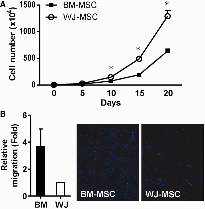
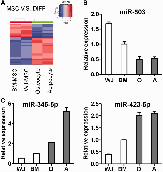
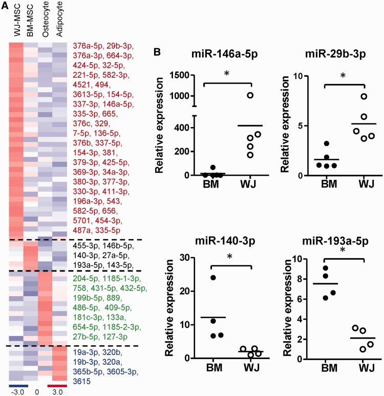
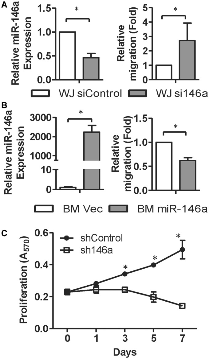
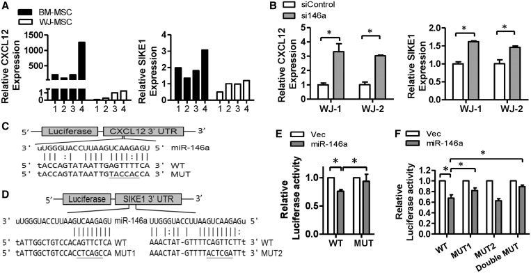
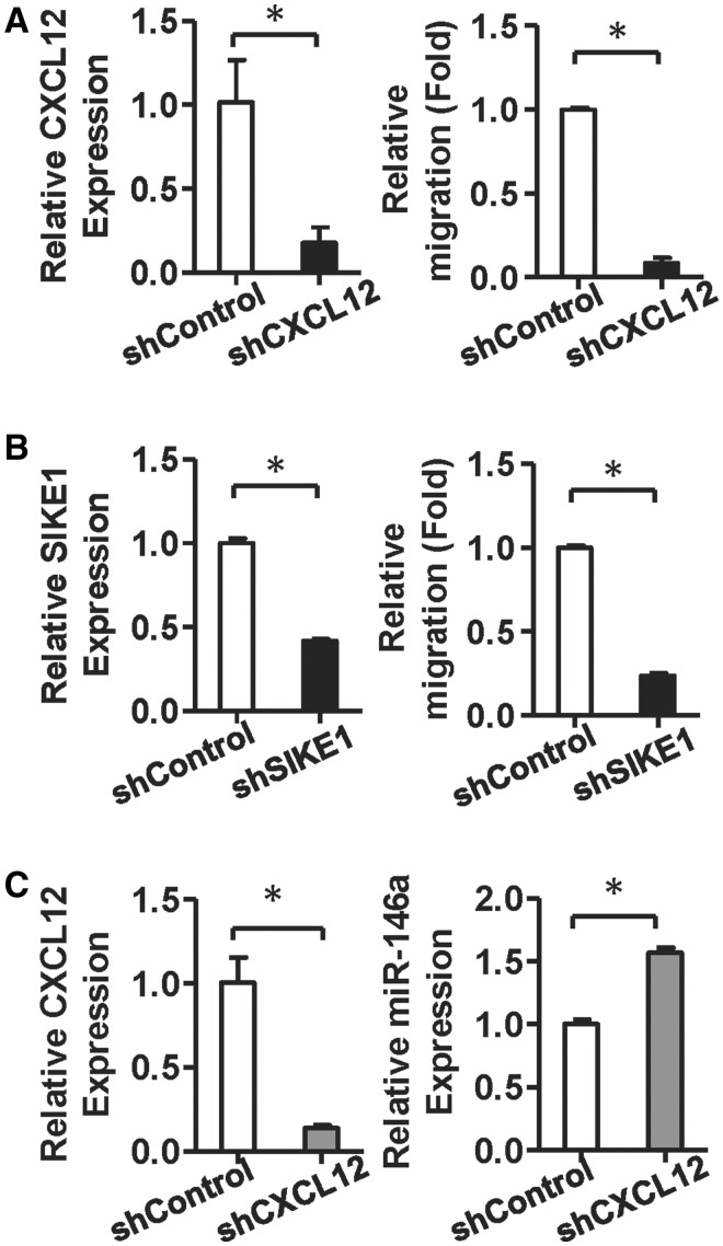
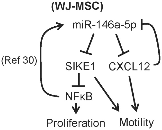
Administration of mesenchymal stem cells (MSCs) has the potential to ameliorate degenerative disorders and to repair damaged tissues. The homing of transplanted MSCs to injured sites is a critical property of engraftment. Our aim was to identify microRNAs involved in controlling MSC proliferation and migration. MSCs can be isolated from bone marrow and umbilical cord Wharton's jelly (BM-MSCs and WJ-MSCs, respectively), and WJ-MSCs show poorer motility yet have a better amplification rate compared with BM-MSCs. Small RNA sequencing revealed that miR-146a-5p is significantly overexpressed and has high abundance in WJ-MSCs. Knockdown of miR-146a-5p in WJ-MSCs inhibited their proliferation yet enhanced their migration, whereas overexpression of miR-146a-5p in BM-MSCs did not influence their osteogenic and adipogenic potentials. Chemokine (C-X-C motif) ligand 12 (CXCL12), together with SIKE1, which is an I-kappa-B kinase epsilon (IKKε) suppressor, is a direct target of miR-146a-5p in MSCs. Knockdown of miR-146a-5p resulted in the down-regulation of nuclear factor kappa-B (NF-κB) activity, which is highly activated in WJ-MSCs and is known to activate miR-146a-5p promoter. miR-146a-5p is also downstream of CXCL12, and a negative feedback loop is therefore formed in MSCs. These findings suggest that miR-146a-5p is critical to the uncoupling of motility and proliferation of MSCs. Our miRNome data also provide a roadmap for further understanding MSC biology.


Figures

Figure 1.
Figure 1.
Differential proliferative and migratory abilities between BM-MSCs and WJ-MSCs. (A) Proliferative potentials of two MSCs. The total cell numbers of cultured WJ-MSCs and BM-MSCs were counted every 5 days till day 20 after plating. Results are presented as mean ± SD in two duplicates. *P < 0.05 by t-test. (B) Migratory ability of WJ-MSCs and BM-MSCs. Migrated cells were counted after 12 h incubation. Fold change in migration ability is compared against the WJ-MSCs. Representative images of migrated MSCs after staining with Hoechst 33342 are shown. Results are presented as mean ± SD from two independent experiments.
Figure 2.
Figure 2.
miRNA patterns in undifferentiated MSCs and differentiated cells derived from BM-MSCs. (A) A heatmap shows the differential expression of miRNAs among stem cells and differentiated progeny cells. miRNAs differentially expressed between undifferentiated and differentiated cells (q < 0.01) were shown. (B and C) Validation of differential expressed miRNAs by RT-qPCR. Highly expressed miRNAs in undifferentiated MSCs (B) or differentiated cells (C) are shown. O: osteocytes; A: adipocytes.
Figure 3.
Figure 3.
Cell type-specific miRNAs. (A) A heatmap shows the specific expression of miRNAs in each cell type. (B) Validation of miRNA levels in different MSCs by RT-qPCR. Each spot represents an independent donor. Data are shown as mean of at least four independent donors. *P < 0.05 by t-test.
Figure 4.
Figure 4.
miR-146a-5p regulates MSC migration and proliferation. (A and B) miR-146a-5p suppresses MSC motility. WJ-MSCs transfected with siRNAs against GFP or miR-146a-5p (siControl or si146a; A), and BM-MSCs transduced with lentiviruses expressing miR-146a-5p or empty lentivirus vector control (B) were used in Transwell assays. miR-146a-5p levels were detected by RT-qPCR (left panels). Results are shown as mean ± SD from four experiments. *P < 0.05 by t-test. (C) miR-146a-5p controls the proliferation of WJ-MSCs. WJ-MSCs were transduced with lentiviruses encoding a short hairpin against miR-146a-5p (sh146a), and cell proliferation rate was measured at the indicated time points using MTT assays. Results are presented as mean ± SD in two duplicates. *P < 0.05 by t-test.
Figure 5.
Figure 5.
CXCL12 and SIKE1 are direct targets of miR-146a-5p. (A) The relative expression levels of CXCL12 and SIKE1 in both MSC types were detected by RT-qPCR (n = 4). (B) CXCL12 and SIKE1 mRNA levels in WJ-MSC transfected with miR-146a-5p antagomirs were analyzed by RT-qPCR. Results are shown as mean ± SD from two duplicates. *P < 0.05 by t-test. (C and D) Putative miR-146a-5p target sites in the CXCL12-3′ UTR (C) and SIKE1-3′ UTR (D). UTR: untranslated region; WT: wild type; MUT: mutant. (E and F) Luciferase reporter assays using the CXCL12-3′ UTR (E) or the SIKE1-3′ UTR (F) in 293T cells transfected with control (Vec) or miR-146a-5p expression vector. Results are presented as mean ± SD of three independent experiments.
Figure 6.
Figure 6.
Knocking down CXCL12 and SIKE1 inhibits WJ-MSC migratory ability, and CXCL12 silencing increases miR-146a-5p levels. (A and B) CXCL12 and SIKE1 control MSC motility. A total of 5 × 10⁴ WJ-MSCs were transduced with CXCL12 (A) or SIKE1 (B) shRNA and then assessed by Transwell migration assay. Migrated cells were counted after 12 h. Results are shown as mean ± SD from two duplicates. *P < 0.05 by t-test. (C) miR-146a-5p expression in WJ-MSCs after knockdown of CXCL12. WJ-MSCs transduced with CXCL12 shRNA for 48 h were harvested. All results are presented as mean ± SD from two duplicates experiments. *P < 0.05 by t-test.
Figure 7.
Figure 7.
The miR146a-5p gene regulatory pathways involve in WJ-MSC proliferation and motility.

References

    1. Prockop DJ. Marrow stromal cells as stem cells for nonhematopoietic tissues. Science. 1997;276:71–74. - PubMed
    1. Horwitz EM, Prockop DJ, Fitzpatrick LA, Koo WW, Gordon PL, Neel M, Sussman M, Orchard P, Marx JC, Pyeritz RE, et al. Transplantability and therapeutic effects of bone marrow-derived mesenchymal cells in children with osteogenesis imperfecta. Nat. Med. 1999;5:309–313. - PubMed
    1. Kawada H, Fujita J, Kinjo K, Matsuzaki Y, Tsuma M, Miyatake H, Muguruma Y, Tsuboi K, Itabashi Y, Ikeda Y, et al. Nonhematopoietic mesenchymal stem cells can be mobilized and differentiate into cardiomyocytes after myocardial infarction. Blood. 2004;104:3581–3587. - PubMed
    1. Battiwalla M, Hematti P. Mesenchymal stem cells in hematopoietic stem cell transplantation. Cytotherapy. 2009;11:503–515. - PMC - PubMed
    1. Borlongan CV, Glover LE, Tajiri N, Kaneko Y, Freeman TB. The great migration of bone marrow-derived stem cells toward the ischemic brain: therapeutic implications for stroke and other neurological disorders. Prog. Neurobiol. 2011;95:213–228. - PMC - PubMed

Publication types

MeSH terms

Associated data