

Neural stem cells grafts decrease neural apoptosis associated with caspase-7 downregulation and BDNF upregulation in rats following spinal cord hemisection
- PMID: 23963709
- PMCID: PMC11497902
- DOI: 10.1007/s10571-013-9969-9
Neural stem cells grafts decrease neural apoptosis associated with caspase-7 downregulation and BDNF upregulation in rats following spinal cord hemisection
Abstract
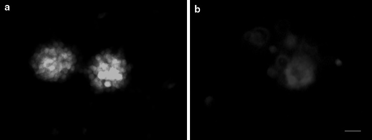
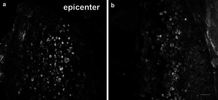
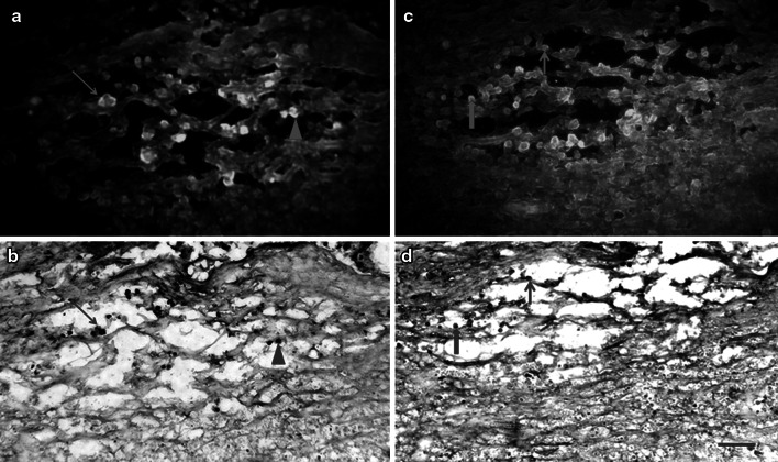
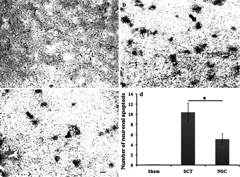
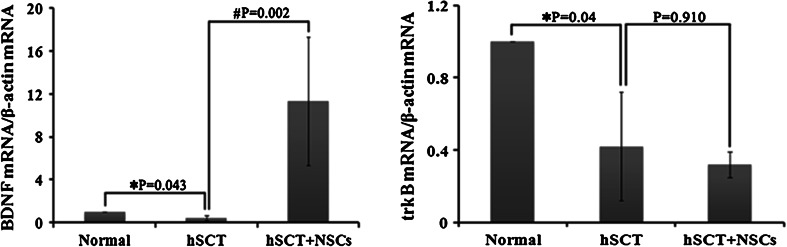
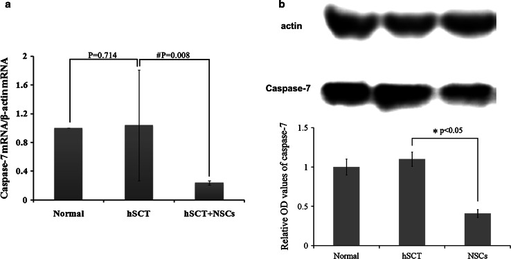
Transplantation of neural stem cells (NSCs) into lesioned spinal cord demonstrated a beneficial effect for neural repair, the underlying mechanism, however, remains to be elusive. Here, we showed that NSCs, possessing the capacity to differentiate toward into neurons and astrocytes, exhibit a neuroprotective effect by anti-apoptosis mechanism in spinal cord hemi-transected rats despite it did not improve behavior. Intravenous NSCs injection substantially upregulated the level of BDNF mRNA but not its receptor TrkB in hemisected spinal cord, while caspase-7, a downstream apoptosis gene of caspase-3, has been largely down-regulated. TUNEL staining showed that the number of apoptosis cells in injured spinal cord decreased significantly, compared with seen in rats with no NSCs administration. The present finding therefore provided crucial evidence to explain neuroprotective effect of NSCs grafts in hemisected spinal cord, which is associated with BDNF upregulation and caspase-7 downregulation.
Conflict of interest statement
We have no conflicts of interest in this study.
Figures

Similar articles
-
Targeted retrograde gene delivery of brain-derived neurotrophic factor suppresses apoptosis of neurons and oligodendroglia after spinal cord injury in rats.Spine (Phila Pa 1976). 2010 Mar 1;35(5):497-504. doi: 10.1097/BRS.0b013e3181b8e89b. Spine (Phila Pa 1976). 2010. PMID: 20190624
-
Neural crest stem cells protect spinal cord neurons from excitotoxic damage and inhibit glial activation by secretion of brain-derived neurotrophic factor.Cell Tissue Res. 2018 Jun;372(3):493-505. doi: 10.1007/s00441-018-2808-z. Epub 2018 Mar 7. Cell Tissue Res. 2018. PMID: 29516218 Free PMC article.
-
Subdural engraftment of serotonergic neurons following spinal hemisection restores spinal serotonin, downregulates serotonin transporter, and increases BDNF tissue content in rat.Brain Res. 2001 Sep 14;913(1):35-46. doi: 10.1016/s0006-8993(01)02749-4. Brain Res. 2001. PMID: 11532245
-
Astrocytes migrate from human neural stem cell grafts and functionally integrate into the injured rat spinal cord.Exp Neurol. 2019 Apr;314:46-57. doi: 10.1016/j.expneurol.2019.01.006. Epub 2019 Jan 15. Exp Neurol. 2019. PMID: 30653967
-
Transplantation of NSCs with OECs alleviates neuropathic pain associated with NGF downregulation in rats following spinal cord injury.Neurosci Lett. 2013 Aug 9;549:103-8. doi: 10.1016/j.neulet.2013.06.005. Epub 2013 Jun 20. Neurosci Lett. 2013. PMID: 23791854
Cited by
-
Effects of brain-derived neurotrophic factor-pretreated neuron stem cell transplantation on Alzheimer's disease model mice.Int J Clin Exp Med. 2015 Nov 15;8(11):21947-55. eCollection 2015. Int J Clin Exp Med. 2015. PMID: 26885166 Free PMC article.
-
Direct lineage conversion of astrocytes to induced neural stem cells or neurons.Neurosci Bull. 2015 Jun;31(3):357-67. doi: 10.1007/s12264-014-1517-1. Epub 2015 Apr 8. Neurosci Bull. 2015. PMID: 25854678 Free PMC article. Review.
-
lncfos/miR-212-5p/CASP7 Axis-Regulated miR-212-5p Protects the Brain Against Ischemic Damage.Mol Neurobiol. 2023 May;60(5):2767-2785. doi: 10.1007/s12035-023-03216-y. Epub 2023 Jan 30. Mol Neurobiol. 2023. PMID: 36715920
-
Co-Transplantation of Human Umbilical Cord Mesenchymal Stem Cells and Human Neural Stem Cells Improves the Outcome in Rats with Spinal Cord Injury.Cell Transplant. 2019 Jul;28(7):893-906. doi: 10.1177/0963689719844525. Epub 2019 Apr 23. Cell Transplant. 2019. PMID: 31012325 Free PMC article.
-
Neural Stem Cell Transplantation Improves Locomotor Function in Spinal Cord Transection Rats Associated with Nerve Regeneration and IGF-1 R Expression.Cell Transplant. 2019 Sep-Oct;28(9-10):1197-1211. doi: 10.1177/0963689719860128. Epub 2019 Jul 4. Cell Transplant. 2019. PMID: 31271053 Free PMC article.
References
-
- Basso DM, Beattie MS, Bresnahan JC (1995) A sensitive and reliable locomotor rating scale for open field testing in rats. J Neurotrauma 12:1–21 - PubMed
-
- Blesch A, Lu P, Tuszynski MH (2002) Neurotrophic factors, gene therapy, and neural stem cells for spinal cord repair. Brain Res Bull 57:833–838 - PubMed
-
- Chen G, Hu YR, Wan H, Xia L, Li JH, Yang F, Qu X, Wang SG, Wang ZC (2010) Functional recovery following traumatic spinal cord injury mediated by a unique polymer scaffold seeded with neural stem cells and Schwann cells. Chin Med J (Engl) 123:2424–2431 - PubMed
-
- Enomoto M, Shinomiya K, Okabe S (2003) Migration and differentiation of neural progenitor cells from two different regions of embryonic central nervous system after transplantation into the intact spinal cord. Eur J Neurosci 17:1223–1232 - PubMed
MeSH terms
Substances
LinkOut - more resources
Full Text Sources
Other Literature Sources
Research Materials
