. 2014 Jan;89(1):1-6.
doi: 10.1002/ajh.23571. Epub 2013 Sep 19.

Vasculopathy, inflammation, and blood flow in leg ulcers of patients with sickle cell anemia

Affiliations

Vasculopathy, inflammation, and blood flow in leg ulcers of patients with sickle cell anemia

Caterina P Minniti et al. Am J Hematol. 2014 Jan.

Abstract

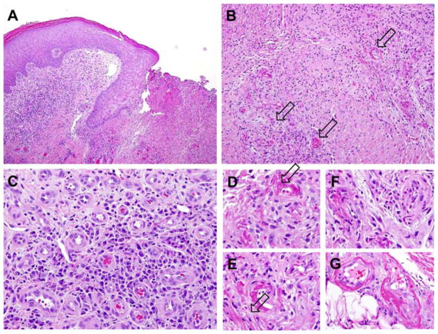
Chronic leg ulcers are frequent and debilitating complications of sickle cell anemia. Inadequate blood supply has been postulated to be an important factor in their occurrence and delayed healing. Little is known about their microcirculatory and histopathological changes. We evaluated the microcirculation of lower extremity ulcers with laser speckle contrast imaging and infrared thermography and obtained clinical and laboratory characteristics in 18 adults with sickle cell anemia and chronic leg ulcers. Skin biopsies were obtained in four subjects. Subjects had markers of severe disease, anemia, high degree of hemolysis, inflammation, and thrombophilia. The highest blood flow was present in the ulcer bed, progressively less in the immediate periwound area, and an unaffected control skin area in the same extremity. Microscopic examination showed evidence of venostasis, inflammation, and vasculopathy. Blood vessels were increased in number, had activated endothelium and evidence of thrombosis/recanalization. High blood flow may be due to chronic inflammation, cutaneous vasodilatation, venostasis, and in situ thrombosis. These changes in skin microcirculation are similar to chronic venous ulcers in the non-sickle cell disease (SCD) population, thus suggesting that leg ulcers may be another end-organ complication with endothelial dysfunction that appears in patients with SCD at a younger age and with higher frequency than in the general population.


Figures

Figure 1
Figure 1
Microvasculature analysis of ulcer area (ROI 1) and surrounding tissue (ROI 2) compared to a distant, nonaffected skin area (ROI 3). (A) Visible light photograph of the imaged ulcer area for a representative subject. Selected regions of interest are indicated with white squares. ROI 1 is the ulcer center, ROI 2 is the peri-wound area, directly adjacent to the ulcer, and ROI 3 is an area >5 cm from ulcer. (B) LSCI image of a representative patient and a graph that represents the average regional blood flows among 11 subjects. There is significantly different blood flow among the three regions of interest, with the highest mean blood flow in the ulcer center (ROI 1) and lowest distant from the ulcer (ROI 3) (n =18, P <0.01; paired Wilcoxon test; P <0.01. (C) Infrared image of the same representative patient. Temperatures as measured by infrared thermography in the ulcer bed (ROI 1), the periwound area (ROI 2) and a distant skin region (ROI 3). Temperature is highest in the periwound area compared to the ulcer bed due to evaporative cooling (P <0.01) and when compared to a distant area (P =0.01). Bar graph indicates mean values, and error bars indicate standard error of the mean, paired Wilcoxon tests.
Figure 2
Figure 2
Microscopic analysis of skin biopsies. Evidence of increase in vascularity, chronic inflammation, vasculopathy with blood vessels occlusion, fibrin deposition in the intima, and microthrombi. Panel A. Scanning magnification view of the skin punch biopsy showing edge of an ulcer from the right ankle of patient MD. The epidermal changes adjacent to the ulcer are characterized by acanthosis, hyperkeratosis, and attenuated rete ridges. There is increased vascularity and inflammation in the dermis. (H&E, 100× original magnification). Panel B. The histologic changes subjacent to the ulcer bed are characterized by chronically inflamed granulation tissue with vasculopathic changes involving some of the small blood vessels. (H&E, 200× original magnification). Panel C. High magnification view of the superficial dermal vessels peripheral to the ulcer show proliferation of thick-walled capillaries and venules, consistent with chronic stasis. There is a lymphoplasmacytic inflammatory infiltrate in the dermis (H&E, 400× original magnification). Panels D, E, F. Very high magnification view of involved vessels subjacent to the ulcer bed reveals eosinophilic fibrin deposits within the vessel wall and partial occlusion of the vascular lumen (H&E, 600× original magnification). Panel G. Scanning magnification view of the skin punch biopsy obtained from the right dorsal foot of patient DD shows vasculopathic changes involving a cluster of small blood vessel in the deep dermis (H&E 40× original magnification). Panel H. High magnification view of the involved vessels reveals eosinophilic fibrin deposits within the vessel wall associated with intimal hyperplasia and narrowing of the vascular lumen (H&E, 400× original magnification).

References

    1. Minniti CP, Taylor JGt, Hildesheim M, et al. Laboratory and echocardiography markers in sickle cell patients with leg ulcers. Am J Hematol. 2011;86:705–708. - PMC - PubMed
    1. Eckman JR. Leg ulcers in sickle cell disease. Hematol Oncol Clin North Am. 1996;10:1333–1344. - PubMed
    1. Borgna-Pignatti C, Marsella M, Zanforlin N. The natural history of thalassemia intermedia. Ann N Y Acad Sci. 2010;1202:214–220. - PubMed
    1. Giraldi S, Abbage KT, Marinoni LP, et al. Leg ulcer in hereditary spherocytosis. Pediatr Dermatol. 2003;20:427–428. - PubMed
    1. Parent F, Bachir D, Inamo J, et al. A hemodynamic study of pulmonary hypertension in sickle cell disease. N Engl J Med. 2011;365:44–53. - PubMed

Publication types