. 2013 Aug;25(8):2831-47.
doi: 10.1105/tpc.113.115287. Epub 2013 Aug 20.

The Arabidopsis metacaspase9 degradome

Affiliations

The Arabidopsis metacaspase9 degradome

Liana Tsiatsiani et al. Plant Cell. 2013 Aug.

Abstract

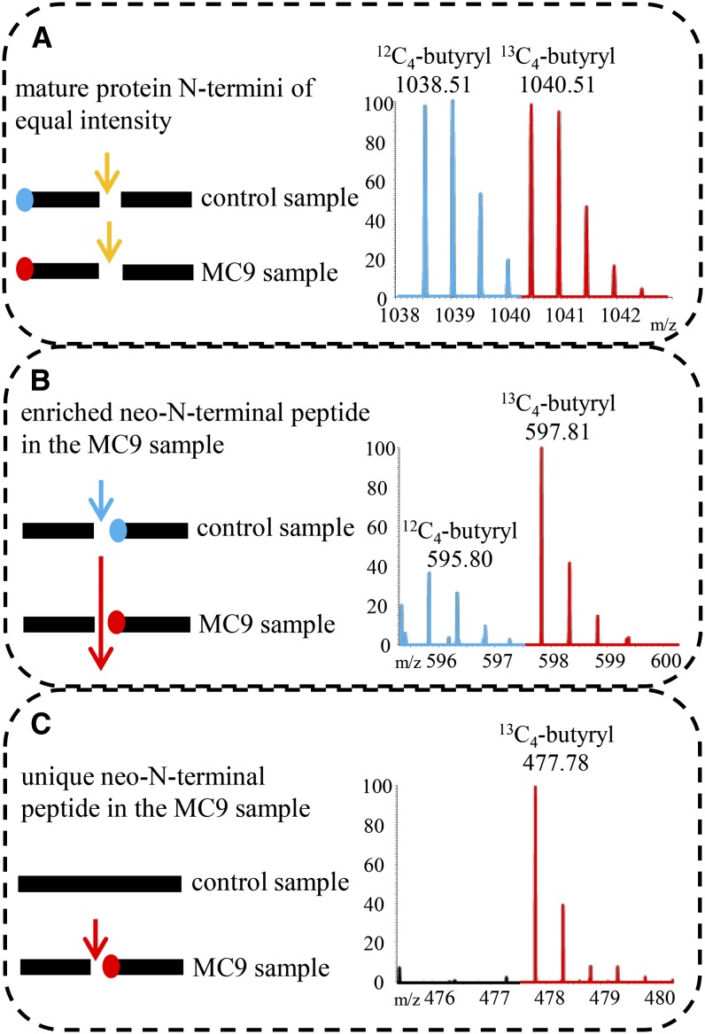
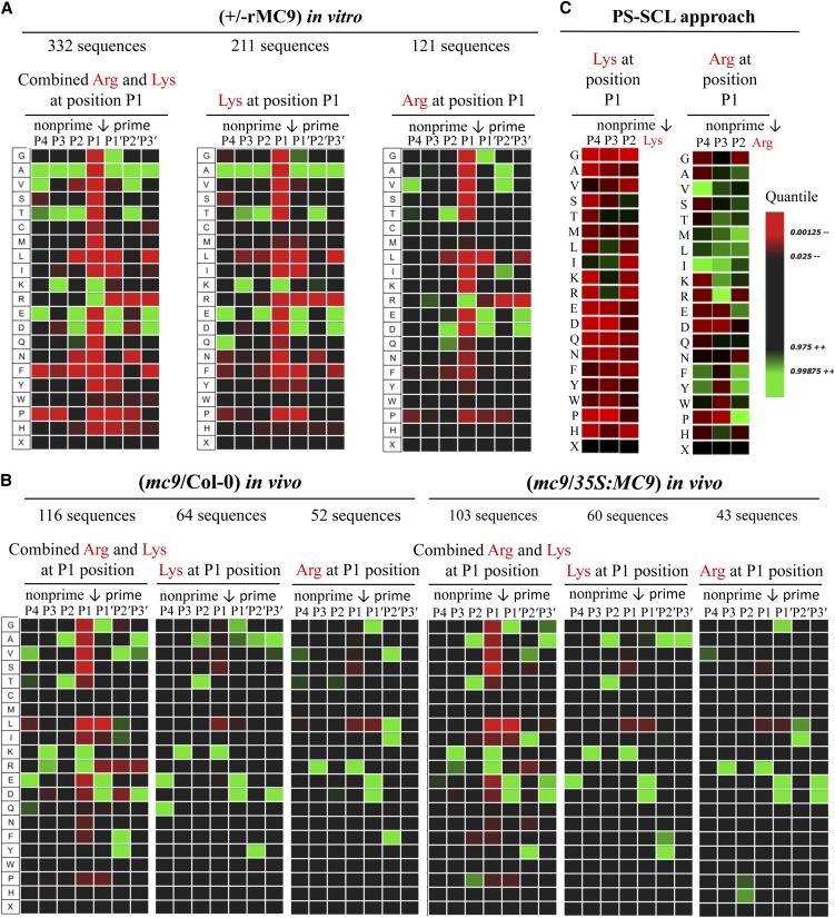
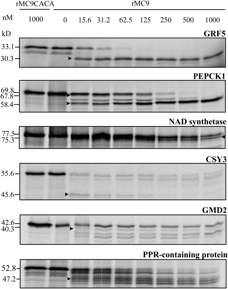
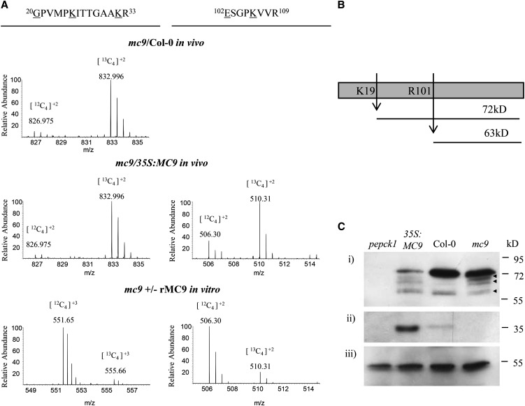
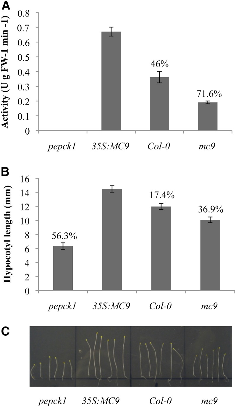
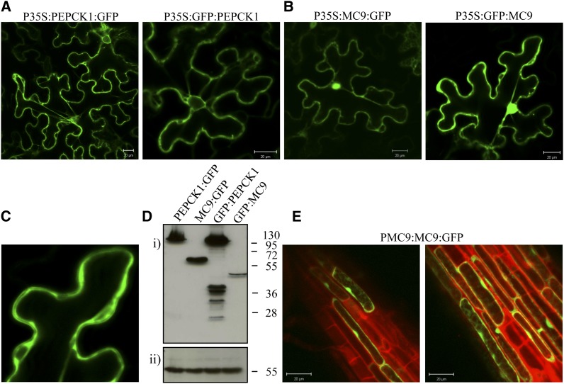
Metacaspases are distant relatives of the metazoan caspases, found in plants, fungi, and protists. However, in contrast with caspases, information about the physiological substrates of metacaspases is still scarce. By means of N-terminal combined fractional diagonal chromatography, the physiological substrates of metacaspase9 (MC9; AT5G04200) were identified in young seedlings of Arabidopsis thaliana on the proteome-wide level, providing additional insight into MC9 cleavage specificity and revealing a previously unknown preference for acidic residues at the substrate prime site position P1'. The functionalities of the identified MC9 substrates hinted at metacaspase functions other than those related to cell death. These results allowed us to resolve the substrate specificity of MC9 in more detail and indicated that the activity of phosphoenolpyruvate carboxykinase 1 (AT4G37870), a key enzyme in gluconeogenesis, is enhanced upon MC9-dependent proteolysis.


Figures

Figure 1.
Figure 1.
Expression Characteristics of the MC9 Gene and Molecular Characterization of the MC9 Transgenic Lines Used in This Study. (A) to (D) GUS staining of root cap of 2-d-old seedlings (A), root cap of 1-week-old seedling (B), tracheary elements in expanding cotyledon (C), and petals prior to abscission (D). (E) Schematic representation of the T-DNA insertion of the GK-540H0 allele. The catalytic His and Cys, the p20 and p10 MC9 protease subunits, and the linker region between the two catalytic subunits are depicted. (F) Immunoblot analysis of Arabidopsis seedlings' proteins with a MC9 polyclonal antibody. Full-length 35.5-kD MC9 is detected in the wild type (Col-0) and 35S:MC9 overexpressor (Vercammen et al., 2006), but not in the mc9 knockout line (GK-540H0 allele). An actin antibody directed against a common epitope of all actin isoforms (Immuno, clone C4) was used as gel loading control. (G) Transcript levels of all Arabidopsis metacaspase genes (MC1 to MC9) in 2-d-old seedlings. Values represent the mean of three measurements ± sd relative to the MC9 gene transcript level in the wild type (Col-0).
Figure 2.
Figure 2.
Illustration of the Different Categories of Isolated (Neo)-N-Terminal Peptides. Mass tagging allowed the differential comparison of N-terminomes derived from samples in which MC9 was active (Col-0 or 35S:MC9 or added rMC9) or absent (control sample mc9). MS spectra of N-terminal peptides were, in most cases, identified as doublet ions of light (in blue) and heavy (in red) isotopes. m/z, mass-to-charge ratio. (A) Protein cleaved in both samples to the same extent (i.e., by trypsin; yellow arrow) and fragments bearing the mature protein N terminus. As the amounts of these peptides were approximately equal, no MC9-driven proteolysis could be deduced. (B) Protein cleaved in both samples, but at a higher frequency in the MC9 sample (large red arrow). The neo-N-terminus fragment was generated in both samples, but in significantly higher amounts in the MC9 sample. Proteolysis of the target in the control sample might have occurred by a redundant protease activity (blue arrow). The fragment is identified as a doublet with the highest intensity ion derived from the MC9 sample. (C) Protein solely cleaved by MC9 (red arrow) in the samples of active MC9 proteases. Thereby, a fragment with a neo-N-terminus was generated, later identified as unique in the MC9 N-terminome and, thus, as a singleton ion.
Figure 3.
Figure 3.
MC9 Specificity Monitored via Its Proteome-Wide Substrate Profiling. Analysis was performed with iceLogo (Colaert et al., 2009) after multiple sequence alignment and statistical correction with the natural occurrence of amino acids in Arabidopsis proteins. Heat maps illustrating the amino acid frequencies at the P4-P3′ positions in the MC9 substrates as observed in the in vivo mc9/Col-0 and mc9/35S:MC9 COFRADIC studies (A), the in vitro COFRADIC (B), and the PS-SCL approach (C). R software was used to build the PS-SCL heat maps with the published values (Vercammen et al., 2006). Arrows mark the cleaved scissile bonds.
Figure 4.
Figure 4.
In Vitro–Transcribed and Translated Proteins Incubated with Wild-Type Recombinant MC9 Protease and the Catalytically Inactive Mutant rMC9CACA. Arrowheads indicate the proteolytic fragments with molecular masses matching those identified by COFRADIC.
Figure 5.
Figure 5.
Cleavage of the PEPCK1 Protein as Deduced from the COFRADIC Data and in Vivo Validated by the PEPCK1 Immunodetection in the Wild Type and the MC9 Gain- and Loss-of-Function Mutant Extracts. (A) Mass spectra of the PEPCK1 proteolysis reporter peptides as identified in mc9/Col-0, mc9/35S:MC9, and the in vitro studies. Mass/charge values for each isotope are given on top of each peptide ion. Butyrylated residues are underlined. (B) Schematic representation of PEPCK1 proteolysis by MC9 at the identified cleavage sites and generation of fragments with variable size. (C) Immunodetection of (i) PEPCK1 protein in extracts of 2-d-old seedlings from pepck1, 35S:MC9, Col-0, and mc9, (ii) full-length MC9 zymogen, and (iii) CAT2 (AT4G35090) loading gel control. The PEPCK1 proteolytic fragments of 57 to 68 kD are marked with arrowheads.
Figure 6.
Figure 6.
Involvement of MC9 in Gluconeogenesis. (A) PEPCK activity as measured in Arabidopsis seedling extracts from pepck1, 35S:MC9, Col-0, and mc9. One unit of PEPCK activity corresponded to the production of 1 μmol product per min at 25°C. Values represent the mean of five replicate measurements ±95% confidence interval. Percentages indicate the observed decrease in PEPCK1 activity relative to 35S:MC9 extracts. FW, fresh weight. (B) Hypocotyl length of the same plant lines grown in the dark without Suc. Values represent the mean length of more than 100 plants per line ±95% confidence interval. Percentages indicate the observed decrease in hypocotyl length relative to the 35S:MC9 plants. (C) Dark-grown pepck1, 35S:MC9, Col-0, and mc9 plants without Suc for 10 d. [See online article for color version of this figure.]
Figure 7.
Figure 7.
Subcellular Localization of MC9 and PEPCK1. (A) Localization of PEPCK1 with C-terminal and N-terminal GFP fusions under the control of the cauliflower mosaic virus 35S promoter. Bars = 20 μm. (B) Localization of MC9 in cytosol and nucleus of tobacco leaf cells under the control of the 35S promoter. Bars = 20 μm. (C) Detail of the MC9 cytosolic localization. (D) Immunodetection of MC9 and PEPCK1 via their GFP tag (i) and of catalase 2 (CAT2) as gel loading control (ii). (E) Localization of MC9 in cytosol and nucleus of Arabidopsis root epidermal cells under the control of its native promoter. Bars = 20 μm.

References

    1. Abramoff M., Magalhães P., Ram S. (2004). Image processing with ImageJ. Biophotonics International 11: 36–42
    1. Agard N.J., Mahrus S., Trinidad J.C., Lynn A., Burlingame A.L., Wells J.A. (2012). Global kinetic analysis of proteolysis via quantitative targeted proteomics. Proc. Natl. Acad. Sci. USA 109: 1913–1918 - PMC - PubMed
    1. Ambit A., Fasel N., Coombs G.H., Mottram J.C. (2008). An essential role for the Leishmania major metacaspase in cell cycle progression. Cell Death Differ. 15: 113–122 - PubMed
    1. Aravind L., Dixit V.M., Koonin E.V. (1999). The domains of death: Evolution of the apoptosis machinery. Trends Biochem. Sci. 24: 47–53 - PubMed
    1. Aravind L., Koonin E.V. (2002). Classification of the caspase-hemoglobinase fold: Detection of new families and implications for the origin of the eukaryotic separins. Proteins 46: 355–367 - PubMed

Publication types

MeSH terms

LinkOut - more resources