. 2013 Sep 3;110(36):14765-70.
doi: 10.1073/pnas.1314137110. Epub 2013 Aug 20.

NR2B subunit of the NMDA glutamate receptor regulates appetite in the parabrachial nucleus

Affiliations

NR2B subunit of the NMDA glutamate receptor regulates appetite in the parabrachial nucleus

Qi Wu et al. Proc Natl Acad Sci U S A. .

Erratum in

Abstract

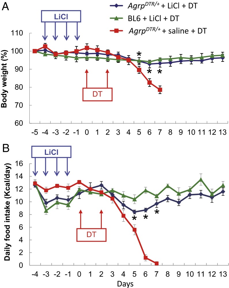
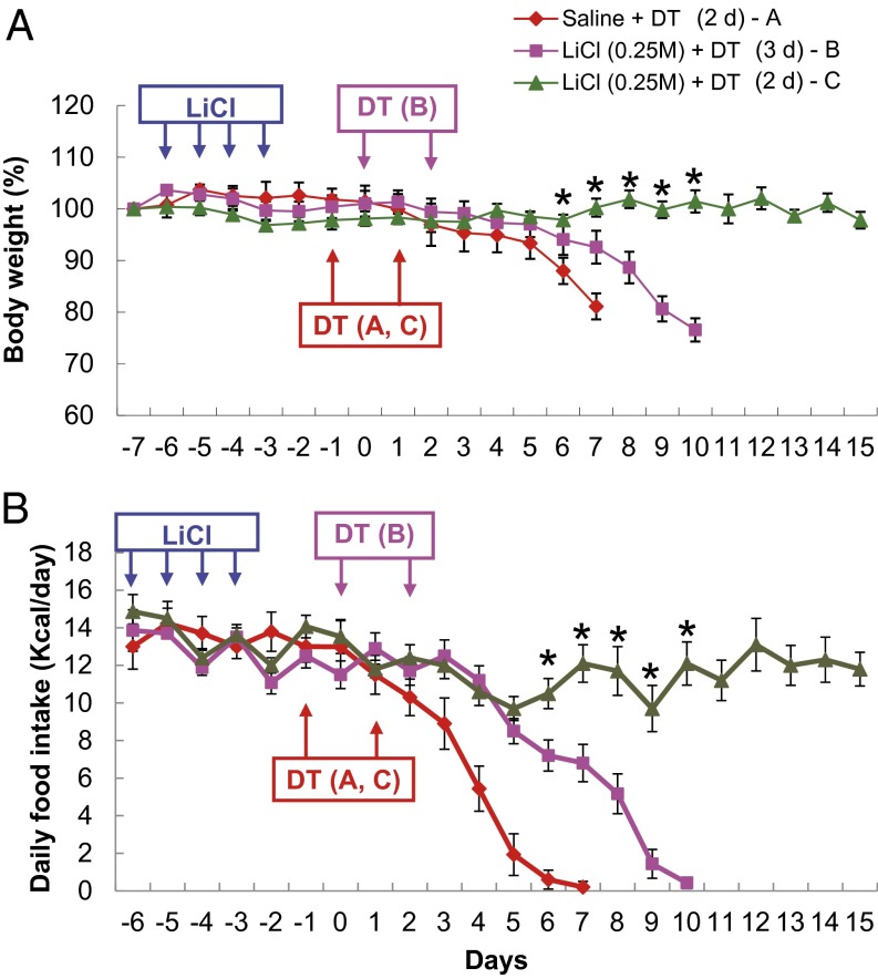
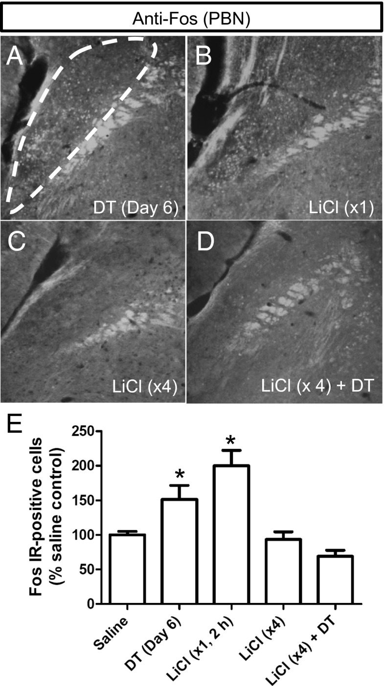
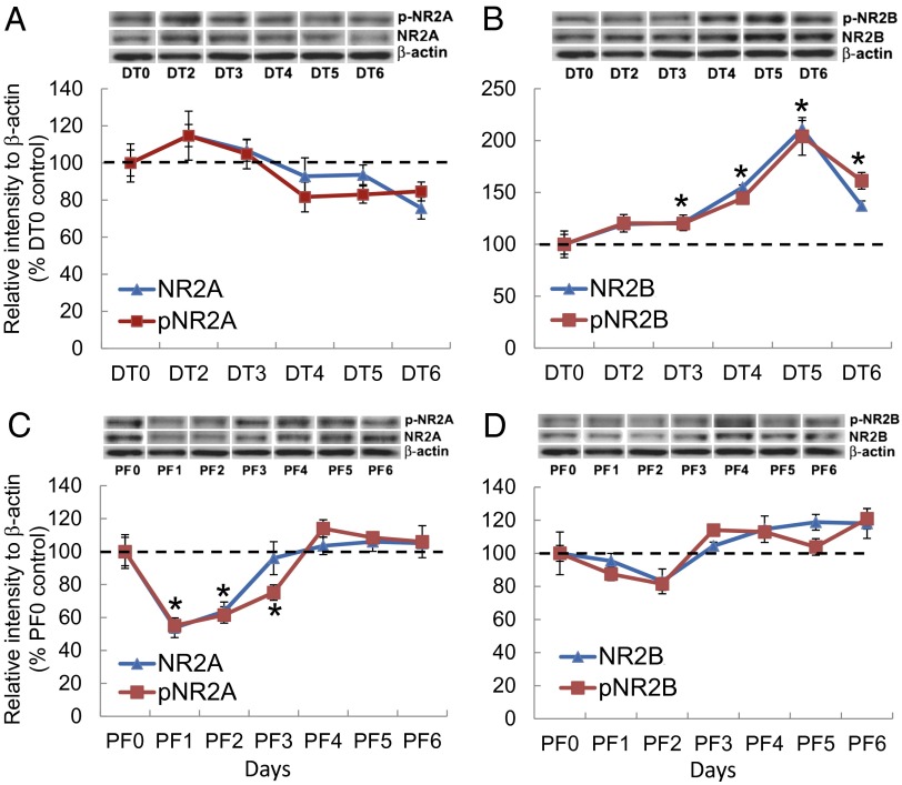
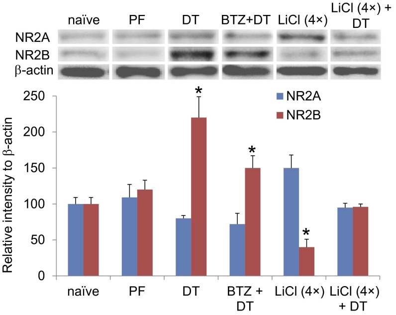
Diphtheria toxin-mediated, acute ablation of hypothalamic neurons expressing agouti-related protein (AgRP) in adult mice leads to anorexia and starvation within 7 d that is caused by hyperactivity of neurons within the parabrachial nucleus (PBN). Because NMDA glutamate receptors are involved in various synaptic plasticity-based behavioral modifications, we hypothesized that modulation of the NR2A and NR2B subunits of the NMDA receptor in PBN neurons could contribute to the anorexia phenotype. We observed by Western blot analyses that ablation of AgRP neurons results in enhanced expression of NR2B along with a modest suppression of NR2A. Interestingly, systemic administration of LiCl in a critical time window before AgRP neuron ablation abolished the anorectic response. LiCl treatment suppressed NR2B levels in the PBN and ameliorated the local Fos induction that is associated with anorexia. This protective role of LiCl on feeding was blunted in vagotomized mice. Chronic infusion of RO25-6981, a selective NR2B inhibitor, into the PBN recapitulated the role of LiCl in maintaining feeding after AgRP neuron ablation. We suggest that the accumulation of NR2B subunits in the PBN contributes to aphagia in response to AgRP neuron ablation and may be involved in other forms of anorexia.

Keywords: NR2B signaling; feeding behavior; nausea.


Conflict of interest statement

The authors declare no conflict of interest.

Figures

Fig. 1.
Fig. 1.
Prolonged administration of LiCl prevents aphagia in AgRP neuron-ablated mice. (A) Body weight of AgrpDTR/+ mice and wild-type (BL6) mice after 4 d of saline or LiCl treatment (0.25 M, 10 µL⋅g−1⋅d−1, i.p.; n = 8 per group). DT-mediated ablation of AgRP neurons was initiated 1 d after LiCl treatment was terminated, and DT was administered i.m. twice as indicated by arrows. (B) Daily food intake was measured for the groups of mice described in A. *P < 0.05 between AgrpDTR/+ mice treated with LiCl and AgrpDTR/+ mice treated with saline for both body weight and food intake; two-way, repeated-measures ANOVA. Results are shown as means ± SEM.
Fig. 2.
Fig. 2.
The effect of LiCl treatment on restoration of appetite in AgRP neuron-ablated mice is transient. (A) Body weight of AgrpDTR/+ mice after 4 d of saline or LiCl treatment (0.25 M, 10 µL⋅g−1⋅d−1, i.p.; n = 6–8 per group). Injection of DT was initiated either 2 d (red line A and green line C traces) or 3 d (purple line B trace) after LiCl treatment was terminated. (B) Daily food intake was measured for the groups of mice described in A. Two-way, repeated-measures ANOVA revealed (i) no effect of LiCl treatment on either body weight or food intake from day −7 to day 0; (ii) a significant effect on both body weight and food intake at days 1–7 in saline-treated and LiCl-treated groups with a 2-d delay before DT treatment (*P < 0.05); and (iii) a significant effect of LiCl on both body weight and food intake from days 1–10 after 2-d vs. 3-d delay in DT treatment (*P < 0.05). Results are shown as means ± SEM.
Fig. 3.
Fig. 3.
Prolonged treatment with LiCl prevents Fos activation in the lateral PBN after ablation of AgRP neurons. (AD) Representative immunohistochemistry images of Fos in neurons from the lateral PBN of AgrpDTR/+ mice (A) 6 d after DT-mediated ablation of AgRP neurons; (B) 2 h after acute LiCl treatment; (C) 4 d after prolonged treatment of LiCl (tissues were collected 2 h after the last LiCl treatment); and (D) 4 d of LiCl treatment followed by DT administration (tissues were collected 6 d after the initiation of DT treatment). (E) Quantified immunoreactivity of Fos+ neurons in the lateral PBN after each of the four treatments. *P < 0.01, (ANOVA); n = 6 mice per group. Results are shown as means ± SEM.
Fig. 4.
Fig. 4.
The NR2B subunit is up-regulated in the PBN neurons after acute ablation of AgRP neurons. (A and C) Western blot analysis showing the progressive change in the relative abundance of total NR2A and pNR2A (Tyr1325) in the PBN of AgrpDTR/+ mice after DT-mediated ablation of AgRP neurons (A) and after pair-feeding (PF) (C). (B and D) Western blot analysis showing the relative abundance of total NR2B and phosphorylated NR2B (Tyr1742) in the PBN of AgrpDTR/+ mice after DT-mediated ablation of AgRP neurons (B) and after pair-feeding (PF) (D). *P < 0.01; ANOVA; n = 5 or 6 mice per group. In all Western blots, β-actin serves as the control. Results are shown as means ± SEM. DT0–DT6, days of DT treatment; PF0–PF6, days of pair-feeding matched to that consumed by the DT-treated group.
Fig. 5.
Fig. 5.
The abundance NR2B in the PBN is attenuated after chronic administration of bretazenil or prolonged treatment with LiCl. Western blot analysis showed the relative abundance of total NR2A and NR2B in the PBN neurons of naive AgrpDTR/+ mice (naive); pair-fed naive mice (PF) and DT-treated AgrpDTR/+ mice (DT); DT-treated AgrpDTR/+ mice treated with chronic infusion of bretazenil (0.3 mg/mL) into the fourth ventricle (BTZ + DT); AgrpDTR/+ mice with prolonged treatment of LiCl [LiCl (4×)]; and AgrpDTR/+ mice with prolonged treatment of LiCl followed by DT [LiCl (4×) + DT]. β-Actin served as the control. *P < 0.01, ANOVA; n = 6–10 mice per group. Results are shown as means ± SEM. Tissue was collected 5 d after initiation of treatment for PF, DT, LiCl (4×) groups; 10 d after initiation of treatment for LiCl (4×)+DT group (5 d after DT); and 14 d after initiation of treatment for BTZ+DT group, i.e. when feeding had been restored.
Fig. 6.
Fig. 6.
Chronic administration of the NR2B subtype-selective antagonist RO25-6981 (RO) into the PBN area prevents aphagia in AgRP neuron-ablated mice. (A) Body weight of AgrpDTR/+ mice after chronic infusion of RO25-6981 or vehicle bilaterally into the PBN (n = 14 per group). DT-mediated ablation of AgRP neurons was initiated 6 d after implantation of minipumps loaded with various doses of RO25-6981 and/or bretazenil (BTZ). (B) Daily food intake was measured for the groups of mice described in A. *P < 0.01; two-way, repeated-measures ANOVA between each of the RO25-6981-treated groups and the vehicle-treated group during days 3–7; n = 6–8 mice per group. Results are shown as means ± SEM.

References

    1. Paoletti P, Neyton J. NMDA receptor subunits: Function and pharmacology. Curr Opin Pharmacol. 2007;7(1):39–47. - PubMed
    1. Traynelis SF, et al. Glutamate receptor ion channels: Structure, regulation, and function. Pharmacol Rev. 2010;62(3):405–496. - PMC - PubMed
    1. Yashiro K, Philpot BD. Regulation of NMDA receptor subunit expression and its implications for LTD, LTP, and metaplasticity. Neuropharmacology. 2008;55(7):1081–1094. - PMC - PubMed
    1. Hung CY, Covasa M, Ritter RC, Burns GA. Hindbrain administration of NMDA receptor antagonist AP-5 increases food intake in the rat. Am J Physiol Regul Integr Comp Physiol. 2006;290(3):R642–R651. - PubMed
    1. Burns GA, Ritter RC. The non-competitive NMDA antagonist MK-801 increases food intake in rats. Pharmacol Biochem Behav. 1997;56(1):145–149. - PubMed

Publication types

MeSH terms

LinkOut - more resources