. 2013 Aug 8:4:93.
doi: 10.3389/fendo.2013.00093. eCollection 2013.

Recent advances in obesity-induced inflammation and insulin resistance

Affiliations

Recent advances in obesity-induced inflammation and insulin resistance

Sanshiro Tateya et al. Front Endocrinol (Lausanne). .

Abstract

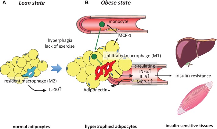
It has been demonstrated in rodents and humans that chronic inflammation characterized by macrophage infiltration occurs mainly in adipose tissue or liver during obesity, in which activation of immune cells is closely associated with insulin sensitivity. Macrophages can be classified as classically activated (M1) macrophages that support microbicidal activity or alternatively activated (M2) macrophages that support allergic and antiparasitic responses. In the context of insulin action, M2 macrophages sustain insulin sensitivity by secreting IL-4 and IL-10, while M1 macrophages induce insulin resistance through the secretion of proinflammatory cytokines, such as TNFα. Polarization of M1/M2 is controlled by various dynamic functions of other immune cells. It has been demonstrated that, in a lean state, TH2 cells, Treg cells, natural killer T cells, or eosinophils contribute to the M2 activation of macrophages by secreting IL-4 or IL-10. In contrast, obesity causes alteration of the constituent immune cells, in which TH1 cells, B cells, neutrophils, or mast cells induce M1 activation of macrophages by the elevated secretion of TNFα and IFNγ. Increased secretion of TNFα and free fatty acids from hypertrophied adipocytes also contributes to the M1 activation of macrophages. Since obesity-induced insulin resistance is established by macrophage infiltration and the activation of immune cells inside tissues, identification of the factors that regulate accumulation and the intracellular signaling cascades that define polarization of M1/M2 would be indispensable. Regulation of these factors would lead to the pharmacological inhibition of obesity-induced insulin resistance. In this review, we introduce molecular mechanisms relevant to the pathophysiology and review the most recent studies of clinical applications targeting chronic inflammation.

Keywords: TNFα; adipose tissue; chronic inflammation; insulin resistance; macrophages; obesity.


Figures

Figure 1
Figure 1
Obesity-induced macrophage infiltration into adipose tissue causes insulin resistance. (A) In adipose tissue in a lean state, most resident macrophages are M2 macrophages that contribute to insulin sensitivity by secreting IL-10. (B) Hyperphagia and lack of exercise cause hypertrophy of adipocytes, which induces MCP-1 secretion to the circulation, leading to the recruitment of circulating monocytes to adipose tissues. These infiltrated monocytes differentiate into activated M1 macrophages, which robustly secrete proinflammatory cytokines such as TNFα, IL-6, and MCP-1, thus contributing to low-grade inflammation in adipose tissue and a decrease of adiponectin. At the same time, these secreted cytokines cause insulin resistance in liver and skeletal muscle by acting as insulin resistance-inducing adipokines.
Figure 2
Figure 2
Accumulation of monocytes/macrophages in adipose tissue and liver, and activation in the tissues. Trafficking: During obesity, adipocytes exhibit hypertrophy, while liver incorporates substantial FFAs, both of which cause tissue inflammation, activation of NF-κB, and AP1 signaling, leading to increased secretion of inflammatory chemokines and cytokines, including CCLs and TNFα. Elevated secretion of CCLs (e.g., MCP-1) elicits the accumulation of CCR-positive monocytes to the site of inflammation, particularly CCR2+ for adipose and liver, but CCR5+ for adipose tissue. In situ activation: In a lean state, resident tissue macrophages display the M2 phenotype, which is achieved and sustained through the JAK/STAT6 pathway in response to IL-4 or IL-13 stimuli. These stimuli are derived from resident TH2 cells, Treg cells, eosinophils, and mast cells. PPARs and KLF4 also induce M2 activation. In turn, obesity and subsequent elevation of tissue FFA or inflammatory cytokines stimulate NF-κB and AP1 signaling, which causes switching of the phenotype to M1, leading to further secretion of TNFα. Signal from inflammasome also activates M1 activation. M1 activation of macrophages can be suppressed by endothelial NO/cGMP signaling. M2 macrophages contribute to insulin sensitivity in neighboring parenchymal cells, while M1 induces insulin resistance, with the M1/M2 balance determining tissue and/or systemic insulin sensitivity.
Figure 3
Figure 3
Control of M1/M2 polarization by neighboring immune cells. In a lean state, resident T cells consist of TH2 cells, Treg cells, and NKT cells. Combined with resident eosinophils, these cells sustain the M2 activation of macrophages through secreting IL-4, IL-10, and IL-13. As obesity progresses, alteration of constituent immune cells occurs, in which the numbers of TH2 cells and Treg cells decline, while in turn, TH1 cells and B cells increase. In addition to these more prevalent cells, neutrophils and mast cells induce M1 activation of macrophages by increased secretion of elastase, TNF, IFNγ, IL-6, and pathogenic IgG. B cells also activate T cells.

References

    1. Ebstein W. Zur therapie des diabetes mellitus, insbesondere uber die anwendeng der salicylauren natron bei demselben. Klin Wochenschr (1876) 13:337–40
    1. Williamson RT. On the treatment of glycosuria and diabetes mellitus with sodium salicylate. Br Med J (1901) 1:760–210.1136/bmj.1.2100.760 - DOI - PMC - PubMed
    1. Reid J, Macdougall AI, Andrews MM. Aspirin and diabetes mellitus. Br Med J (1957) 2:1071–410.1136/bmj.2.5053.1071 - DOI - PMC - PubMed
    1. Reid J, Lightbody TD. The insulin equivalence of salicylate. Br Med J (1959) 1:897–90010.1136/bmj.1.5126.897 - DOI - PMC - PubMed
    1. Hotamisligil GS, Shargill NS, Spiegelman BM. Adipose expression of tumor necrosis factor-alpha: direct role in obesity-linked insulin resistance. Science (1993) 259:87–9110.1126/science.7678183 - DOI - PubMed