. 2013 Aug 6:4:297.
doi: 10.3389/fpls.2013.00297. eCollection 2013.

A tale of two neglected systems-structure and function of the thin- and thick-walled sieve tubes in monocotyledonous leaves

Affiliations

A tale of two neglected systems-structure and function of the thin- and thick-walled sieve tubes in monocotyledonous leaves

C E J Botha. Front Plant Sci. .

Abstract

There is a large body of information relating to the ontogeny, development and the vasculature of eudicotyledonous leaves. However, there is less information available concerning the vascular anatomy of monocotyledonous leaves. This is surprising, given that there are two uniquely different phloem systems present in large groups such as grasses and sedges. Monocotyledonous leaves contain marginal, large, intermediate, and small longitudinal veins that are interconnected by numerous transverse veins. The longitudinal veins contain two metaphloem sieve tube types, which, based upon their ontogeny and position within the phloem, are termed early (thin-walled) and late (thick-walled) sieve tubes. Early metaphloem comprises sieve tubes, companion cells and vascular parenchyma (VP) cells, whilst the late metaphloem, contains thick-walled sieve tubes (TSTs) that lack companion cells. TSTs are generally adjacent to, or no more than one cell removed from the metaxylem. Unlike thin-walled sieve tube (ST) -companion cell complexes, TSTs are connected to parenchyma by pore-plasmodesma units and are generally symplasmically isolated from the STs. This paper addresses key structural and functional differences between thin- and thick-walled sieve tubes and explores the unique advantages of alternate transport strategies that this 5-7 million years old dual system may offer. It would seem that these two systems may enhance, add to, or play a significant role in increasing the efficiency of solute retrieval as well as of assimilate transfer.

Keywords: early and late metaphloem; monocotyledon; thick-walled and thin-walled sieve tubes; vascular bundle structure.


Figures

Figure 1
Figure 1
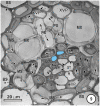
Transmission electron micrograph of a large intermediate vascular bundle in the rice leaf blade surrounded by a parenchymatous bundle sheath (BS). The mestome sheath (MS) is only associated with the phloem pole of this vascular bundle. Metaxylem vessels (MX, red cell walls) are associated with two xylem vascular parenchyma cells (XVP, walls outlined in red and with a pale blue fill). Two late-formed, thick-walled metaphloem sieve tubes (unlabeled arrows and blue fill) abut XVPs. The early metaphloem (outlined in white) contains thin-walled sieve tubes (ST) associated with companion cells (CC) and several parenchyma cells (VP).
Figure 2
Figure 2
Detail of the phloem within a large intermediate vascular bundle in the rice leaf blade. Two thick-walled (TSTs, arrows, blue outline, pale blue fill) and numerous thin-walled (STs) sieve tubes are visible. All STs are associated with companion cells (CC, not all are labeled) and surrounding the ST-CC's are peripherally-located vascular parenchyma (VP), external to which is the bundle sheath (BS). Note that the lower ST are larger than the upper ST.
Figure 3
Figure 3
Detail from a small-intermediate bundle in the rice leaf blade. Pore-plasmodesma units (paired arrowheads, left) connect TST at left, to the VP cell above. The ST (upper right) is connected via PPUs to a companion cell (CC) via PPU (paired arrowheads, upper right) and the two ST (right) are connected via a lateral sieve area (LS). Plasmodesmata (Pd, left) interface VP cells. No connections between thick-and thin-walled sieve tubes are visible in this TEM image.
Figure 4
Figure 4
Detail showing a field containing several thin-walled sieve tubes, associated vascular parenchyma (VP) and companion cells (CC). Thin-walled sieve tubes (ST) are connected to CC by pore-plasmodesmata units (PPU).
Figure 5
Figure 5
Aspects of anatomy and cell associations in a small transverse vein in the leaf blade of Saccharum officinarum (NCO 376). This vein contains one metaxylem vessel (MX) coupled via prominent half-bordered pit pairs (arrowheads) to xylem vascular parenchyma cells (XVP), which, in turn, are symplasmically associated with several vascular parenchyma cells (VP). A solitary sieve tube is next to the MX. Judging by the thin wall it is in all probability an ST.
Figure 6
Figure 6
Aspects of anatomy and cell associations in a transverse section through part of an intermediate vascular bundle (left) from a mature Panicum maximumleaf blade, cut at the level of an emerging transverse vein sieve tube (ST, to right). Based on its position, this is also thin-walled. Two TST (arrowheads) and several ST are visible in the longitudinal bundle.
Figure 7
Figure 7
Aspects of anatomy and cell associations in a transverse vein in a Bromus unioloides leaf. A single sieve tube is adjacent to a solitary xylem vessel, which is in contact with (XVP) that are in contact with the single vessel (XV). As is common in many grasses, the bundle sheath is incomplete, and the BS is interspersed with prominent intercellular spaces.
Figure 8
Figure 8
Aspects of anatomy and cell associations in a small intermediate vein in Eragrostis plana. Part of two metaxylem vessels (MX, extreme left top and right top) are separated from the phloem by XVP. This vein contains a single TST and several ST. Lateral sieve area pores (paired arrowheads) connect the two central STs.
Figure 9
Figure 9
Intermediate vascular bundle in the leaf blade of Mariscus congestus (Cyperaceae). The vascular tissue is surrounded by two sheaths—an outer parenchymatous bundle sheath (BS) and an inner lignified mestome sheath (MS). Note extreme thickening of inner tangential walls of MS cells. Only two thick-walled sieve tubes (pale blue fill), that are separated from the MX by a single row of XVP. MX are associated with smaller protoxylem vessels (PX). STs are associated with companion cells (CC) and are bordered by VP.
Figure 10
Figure 10
Microautoradiographs of small vascular bundles in a Zea mays leaf and portions of transverse veins after 5 min feeding with 14CO2, followed by a 10-min 12CO2 chase showing accumulation of the 14C label in thick- and thin-walled sieve tubes. Label has accumulated in phloem cells as well as being transferred to an emergent thick-walled transverse vein sieve tube to the right, which enters the small bundle at the level of the thick-walled sieve tube.
Figure 11
Figure 11
Microautoradiographs of small vascular bundles in a Zea mays leaf and portions of transverse veins after 5 min feeding with 14CO2, followed by a 10-min 12CO2 chase showing accumulation of the 14C label in thick- and thin-walled sieve tubes. The sieve tube of transverse vein enters the bundle at the level of the thin-walled sieve tubes (STs). Label has accumulated in STs and has been exported to a transverse vein ST. b, bundle-sheath cell; m, mesophyll cell; s, thin-walled sieve tube; v, vessel; unlabeled arrows point to thick-walled sieve tubes; bars = 16μm. Adapted and used with permission of the publishers (Springer; License No 2795360987331) from Fritz et al. (1983). Labeling as in original paper and for details, see Fritz et al. (1983).
Figure 12
Figure 12
Diagram redrawn and interpreted from Figure 10, showing a thick-walled sieve tube (blue fill, solid dot) connection to a transverse vein TST (left). The emergences of a transverse vein ST from its longitudinal vein connection. XVPs (with half-bordered pit-pairs) about the xylem (MX) and vascular parenchyma (VP) are in association with the TSTs.
Figure 13
Figure 13
Diagrams redrawn and interpreted from Figure 11, showing a thin-walled sieve tube (S) connection to a transverse vein ST. The emergences of a transverse vein ST from its longitudinal vein connection. XVPs (with half-bordered pit-pairs) about the xylem (MX) and vascular parenchyma (VP) are in association with the TSTs.
Figure 14
Figure 14
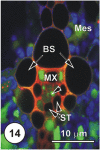
LSCM imaged transections through a small (Figure 14), an intermediate (Figure 15) and a large (Figure 16) longitudinal bundles, showing distribution of 5,6-CF, after uptake of 5,6-CFDA (green fluorescence) co-transported with propidium iodide (red fluorescence; PI stains lignin and cellulosic walls) after 45 min uptake and 6 cm above the cut end of a rice leaf blade [full methodology in Botha et al. (2008)]. Chloroplast- containing mesophyll cells (Mes) have been false colored to blue to prevent interference with propidium iodide or the 5,6-CF dyes. Serial sections were cut directly into silicone oil and sections were covered with coverslips prior to examination using a Leica SP2 LCSM. Propidium iodide selectively stained lignified walls (red fluorescence). Small vein, showing that 5,6-CF had been offloaded into xylem parenchyma, either side of the solitary metaxylem vessel (MX) Note that the two thin-walled sieve tubes (ST, abaxial) contain no evidence of the probe, but thick-walled sieve tubes (dart below the metaxylem vessels). There is no evidence that 5,6-CF has offloaded to the bundle sheath (BS), but it is evident as well as in mesophyll (Mes), indicating symplasmic transport outwards into the mesophyll.
Figure 15
Figure 15
LSCM imaged transections through a small (Figure 14), an intermediate (Figure 15) and a large (Figure 16) longitudinal bundles, showing distribution of 5,6-CF, after uptake of 5,6-CFDA (green fluorescence) co-transported with propidium iodide (red fluorescence; PI stains lignin and cellulosic walls) after 45 min uptake and 6 cm above the cut end of a rice leaf blade [full methodology in Botha et al. (2008)]. Again, there is no evidence of 5,6-CF in ST, but TST and concomitant VP contain the green fluorescence (arrowheads) 5,6-CF is also present in mesophyll cells (Mes).
Figure 16
Figure 16
LSCM imaged transections through a small (Figure 14), an intermediate (Figure 15) and a large (Figure 16) longitudinal bundles, showing distribution of 5,6-CF, after uptake of 5,6-CFDA (green fluorescence) co-transported with propidium iodide (red fluorescence; PI stains lignin and cellulosic walls) after 45 min uptake and 6 cm above the cut end of a rice leaf blade [full methodology in Botha et al. (2008)]. Vascular parenchyma associated with the protoxylem (PX) contains 5,6-CF, as do cells immediately below the MX, where VP and TST contain 5,6-CF (green fluorescence). The ST and below these, large diameter early ST (arrowheads) contain no 5,6-CF. Note that some 5,6-CF is present in the hypodermal fibers associated with this vascular bundle. As is common in grasses, vascular bundles are surrounded by a bundle sheath (BS).
Figure 17
Figure 17
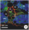
Transection of a small intermediate transverse vein in a rice leaf, showing the distribution of 5,6-CF after uptake of 5,6-CFDA in the transpiration stream. 5,6-CFDA was co-transported with a weak solution of propidium iodide (red fluorescence) via the xylem for 30 min. An emergent thick-walled sieve tube (paired darts), together with two longitudinal TST (arrows), contains 5,6-CF (green fluorescence). Thin walled sieve tubes (asterisks) do not contain 5,6-CF. Note that some 5,6-CF has transported symplasmically, through the bundle sheath (B) to the mesophyll (Mes). Preparation as per Figures 14–16.
Figure 18
Figure 18
Plasmodesmograms showing the distribution of plasmodesmata, expressed as percent plasmodesmata/μm cell-wall interface for the C3 grass, Bromus unioloidesand the C4 grass Panicum maximum expressed as percent plasmodesmata/μm vein. B. unioloides has a double sheath—an outer parenchyma sheath (PS) and an inner thick-walled mestome sheath (MS). In Panicum maximum the Kranz mesophyll sheath (KMS) surrounds the inner bundle sheath (BS). Note that thick-walled sieve tubes (TSTs, solid dots) do not have connections to the ST-CC (open circles) complex. STs and TSTs have low frequency connections to vascular parenchyma cells (VP). Redrawn from Botha and van Bel (1992).

References

    1. Aoki N., Hirose T., Scofield G. N., Whitfield P. R., Furbank R. T. (2003). The sucrose transporter gene family in rice. Plant Cell Physiol. 44, 223–232 10.1093/pcp/pcg030 - DOI - PubMed
    1. Aoki N., Hirose T., Takahashi S., Ono K., Ishimaru K., Ohsigi R. (1999). Molecular cloning and expression analysis of a gene for a sucrose transporter in maize (Zea mays L.). Plant Cell Physiol. 40, 1072–1078 10.1093/oxfordjournals.pcp.a029489 - DOI - PubMed
    1. Aoki N., Scofield G. N., Wang X. D., Patrick J. W., Offler C. E., Furbank R. T. (2004). Expression and localisation analysis of the wheat sucrose transporter TaSUT1 in vegetative tissues. Planta 219, 176–184 - PubMed
    1. Aoki N., Withfield P., Hoeren F., Scofield G., Newell K., Patrick J., et al. (2002). Three sucrose transporter genes are expressed in the developing grain of hexaploid wheat. Plant Mol. Biol. 50, 453–462 10.1023/A:1019846832163 - DOI - PubMed
    1. Bosabalidis A. M., Evert R. F., Russin W. A. (1994). Ontogeny of the vascular bundles and contiguous tissues in the maize leaf blade. Am. J. Bot. 81, 745–752 10.2307/2445653 - DOI

LinkOut - more resources