. 2013 Jul 24;7(4):41301.
doi: 10.1063/1.4816835. eCollection 2013 Jul.

Micro- and nanofluidic technologies for epigenetic profiling

Affiliations

Micro- and nanofluidic technologies for epigenetic profiling

Toshiki Matsuoka et al. Biomicrofluidics. .

Abstract

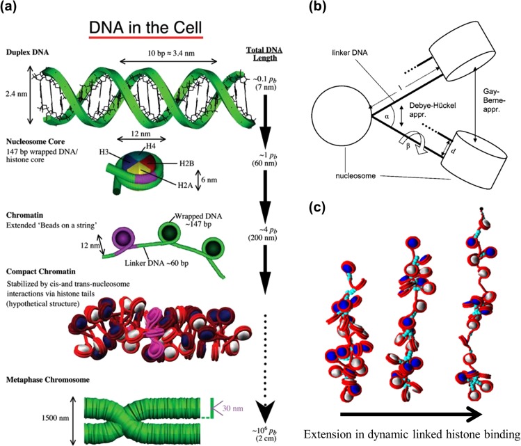
This short review provides an overview of the impact micro- and nanotechnologies can make in studying epigenetic structures. The importance of mapping histone modifications on chromatin prompts us to highlight the complexities and challenges associated with histone mapping, as compared to DNA sequencing. First, the histone code comprised over 30 variations, compared to 4 nucleotides for DNA. Second, whereas DNA can be amplified using polymerase chain reaction, chromatin cannot be amplified, creating challenges in obtaining sufficient material for analysis. Third, while every person has only a single genome, there exist multiple epigenomes in cells of different types and origins. Finally, we summarize existing technologies for performing these types of analyses. Although there are still relatively few examples of micro- and nanofluidic technologies for chromatin analysis, the unique advantages of using such technologies to address inherent challenges in epigenetic studies, such as limited sample material, complex readouts, and the need for high-content screens, make this an area of significant growth and opportunity.


Figures

Figure 1
Figure 1
The concept of epigenetic regulation and reprogramming. Fertilized egg represents totipotency or iPS and ES represents pluripotency, they have the potential to differentiate down all pathways through dynamic epigenetic regulation. Bottom roots represent differentiated cells such as skin, neurons, liver, lung, muscle, blood cells, etc. Alternatively, differentiated cell types in an individual can reverse their fate through epigenetic reprogramming. These induced pluripotent cells can be re-differentiated to various cell types through dynamic epigenetic regulation, but cannot develop into an individual.
Figure 2
Figure 2
Schematic of DNA folding in the cell and higher coarse-grained models of chromatin. (a) Bare DNA is folded to form highly condensed chromatin structures such as a chromosome. The structure of compact chromatin is still uncertain but a compact chromatin fiber is a likely candidate. Reprinted with permission from Schlick et al., J. Biol. Chem. 287, 5183 (2012). Copyright 2012 American Society for Biochemistry and Molecular Biology. (b) A chromatin fiber model can be defined by the opening angle between the incoming and outgoing linker DNA of length l, torsional angle of the linker DNA, and the interactions between nucleosomes and linker DNA. Reprinted with permission from Aumann et al., Phys. Rev. E 73, 041927 (2006). Copyright 2006 American Physical Society. (c) A heteromorphic-chromatin stretching simulation where the components such as linker histone and the nucleosome surface charge distribution are explicitly modeled. Reprinted with permission from Schlick et al., J. Biol. Chem. 287, 5183 (2012). Copyright 2012 American Society for Biochemistry and Molecular Biology.
Figure 3
Figure 3
Transcriptional relationship between chromatin structure and epigenetic modifications on histones. Gene transcriptional activity can be controlled by chromatin structure such as heterochromatin and euchromatin. These chromatin structures can also be controlled by histone modifications, as demonstrated in the diagram which shows post-translational histone modifications on each histone (Ac, acetylation; Me, Methylation).
Figure 4
Figure 4
Micro/nanofluidic technologies for ChIP assays. (a) Schematic of a DNA-enrichment microfluidic chip. Reprinted with permission from Oh et al., Anal. Chem. 81, 2832 (2009). Copyright 2009 American Chemical Society. (b) Automated microfluidic ChIP assay device. Reprinted with permission from Wu et al., Lab Chip 9, 1365 (2009). Copyright 2009 Royal Society of Chemistry. (c) High throughput automated ChIP assay platform. Reprinted with permission from Wu et al., Lab Chip 12, 2190 (2012). Copyright 2012 Royal Society of Chemistry. (d) ChIP assay platform for histone modification analysis of a low number of cells. Reprinted with permission from Geng et al., Lab Chip 11, 2842 (2011). Copyright 2011 Royal Society of Chemistry.
Figure 5
Figure 5
Chromatin linearization in micro/nanofluidic systems. (a) Schematics of reconstituted chromatin linearization in nanochannels. (b) Linearization of chromatin assembled from lambda-DNA and bare lambda DNA in nanochannels. Reprinted with permission from Streng et al., Lab Chip 9, 2772 (2009). Copyright 2009 Royal Society of Chemistry. (c) Experimental platform for single chromatin analysis at the nanoscale. (d) Coincidence two color measurements of stained DNA and GFP expressed histone on a single chromatin. Reprinted with permission from Cipriany et al., Anal. Chem. 82, 2480 (2010). Copyright 2010 American Chemical Society. (e) Chromatin elongation induced by hydrodynamic squeezing flow as well as confinement effect as channel narrows. (f) Multi-color epigenetic marker mapping for histone-H3K9me3 or histone-H4Ac on linearized chromatin. Reprinted with permission from Matsuoka et al., Nano Lett. 12, 6480 (2012). Copyright 2012 American Chemical Society. (g) Individual chromatin molecules contain valuable genetic and epigenetic information. Reprinted with permission from Cerf et al., ACS Nano 6, 7928 (2012). Copyright 2012 American Chemical Society.

References

    1. Papp B. and Plath K., Cell 152, 1324 (2013).10.1016/j.cell.2013.02.043 - DOI - PMC - PubMed
    1. Lim J. P. and Brunet A., Trends Genet. TIG 29, 176 (2013).10.1016/j.tig.2012.12.008 - DOI - PMC - PubMed
    1. Bohacek J. and Mansuy I. M., Neuropsychopharmacology 38, 220 (2013).10.1038/npp.2012.110 - DOI - PMC - PubMed
    1. Anway M. D., Cupp A. S., Uzumcu M., and Skinner M. K., Science 308, 1466 (2005).10.1126/science.1108190 - DOI - PMC - PubMed
    1. Guerrero-Bosagna C., Settles M., Lucker B., and Skinner M. K., PloS ONE 5, e13100 (2010). - PMC - PubMed

LinkOut - more resources