. 2013;19(9):1385-92.
doi: 10.3201/eid1909.130682.

Divergent astrovirus associated with neurologic disease in cattle

Affiliations

Divergent astrovirus associated with neurologic disease in cattle

Linlin Li et al. Emerg Infect Dis. 2013.

Abstract

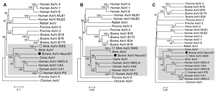
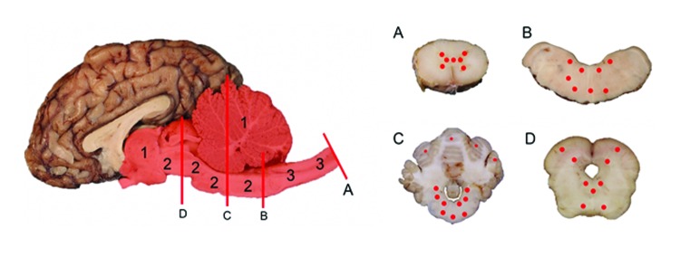
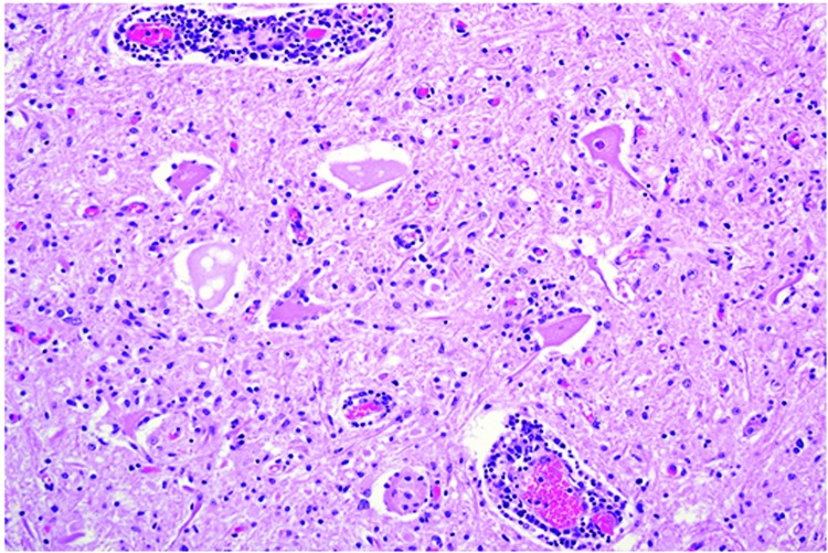
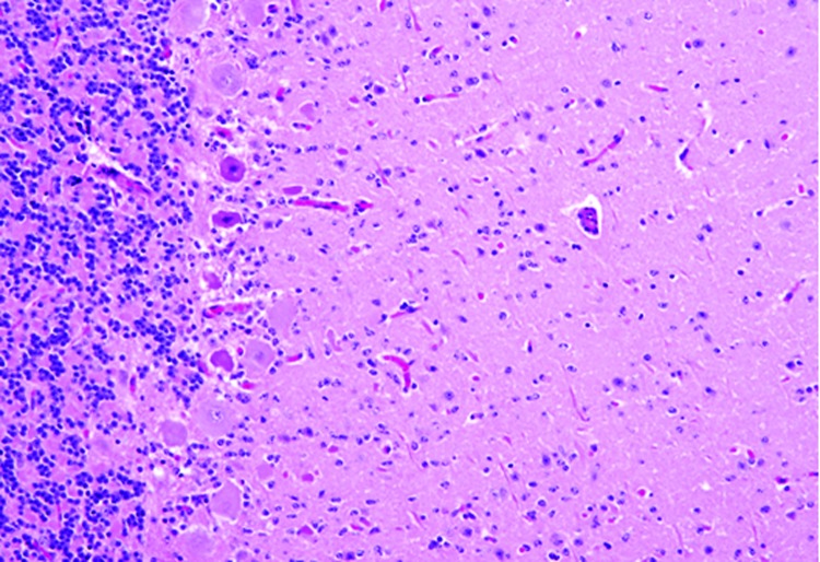
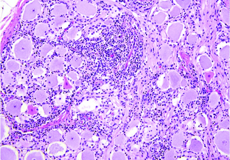
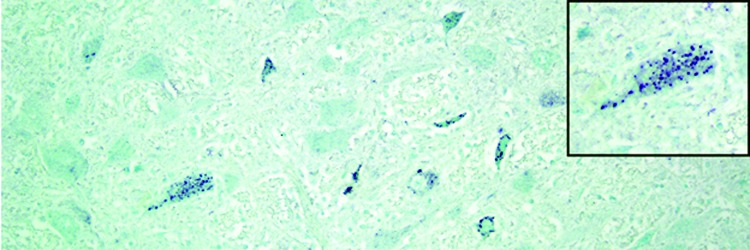
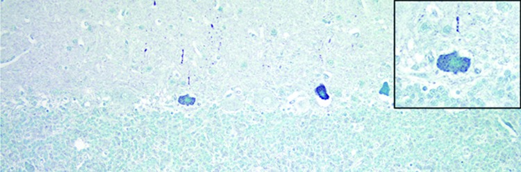
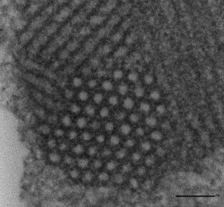
Using viral metagenomics of brain tissue from a young adult crossbreed steer with acute onset of neurologic disease, we sequenced the complete genome of a novel astrovirus (BoAstV-NeuroS1) that was phylogenetically related to an ovine astrovirus. In a retrospective analysis of 32 cases of bovine encephalitides of unknown etiology, 3 other infected animals were detected by using PCR and in situ hybridization for viral RNA. Viral RNA was restricted to the nervous system and detected in the cytoplasm of affected neurons within the spinal cord, brainstem, and cerebellum. Microscopically, the lesions were of widespread neuronal necrosis, microgliosis, and perivascular cuffing preferentially distributed in gray matter and most severe in the cerebellum and brainstem, with increasing intensity caudally down the spinal cord. These results suggest that infection with BoAstV-NeuroS1 is a potential cause of neurologic disease in cattle.

Keywords: Astrovirus; bovine; brain; cattle; in situ hybridization; neurologic disease; next-generation sequencing; viruses.


Figures

Figure 1
Figure 1
Phylogenetic tree based on aligned amino acid sequences of the full length of the protease (open reading frame [ORF] 1a) (A), RNA-dependent RNA polymerase (ORF1b) (B), and capsid (ORF2) region (C) of representative astrovirus (AstV) species. BoAstV-NeuroS1 labeled with filled circle. AstVs with neurotropic potential labeled by empty circles. Clade containing these 3 viruses is shaded. Scale bar indicatesestimated protein sequence phylogenetic distance. GenBank accession numbers for astrovirus used in the analysis are as follows: bovine BoAstV-NeuroS1 (KF233994), human AstV-1 (JN887820), human AstV-4 (DQ344027), human AstV-6 (HM237363), human AstV MLB1 (JQ086552), human AstV MLB2 (NC_016155), human AstV-VA1 (FJ973620), human AstV-VA4 (JX857869), human AstV-HMO-A (NC_013443), human AstV-HMO-B (GQ415661), human AstV-HMO-C (GQ415662), human AstV-PS (GQ891990), ovine AstV (NC_002469), ovine AstV-2 (JN592482), bovine AstV B18 (HQ916313), bovine AstV-B34 (HQ916315), bovine AstV-B76 (HQ916316), bovine AstV-B170 (HQ916314), porcine AstV-2 (JF713712), porcine AstV-5 (JF713711), mink AstV (AY179509), mink AstV-SMS (GU985458), rabbit AstV (JF729316), and chicken AstV (JF414802).
Figure 2
Figure 2
Yearling steer with encephalomyelitis. Midsagittal section of brain and multiple transverse sections of cerebellum, brainstem, and spinal cord depicting the location and severity of microscopic lesions. Midsagittal section of the brain: red highlight indicates areas of the central nervous system affected, numbers indicate severity of the lesions (1 = least severe; 2 = more severe; 3 = most severe), and red lines (A, B, C, D) indicate the levels where transverse sections were cut. A, spinal cord. B, medulla oblongata. C, cerebellum and cerebellar peduncles. D, midbrain (superior colliculus). Cross-sections of brainstem, cerebellum, and spinal cord: red dots indicate sites and relative intensity of microscopic lesions.
Figure 3
Figure 3
Spinal cord at the L5 segment of a heifer with encephalomyelitis (animal 2). Note the nonsuppurative encephalomyelitis with lymphocytic perivascular cuffs, neuronal degeneration and necrosis, spheroids from necrotic neurons, and neuronophagia with widespread microgliosis. Hematoxylin and eosin stain. Original magnification ×400.
Figure 4
Figure 4
Cerebellum of a yearling steer with encephalomyelitis (animal 1). Note the selective extensive acute necrosis and degeneration of Purkinje cells. Numerous necrotic dendritic spheroids in the molecular layer with a cellular proliferation of Bergmann glia and of microgliosis. Hematoxylin and eosin stain. Original magnification ×400.
Figure 5
Figure 5
Dorsal root ganglion of a heifer with encephalomyelitis (animal 2). Multifocal marked interstitial lymphocyte, macrophage, and plasma cell infiltrates with multifocal neuronal degeneration and necrosis can be seen. Hematoxylin and eosin stain. Original magnification ×400.
Figure 6
Figure 6
Medulla oblongata of a heifer with encephalomyelitis (animal 2). Punctate cytoplasmic staining (green) in multiple neurons within a nucleus can be seen; inset shows a higher magnification of a positive brainstem neuron. In situ hybridization for viral RNA. Original magnification ×400.
Figure 7
Figure 7
Cerebellum of a yearling steer with encephalomyelitis (animal 1). Punctate to diffuse positive (green) staining of Purkinje cells cytoplasm and dendritic processes can be seen; inset shows a higher magnification of a positive Purkinje cell. In situ hybridization for viral RNA. Original magnification ×400.
Figure 8
Figure 8
Transmission electron microscopic image of necrotic neuron in the lumbar region of a heifer with encephalomyelitis. Intracytoplasmic paracrystalline array of 27–28-nm diameter viral-like particles. Scale bar indicates 100 nm.

References

    1. Appleton H, Higgins PG. Viruses and gastroenteritis in infants. Lancet. 1975;1:1297 . 10.1016/S0140-6736(75)92581-7 - DOI - PubMed
    1. De Benedictis P, Schultz-Cherry S, Burnham A, Cattoli G. Astrovirus infections in humans and animals—molecular biology, genetic diversity, and interspecies transmissions. Infect Genet Evol. 2011;11:1529–44 . 10.1016/j.meegid.2011.07.024 - DOI - PMC - PubMed
    1. Walter JE, Mitchell DK. Astrovirus infection in children. Curr Opin Infect Dis. 2003;16:247–53 . 10.1097/00001432-200306000-00011 - DOI - PubMed
    1. Moser LA, Schultz-Cherry S. Pathogenesis of astrovirus infection. Viral Immunol. 2005;18:4–10 . 10.1089/vim.2005.18.4 - DOI - PubMed
    1. Blomström AL, Widén F, Hammer AS, Belák S, Berg M. Detection of a novel astrovirus in brain tissue of mink suffering from shaking mink syndrome by use of viral metagenomics. J Clin Microbiol. 2010;48:4392–6 . 10.1128/JCM.01040-10 - DOI - PMC - PubMed

Publication types

MeSH terms

Associated data