. 2013 Aug 20;18(8):10056-80.
doi: 10.3390/molecules180810056.

Modeling the interaction of dodecylphosphocholine micelles with the anticoccidial peptide PW2 guided by NMR data

Affiliations

Modeling the interaction of dodecylphosphocholine micelles with the anticoccidial peptide PW2 guided by NMR data

Francisco Gomes-Neto et al. Molecules. .

Abstract

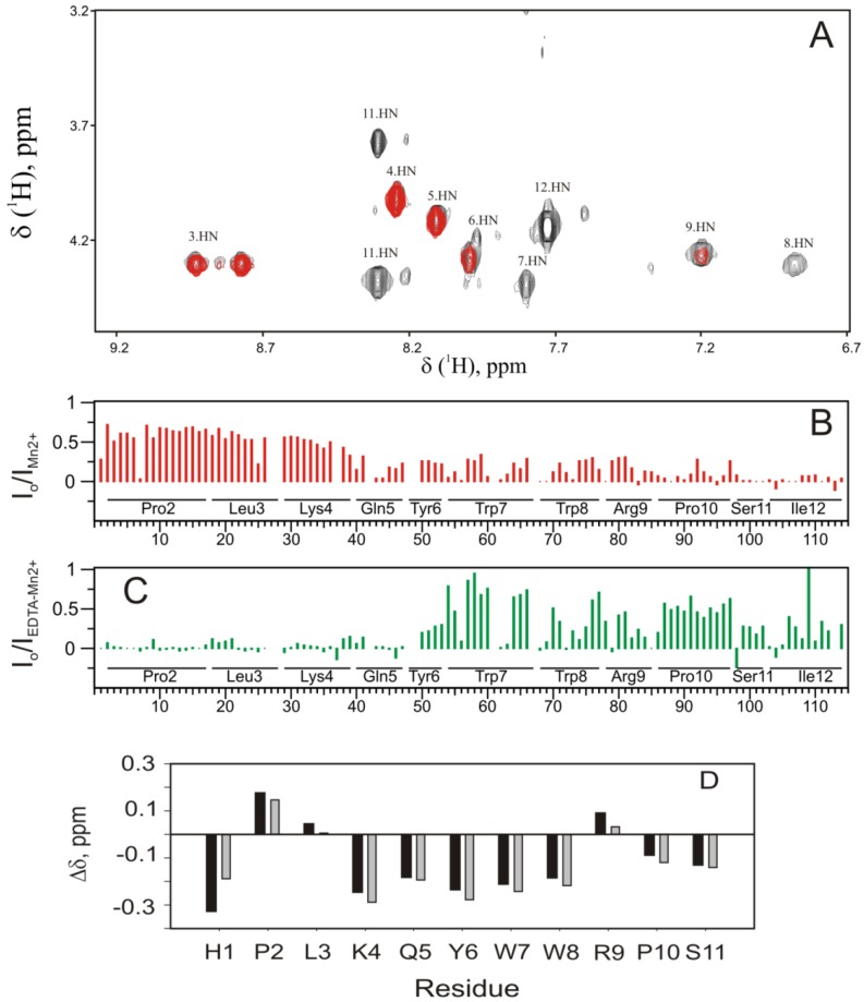
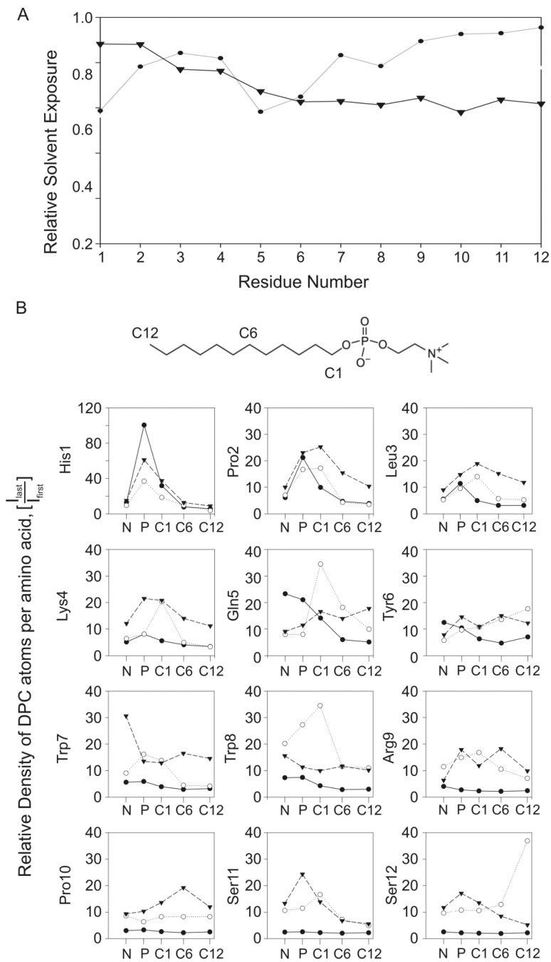
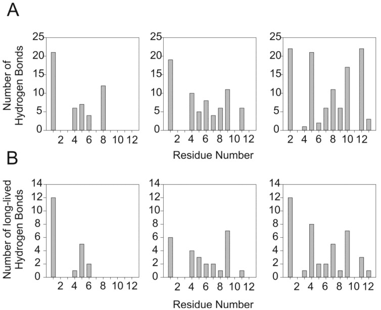
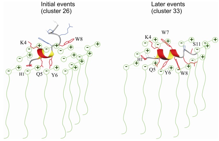
Antimicrobial peptides are highly dynamic entities that acquire structure upon binding to a membrane interface. To better understand the structure and the mechanism for the molecular recognition of dodecylphosphocholine (DPC) micelles by the anticoccidial peptide PW2, we performed molecular dynamics (MD) simulations guided by NMR experimental data, focusing on strategies to explore the transient nature of micelles, which rearrange on a millisecond to second timescale. We simulated the association of PW2 with a pre-built DPC micelle and with free-DPC molecules that spontaneously forms micelles in the presence of the peptide along the simulation. The simulation with spontaneous micelle formation provided the adequate environment which replicated the experimental data. The unrestrained MD simulations reproduced the NMR structure for the entire 100 ns MD simulation time. Hidden discrete conformational states could be described. Coulomb interactions are important for initial approximation and hydrogen bonds for anchoring the aromatic region at the interface, being essential for the stabilization of the interaction. Arg9 is strongly attached with phosphate. We observed a helix elongation process stabilized by the intermolecular peptide-micelle association. Full association that mimics the experimental data only happens after complete micelle re-association. Fast micelle dynamics without dissociation of surfactants leads to only superficial binding.


Figures

Figure 1
Figure 1
Restrained MD simulation of PW2 interacting with pre-built DPC micelle. (A) Cluster index as a function of MD simulation time. Snapshots of interaction are presented at particular simulation times. In the beginning of the MD simulation, there was a rapid increase in the number of detected clusters (0 to 1.5 ns) while the peptide was approaching the membrane surface. The period after this (1.5 and 7 ns) showed binding instability where PW2 was partially bound by the N-terminal His 1 residue. After 7 ns, there was complete interaction of PW2 with the micelle surface, stabilizing cluster 26, and followed by partial dissociation events, indicated by new cluster index increments. Cluster 26 refers to the PW2 structures fully associated with the DPC micelle. (B) NMR structure ensemble (PDB ID: 2JQ2), superposition of the 20 lowest energy structures. (right) superposition of 20 representative structures of cluster 26. The backbone is in black and the side chains in blue lines. The second Trp 7 rotamer is in red lines. (C) Ribbon representation of clusters 26 (fully bound state, left) and 124 (partially dissociated state, middle) and superposition of both (right), as indicated in the figure. Side chains are represented by dark blue lines for cluster 26 and blue lines for cluster 124.
Figure 2
Figure 2
Restrained MD simulation of PW2 interacting with free-DPC during spontaneous micelle formation. (A) Cluster index as a function of MD simulation time. Snapshots of interaction are presented at particular simulation times. Initially (0 to 5 ns) there was a fast increase in the number of detected clusters. After 5 ns cluster 33 was stabilized and remained for the entire simulation. Cluster 33 refers to the PW2 structures fully associated with the DPC micelle. (B) Superposition of 20 representative structure of cluster 33: backbone (left), heavy atoms (middle) and ribbon representation of the 310-helix (right), spanning from Leu 3 to Trp 8.
Figure 3
Figure 3
Ramanchadran plot of NMR ensemble, cluster 26 and cluster 33. (A) The Ramachandran plot is shown for each amino acid residue of PW2 in the NMR ensemble (top), cluster 26 (middle) and cluster 33 (bottom). The 310-helix spans from Leu 3 to Tyr 6 for NMR ensemble, while in cluster 26 and cluster 33, from Leu 3 to Trp 8. (B) Ribbon representation of clusters 26 (left) showing the side chains in black and cluster 33 (middle) in dark blue. In the right, superposition of clusters 26 and 33 showing the same spatial orientation of residues Trp 7 and Trp 8. The arrows indicate the two allowed region of Trp 7 observed for the two clusters.
Figure 4
Figure 4
Unrestrained MD simulation of PW2 interacting with free-DPC during spontaneous micelle formation. (A) Cluster index as a function of MD simulation time. From 0 to 7 ns we observed an increasing number of clusters. After 7 ns, five long-lived clusters were detected (104, 193, 234, 259 and 265) and representative snapshots of the interaction are shown for each of them. (B) Ramachandran plot of all five stable clusters for each amino acid residue. Note that the shift of the Ramanchadran position of Trp 7 from generously allowed region to helix position, indicated by arrows. (C) Superposition of 20 representative structures of cluster 265 (left) and ribbon representation of cluster 265 structure (right).
Figure 5
Figure 5
Interaction of the PW2 with DPC micelles. (A) Paramagnetic relaxation enhancement (PRE) induced by Mn2+ ions. Amide region of a TOCSY spectrum in the absence (black) and presence (gray) of 2 mM Mn2+ ions. The resonances of Trp 7, Trp 8, Ser 11 and Ile 12 disappeared and Arg 9 was partially affected, while Tyr 6, Gln 5, Lys 4, Leu 3 were less affected or unaffected. (B) Ratio between the TOCSY cross-peaks intensity in presence (IMn2+) and the absence (Io) of 2 mM Mn2+, expressed as a function of the hydrogen resonance cross-peak assignment (indicated by numeric index and detailed in Table S1). The Mn2+ PRE effect was strong for the aromatic region and C-terminal region. (C) Ratio between the TOCSY cross-peaks intensity in presence (IMn2+/EDTA) of 2 mM Mn2+and 3 mM EDTA and the absence (Io), expressed as a function of the hydrogen resonance cross-peak assignment (indicated by numeric index and detailed in Table S1). The Mn2+/EDTA PRE effect was strong for the N-terminal region. (D) Hα chemical shift difference between PW2 in DPC and random coil chemical shifts. Random coil values were according to Wüthrich and coworkers [25,26] and Dyson and coworkers [27,28]. A negative difference suggests the tendency to form a helix from Lys 4 to Trp 8.
Figure 6
Figure 6
Analysis of PW2 interaction with DPC micelles. (A) Relative water exposure of PW2 as function of amino acid residues, for the MD simulations in pre-build DPC micelles (closed circles) and restrained with spontaneous micelle formation (inverted triangle). (B) A DPC density profile, g(r), was generated for the center of mass of each amino acid side chain as a function of selected DPC atoms: choline nitrogen (N), polar head phosphate (P), and aliphatic chain atoms C1, C6 and C12. The intensity of the first density peak was plotted as a function of the DPC atom for each MD simulation. The symbols are the same for panel A. The DPC structure illustrates the position of each DPC atom.
Figure 7
Figure 7
The PW2/DPC interaction depicted by hydrogen bond profile. (A) Number of intra-molecular hydrogen bonds (PW2-DPC) for the entire MD simulation as a function of the amino acid residue. The left side is for the simulation in pre-build DPC micelles, the middle for the restrained MD simulation during spontaneous micelle formation and the right for the unrestrained MD simulation during spontaneous micelle formation. (B) Number of long-lived intra-molecular hydrogen bonds (PW2-DPC, >1 ns) as a function of the amino acid residue. The left side is for the simulation in pre-build DPC micelles, the middle for the restrained MD simulation during spontaneous micelle formation and the right for the unrestrained MD simulation during spontaneous micelle formation.
Figure 8
Figure 8
Schematic representation of the proposed model of two events for PW2 association with DPC interface. In the left is a static representation of the association of a cluster 26 structure with the micelle. In red are the residues that are hydrogen bonded with DPC head group during simulation. Note that residues His 1, Lys 4, Gln 5 and Tyr 6 forms long-lived hydrogen bonds, while Trp 8 forms only short-lived hyrogens bonds. Cluster 26 structures interact superficially with the DPC head groups and the C-terminal residues are weakly bound. In the right we represent the association of a cluster 33 structure with DPC micelles. Note that that the helix was elongated if compared with cluster 26. This elongation was only possible in the simulation with free-DPC with spontaneous micelles formation. Our model assumes that this deeper association was only possible after micelle re-equilibration, event that happens in millisecond to second timescale. In red are the residues that are hydrogen bonded with DPC micelles. Note that most of the residues show long-lived hydrogen bonds. Number of intra-molecular hydrogen bonds (PW2-DPC) increased consistently when compared for the entire MD simulation as a function of the amino acid residue.

Similar articles

Cited by

References

    1. Bulet P., Stocklin R., Menin L. Anti-microbial peptides: From invertebrates to vertebrates. Immunol. Rev. 2004;198:169–184. doi: 10.1111/j.0105-2896.2004.0124.x. - DOI - PubMed
    1. Thomma B.P.H.J., Cammue B.P.A., Thevissen K. Mode of action of plant defensins suggests therapeutic potential. Curr. Drug Targets Infect. Disord. 2003;3:1–8. - PubMed
    1. Zasloff M. Antimicrobial peptides of multicellular organisms. Nature. 2002;415:389–395. doi: 10.1038/415389a. - DOI - PubMed
    1. Nicolas P., Mor A. Peptides as weapons against microorganisms in the chemical defense system of vertebrates. Annu. Rev. Microbiol. 1995;49:277–304. doi: 10.1146/annurev.mi.49.100195.001425. - DOI - PubMed
    1. Wu M.H., Maier E., Benz R., Hancock R.E.W. Mechanism of interaction of different classes of cationic antimicrobial peptides with planar bilayers and with the cytoplasmic membrane of Escherichia coli. Biochemistry. 1999;38:7235–7242. doi: 10.1021/bi9826299. - DOI - PubMed

Publication types

MeSH terms

LinkOut - more resources