. 2013 Sep 1;12(17):2781-93.
doi: 10.4161/cc.25870. Epub 2013 Aug 6.

Mitochondrial p53 mediates a transcription-independent regulation of cell respiration and interacts with the mitochondrial F₁F0-ATP synthase

Affiliations

Mitochondrial p53 mediates a transcription-independent regulation of cell respiration and interacts with the mitochondrial F₁F0-ATP synthase

Marie Bergeaud et al. Cell Cycle. .

Abstract

We and others previously reported that endogenous p53 can be located at mitochondria in the absence of stress, suggesting that p53 has a role in the normal physiology of this organelle. The aim of this study was to characterize in unstressed cells the intramitochondrial localization of p53 and identify new partners and functions of p53 in mitochondria. We find that the intramitochondrial pool of p53 is located in the intermembrane space and the matrix. Of note, unstressed HCT116 p53(+/+) cells simultaneously show increased O₂ consumption and decreased mitochondrial superoxide production compared with their p53-null counterpart. This data was confirmed by stable H1299 cell lines expressing low levels of p53 specifically targeted to the matrix. Using immunoprecipitation and mass spectrometry, we identified the oligomycin sensitivity-conferring protein (OSCP), a subunit of the F₁F₀-ATP synthase complex, as a new partner of endogenous p53, specifically interacting with p53 localized in the matrix. Interestingly, this interaction seems implicated in mitochondrial p53 localization. Moreover, p53 localized in the matrix promotes the assembly of F₁F₀-ATP synthase. Taking into account that deregulations of mitochondrial respiration and reactive oxygen species production are tightly linked to cancer development, we suggest that mitochondrial p53 may be an important regulator of normal mitochondrial and cellular physiology, potentially exerting tumor suppression activity inside mitochondria.

Keywords: OSCP; cancer; energetic metabolism; mitochondria; oxidative phosphorylation; p53; reactive oxygen species; respiration; tumor suppressor.


Figures

None
Figure 1. Endogenous p53 resides in different mitochondrial compartments in unstressed cells. (A) Left panel: 15 μg of whole-cell lysates (Wcl), nuclei plus unbroken cells (N*), cytosolic fraction (Cy), and mitochondrial fraction (M) of HCT116 cells treated or untreated with 125 μg/ml of etoposide for 24 h were subjected to immunoblot analysis for p53, PCNA, Enolase, and Tom40. Right panel: Densitometry of blot in the left panel. The ratio of mitochondrial p53 /total cellular p53 levels was calculated to determine the relative amount of the mitochondrial p53. (B) Left panel: 300 µg of mitochondrial protein isolated from HCT116 cells were treated with sodium carbonate (pH 11.5) and ultracentrifuged. The soluble proteins were recovered in the supernatant (S), while the integral membrane proteins remained in the pellet (P). Proteins were subjected to immunoblot analysis for p53, PCNA, actin, Bak, and Tom40. Right panel: The level of p53, actin, Tom40, and Bak proteins was quantified in soluble and membrane fractions after alkali treatment by densitometry of blot in the left panel. (C) Left panel: 50 μg of mitochondrial proteins isolated from mitochondria of unstressed HCT116 cells were treated with increasing quantities of proteinase K (K; 150 ng, 300 ng, and 450 ng) prior to (mitochondria, lanes 2–4) or after swelling (mitoplasts, lanes 5–7). Swelling disrupts the outer membrane while keeping the inner membrane intact. Untreated mitochondrial extracts were used as control (Ctrl). Samples were subjected to immunoblot analysis for p53 and marker proteins of different mitochondrial subcompartments: OSCP (matrix), mitofilin (IMS), actin (mitochondrial periphery). Right panel: Densitometry of blot in the left panel. The mitochondrial matrix protein OSCP was used as control for loading and mitochondrial membrane integrity. The levels of p53, actin and mitofilin were normalized to OSCP in each condition (lanes 1, 4, and 7) and compared with intact mitochondria (M) or mitoplasts (Mitopl) treated or not treated (Ctrl) with proteinase K (K).
None
Figure 2. Targeting of p53 to the mitochondrial matrix and intermembrane space in H1299 cells. (A) Characterization of the stable H1299-p53-Mx cell line. Left upper panel: Schematic representation of the cDNA construct to target p53 to the mitochondrial matrix (p53-Mx): the mitochondrial import leader of ornithin transcarbamylase (OTC) is fused to human wild type p53 in pcDNA3 expression vector. Left lower panel: Low-level expression of p53-Mx was assessed by immunoblot of whole-cell lysates of H1299-p53-Mx vs. H1299 pcDNA3 control line. Actin served as a loading control. Right panel: Mitochondrial purification and subfractionation. Fifteen μg of all fractions: whole-cell lystates (Wcl), nuclei plus unbroken cells (N*), mitochondria and mitoplasts treated with increasing concentrations of proteinase K (K), or left untreated (Ctrl) of H1299-p53-Mx cells were loaded on the same gel. The blot was subjected to immunoblot analysis with anti-p53, anti-lamins, anti-mitofilin, and anti-OSCP antibodies. (B) Characterization of the stable H1299-p53-IMS clone (C1) cell line. Left upper panel: Schematic representation of the cDNA construct to target p53 to the intermembrane space (p53-IMS): mitochondrial import leader of cytochrome B2 (CytB2), composed of 2 cleavable sorting signals, fused to human wild-type p53. Left lower panel: Expression of p53-IMS was assessed by immunoblot on the H1299-p53-IMS C1 vs. the H1299 pcDNA3 control line. Right panel: The same treatments and immunoblot were performed as for H1299-p53-Mx cells.
None
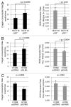
Figure 3. Mitochondrial matrix p53 promotes oxygen consumption and decreases ROS production. Average values of oxygen consumption rate and mitochondrial ROS generation in unstressed (A) HCT116 p53+/+ vs. HCT116 p53−/− cells, (B) H1299-p53-Mx vs. H1299 pcDNA3 cells, (C) H1299-p53-IMS vs. H1299 pcDNA3 cells. Oxygen consumption was measured with a Clark electrode. Mitochondrial superoxide anion level was determined with the MitoSOX probe.
None
Figure 4. p53 interacts with the OSCP subunit of the F1F0-ATP synthase complex of mitochondria. (A) Extracts from isolated mitochondria from HCT116 cells, untreated, or treated with etoposide for 24 h, were immunoprecipitated with p53 antibodies (FL393) or control antibody (IgG). p53 and control immunoprecipitations were subjected to mass spectrometry analysis. Three peptides were specifically identified in p53 immunoprecipitations under stress conditions (upper panel). These peptides (bold underlined) correspond to the OSCP protein sequence. (B) The same immunoprecipitations (IP) and mitochondrial extracts (input) were loaded on the same gel and analyzed by immunoblot using antibodies to p53 (DO1) and OSCP. Tom40 serves as negative control. (C) Mitochondrial extracts from H1299 p53-Mx, p53-IMS, and pcDNA3 as control were immunoprecipitated with p53 antibodies (FL393) or not (input), loaded on the same gel and analyzed by immunoblot using antibodies to p53 (DO1) and OSCP, and MnSOD as negative control.
None
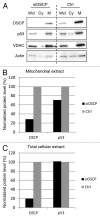
Figure 5. Knockdown of OSCP results in a decrease of mitochondrial p53 levels. HCT116 p53+/+ cells were transfected with OSCP siRNA or control siRNA (ctrl). After 72 h, cells were harvested, and whole-cell extracts (Wcl), cytosol (Cy), and mitochondrial (M) fractions were prepared and loaded on the same gel. (A) Immunoblot analysis. Membrane was probed for OSCP, p53, VDAC1, and actin. These latter 2 proteins were used as loading control of mitochondria and total cellular extracts, respectively. (B and C) Densitometry of blot in (A). (B) OSCP and p53 levels in mitochondrial fraction were normalized to their respective VDAC1 levels. (C) OSCP and p53 levels in total cellular extract were normalized to their respective actin levels in knockdown and Ctrl condition.
None
Figure 6. Mitochondrial p53 increases mature F1F0-ATP synthase steady-state levels. Mitochondrial proteins isolated from isogenic HCT116 (+/− endogenous wtp53) and H1299 cells (+/− matrix or IMS p53) were solubilized with 1% digitonin, analyzed by BN-PAGE and immunoblotted for OSCP and Tom40. Under the conditions employed, an antibody to OSCP recognizes the F1F0-ATP synthase holoenzyme (at approximately 600kDa probably in dimeric form) and no individual F1-ATP synthase subcomplexes. The TOM complex detected by anti-Tom40 antibody runs at 440 kDa and served as loading control of mitochondrial protein complexes. (A) Example of BN-PAGE immunoblotting. (B–D) Densitometry of BN-PAGE blots. Quantification of F1F0-ATP synthase level was normalized to Tom40 in (B) HCT116 p53+/+ vs. HCT116 p53−/− cells, in (C) H1299-p53-Mx vs. H1299 pcDNA3 cells, in (D) H1299-p53-IMS vs. H1299-pcDNA3 cells.

References

    1. Hollstein M, Sidransky D, Vogelstein B, Harris CC. p53 mutations in human cancers. Science. 1991;253:49–53. doi: 10.1126/science.1905840. - DOI - PubMed
    1. Fields S, Jang SK. Presence of a potent transcription activating sequence in the p53 protein. Science. 1990;249:1046–9. doi: 10.1126/science.2144363. - DOI - PubMed
    1. Raycroft L, Wu HY, Lozano G. Transcriptional activation by wild-type but not transforming mutants of the p53 anti-oncogene. Science. 1990;249:1049–51. doi: 10.1126/science.2144364. - DOI - PMC - PubMed
    1. Weintraub H, Hauschka S, Tapscott SJ. The MCK enhancer contains a p53 responsive element. Proc Natl Acad Sci U S A. 1991;88:4570–1. doi: 10.1073/pnas.88.11.4570. - DOI - PMC - PubMed
    1. el-Deiry WS, Kern SE, Pietenpol JA, Kinzler KW, Vogelstein B. Definition of a consensus binding site for p53. Nat Genet. 1992;1:45–9. doi: 10.1038/ng0492-45. - DOI - PubMed

MeSH terms

Substances

LinkOut - more resources