. 2013 Nov;87(21):11462-75.
doi: 10.1128/JVI.01681-13. Epub 2013 Aug 21.

A functional interaction between gp41 and gp120 is observed for monomeric but not oligomeric, uncleaved HIV-1 Env gp140

Affiliations

A functional interaction between gp41 and gp120 is observed for monomeric but not oligomeric, uncleaved HIV-1 Env gp140

Miklos Guttman et al. J Virol. 2013 Nov.

Abstract

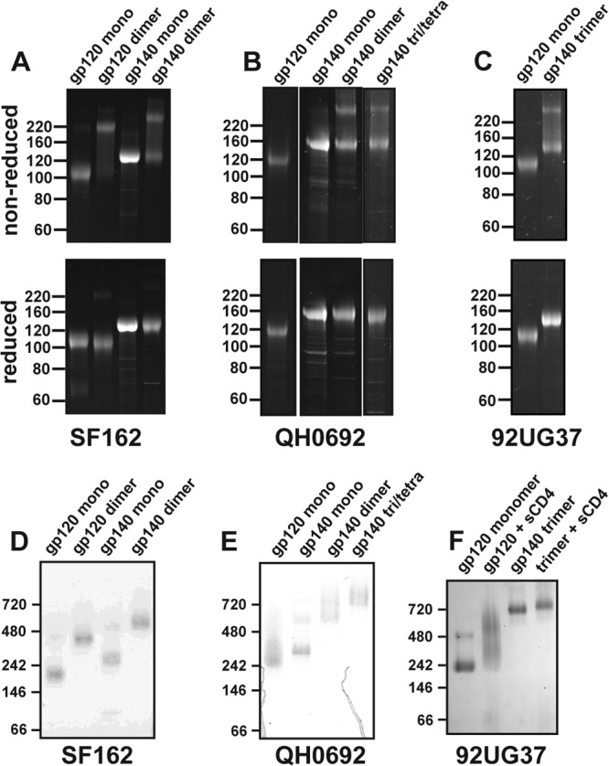
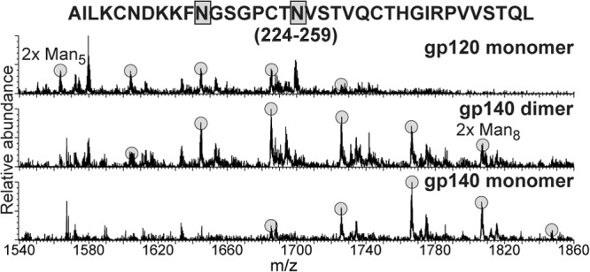
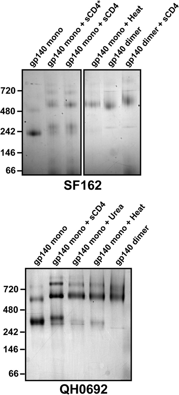
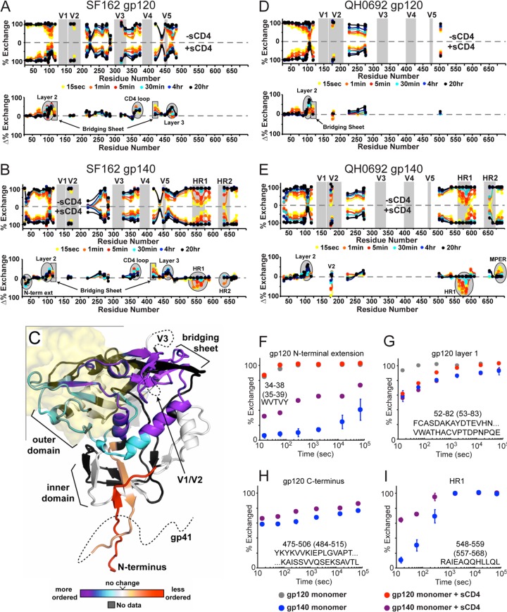
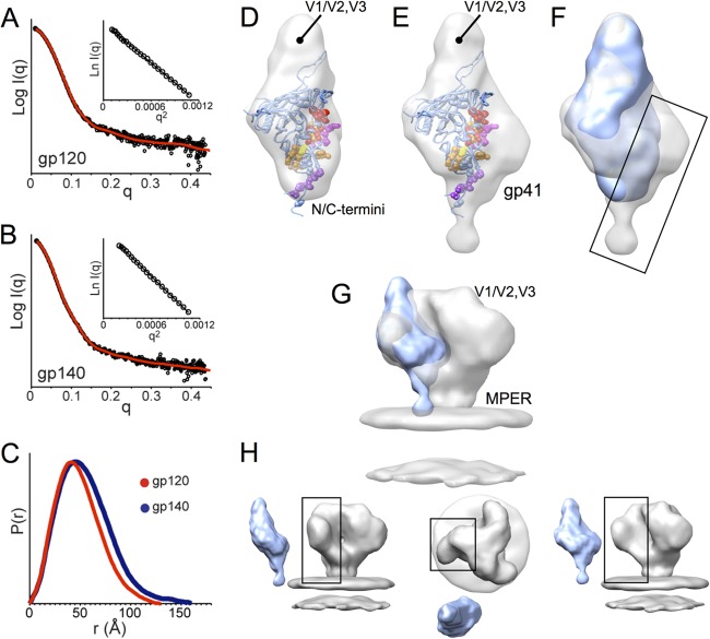
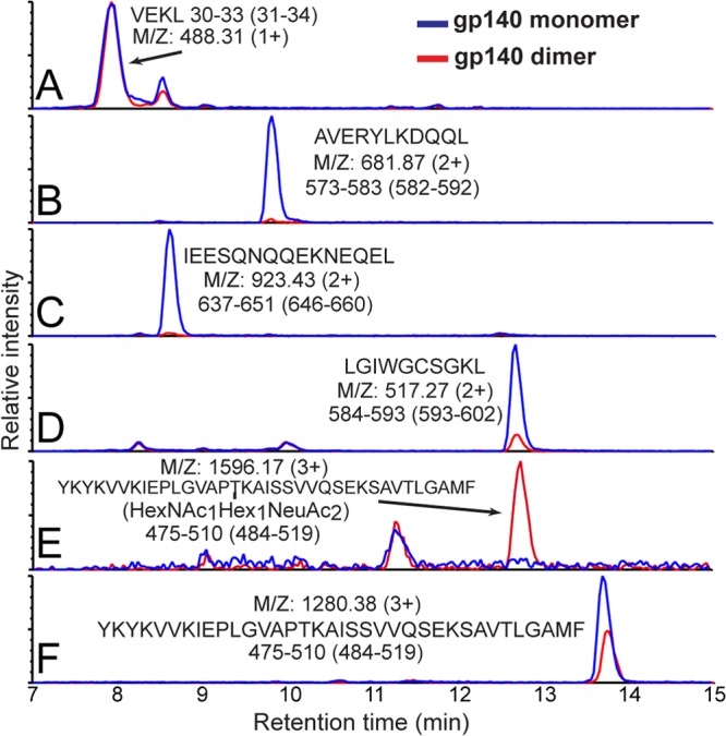
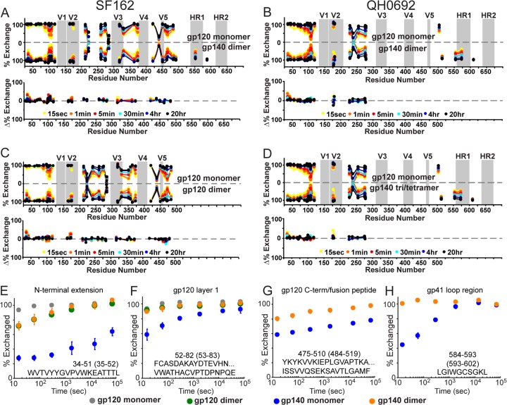
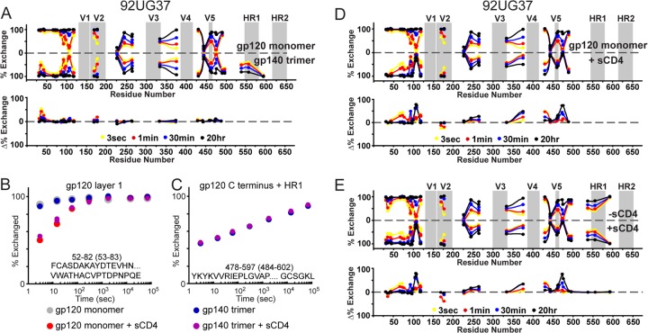
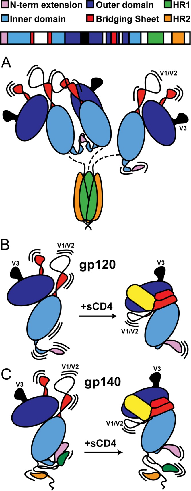
The envelope glycoprotein (Env) is the sole antigenic feature on the surface of HIV and the target for the humoral immune system. Soluble, uncleaved gp140 Env constructs truncated at the transmembrane domain are being investigated intensively as potential vaccine immunogens by many groups, and understanding their structural properties is essential. We used hydrogen/deuterium-exchange mass spectrometry and small-angle X-ray scattering to probe structural order in a panel of commonly used gp140 constructs and matched gp120 monomers. We observed that oligomeric forms of uncleaved gp140, generally presumed to be trimeric, contain a protease-resistant form of gp41 akin to the postfusion, helical bundle conformation and appear to lack specific interactions between gp120 and gp41. In contrast, the monomeric form of gp140 shows significant stabilization of the gp120 inner domain imparted by the gp41 region, demonstrating excellent agreement with past mutagenesis studies. Moreover, the gp140 monomers respond to CD4 binding in manner that is consistent with the initial stages of Env activation: CD4 binding induces structural ordering throughout gp120 while loosening its association with gp41. The results indicate that uncleaved gp140 oligomers do not represent an authentic prefusion form of Env, whereas gp140 monomers isolated from the same glycoprotein preparations in many ways exhibit function and internal structural order that are consistent with expectations for certain aspects of native Env. gp140 monomers may thus be a useful reagent for advancing structural and functional studies.


Figures

Fig 1
Fig 1
SDS-PAGE and BN-PAGE analysis of gp140 constructs. (A to C) SDS-PAGE gels under nonreducing and reducing conditions for SF162 (A), QH0692 (B), and 92UG37 (C) constructs. In gp140 dimer and trimer materials, bands corresponding to disulfide bonded protomers are evident. Gels were stained with SYPRO Ruby (Invitrogen, Carlsbad, CA). (D to F) BN-PAGE gels are shown for the respective constructs.
Fig 2
Fig 2
Glycoform distributions at positions 234 and 241 in gp120, gp140 dimer, and gp140 monomer. The peptic 224-259 fragment was found to contain two high-mannose type N-linked glycans. Each glycoform resulting from different degrees of mannose trimming is highlighted.
Fig 3
Fig 3
Butterfly plots comparing the H/D-exchange profiles of monomeric gp120 and gp140. The upper plot shows the exchange profiles of all observable fragments for gp120 (top) and monomeric gp140 (bottom) for isolates SF162 (A) and QH0692 (B). The percent exchange is shown each fragment at the midpoint in primary sequence. For example the exchange profile of fragment 104-110 will be plotted at position 107. The positions of the variable loops and heptad repeats are highlighted in gray. The difference plots under each butterfly plot show the difference in percent exchange at each time point. The exchange plots of each individual fragment are shown in Fig. S1 and S2 in the supplemental material. (C) Differences in exchange profiles between gp120 and monomeric gp140 are shown on the core structure of gp120 (hybrid of PDB 2NY7 and 3JWD as described previously [35]). Regions are colored from purple (more protected/more ordered) to red (less protected/more disordered relative to gp120).
Fig 4
Fig 4
Dimerization upon destabilization of monomeric gp140. BN-PAGE analysis of monomeric gp140 SF162 (top) and QH0692 (bottom) after sCD4 binding, chemical perturbation (4 M Urea for 10 min), or heat denaturation (50°C for 10 min). *, gp140 was incubated with sCD4 for 2 h, rather than added immediately before BN-PAGE.
Fig 5
Fig 5
Effect of CD4 binding to gp120 and gp140 monomers as monitored by H/D exchange. (A, B, D, and E) Butterfly plots for monomeric gp120 with or without sCD4 (A and D) and gp140 monomer with or without sCD4 (B and E). Plot descriptions are described in Fig. 3. (C) Changes in monomeric gp140 upon sCD4 binding shown on the CD4-bound core crystal structure (PDB 3JWD). Regions are colored from purple (more protected/more ordered) to red (less protected/more disordered relative to gp120). The position of CD4 is illustrated as a transparent yellow surface. (F to I) Individual deuterium uptake profiles are shown for peptides (isolate SF162) from the N-terminal extension (F), layer 1 of the gp120 inner domain (G), the C-terminal end of gp120 (H), and HR1 from gp41 (I). Numbering in parentheses refers to HXB2 numbering.
Fig 6
Fig 6
SAXS of monomeric gp120 and gp140 SF162 suggests gp41 is positioned alongside the gp120 inner domain. (A and B) Experimental SAXS patterns (black) measured for gp120 (A) and gp140 (B) monomers compared to fits obtained from ab initio reconstructions using DAMMIN (red) (47). Insets show the linearity of the Guinier plots. (C) Distance distribution [P(r)] plots for gp120 (red) and gp140 (blue) obtained from GNOM (47). (D and E) Shape reconstructions for monomeric gp120 (D) and monomeric gp140 (E) are shown with the structural model (PDB 3JWD and 2B4C to show approximate locations for N/C-terminal extensions and V3) docked into the SAXS envelope; gp120 core residues that have been identified as playing a role in association with gp41 are shown in space-filling balls and colored as C-terminal extenions (purple), β-sheet “platform” (yellow), layer 1 (magenta), and layer 2 (red) (60). (F) Comparison of SAXS reconstructions for gp120 (blue) and gp140 monomers (gray) reveals additional density in the gp140 monomer localized around the putative gp41 interactive face of gp120 (box). (G and H) Comparison of a gp140 monomer SAXS model with cryo-EM reconstruction for the Env trimer (gray, EMD-5019 [9]) shows good agreement, suggesting that the protomer retains large-scale organization similar to what exists in the membrane-presented Env trimer.
Fig 7
Fig 7
Ion-extracted traces for various peptic fragments. The intensities for several peptides from monomeric (blue) and dimeric (red) gp140 (SF162) after pepsin digestion are shown. The residue numbers for each segment are shown below the amino acid sequence, with HXB2 numbering in parentheses. Although gp120 segment intensities (see, for example, section A) were nearly identical in various constructs, as shown in sections B to F, gp41 fragments in dimeric gp140 were not observed except for a moderate signal for LGIWGCSGKL (section D). A large fragment from the C terminus of gp120 was observed bearing an O-linked glycan at T499 only in dimeric gp140 (sections E and F).
Fig 8
Fig 8
H/D-exchange profiles of monomeric gp120 and oligomeric gp140s reveals no gp120-gp41 interactions in the oligomers. Butterfly plots for SF162 monomeric gp120 versus dimeric gp140 (A), QH0692 monomeric gp120 versus dimeric gp140 (B), SF162 monomeric gp120 versus dimeric gp120 (C), and QH0692 monomeric gp120 versus trimeric/tetrameric gp140 (D) are shown. An explanation for data representation in these types of plots is provided in the legend for Fig. 3. (E to H) Deuterium-uptake plots of selected peptides from both monomeric and dimeric gp120 and gp140 (SF162) constructs; numbering in parentheses refers to HXB2 numbering.
Fig 9
Fig 9
Uncleaved gp140 trimer from isolate 92UG37 does not exhibit functional gp120-gp41 interactions. (A) Plots comparing the exchange profiles of 92UG37 monomeric gp120 versus foldon-stabilized, trimeric gp140. Individual exchange plots are shown for a fragment from gp120 layer 1 (B) and the large fragment spanning the C-terminal end of gp120 to the end of HR1 (C). Numbering in parentheses refers to HXB2 numbering. Plots comparing the exchange profile with or without sCD4 are shown for monomeric gp120 (D) and trimeric gp140 (E) are also shown. Individual exchange plots for each fragment are shown in Fig. S3 in the supplemental material.
Fig 10
Fig 10
Structural models of various Env constructs. (A) gp140 trimers (and dimers) show no evidence of gp41/gp120 interactions. Instead, gp41 adopts a post-fusion-like state, while a portion of gp120s form dimers, presumably through contacts in V1/V2 and the N-terminal extension. (B) With monomeric gp120, CD4 (shown in yellow) binding stabilizes the gp120 inner domain, along with the elements that form the bridging sheet. (C) With monomeric gp140, CD4 has a similar effect on the bridging sheet but at the same time weakens the interface between gp120 and gp41, which involves the N- and C-terminal extensions of gp120 and HR1 of gp41. A color-coded sequence of gp140 is shown at the top, and the structural regions of interest are annotated in Fig. S1 to S3 in the supplemental material.

References

    1. Lu M, Blacklow SC, Kim PS. 1995. A trimeric structural domain of the HIV-1 transmembrane glycoprotein. Nat. Struct. Biol. 2:1075–1082 - PubMed
    1. Chan DC, Fass D, Berger JM, Kim PS. 1997. Core structure of gp41 from the HIV envelope glycoprotein. Cell 89:263–273 - PubMed
    1. Buzon V, Natrajan G, Schibli D, Campelo F, Kozlov MM, Weissenhorn W. 2010. Crystal structure of HIV-1 gp41 including both fusion peptide and membrane proximal external regions. PLoS Pathog. 6:e1000880.10.1371/journal.ppat.1000880 - DOI - PMC - PubMed
    1. Kwong PD, Wyatt R, Robinson J, Sweet RW, Sodroski J, Hendrickson WA. 1998. Structure of an HIV gp120 envelope glycoprotein in complex with the CD4 receptor and a neutralizing human antibody. Nature 393:648–659 - PMC - PubMed
    1. Zhou T, Xu L, Dey B, Hessell AJ, Van Ryk D, Xiang SH, Yang X, Zhang MY, Zwick MB, Arthos J, Burton DR, Dimitrov DS, Sodroski J, Wyatt R, Nabel GJ, Kwong PD. 2007. Structural definition of a conserved neutralization epitope on HIV-1 gp120. Nature 445:732–737 - PMC - PubMed

Publication types

MeSH terms

Substances