Alpha-synuclein post-translational modifications as potential biomarkers for Parkinson disease and other synucleinopathies
- PMID: 23966418
- PMCID: PMC3861707
- DOI: 10.1074/mcp.R113.032730
Alpha-synuclein post-translational modifications as potential biomarkers for Parkinson disease and other synucleinopathies
Abstract
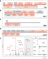
The development of novel therapies against neurodegenerative disorders requires the ability to detect their early, presymptomatic manifestations in order to enable treatment before irreversible cellular damage occurs. Precocious signs indicative of neurodegeneration include characteristic changes in certain protein levels, which can be used as diagnostic biomarkers when they can be detected in fluids such as blood plasma or cerebrospinal fluid. In the case of synucleinopathies, cerebrospinal alpha-synuclein (α-syn) has attracted great interest as a potential biomarker; however, there is ongoing debate regarding the association between cerebrospinal α-syn levels and neurodegeneration in Parkinson disease and synucleinopathies. Post-translational modifications (PTMs) have emerged as important determinants of α-syn's physiological and pathological functions. Several PTMs are enriched within Lewy bodies and exist at higher levels in α-synucleinopathy brains, suggesting that certain modified forms of α-syn might be more relevant biomarkers than the total α-syn levels. However, the quantification of PTMs in bodily fluids poses several challenges. This review describes the limitations of current immunoassay-based α-syn quantification methods and highlights how these limitations can be overcome using novel mass-spectrometry-based assays. In addition, we describe how advances in chemical synthesis, which have enabled the preparation of α-syn proteins that are site-specifically modified at single or multiple residues, can facilitate the development of more accurate assays for detecting and quantifying α-syn PTMs in health and disease.
Figures


Similar articles
-
Phosphorylated α-synuclein as a potential biomarker for Parkinson's disease and related disorders.Expert Rev Mol Diagn. 2012 Mar;12(2):115-7. doi: 10.1586/erm.12.5. Expert Rev Mol Diagn. 2012. PMID: 22369370 No abstract available.
-
Posttranslational Modifications of α-Synuclein, Their Therapeutic Potential, and Crosstalk in Health and Neurodegenerative Diseases.Pharmacol Rev. 2024 Oct 16;76(6):1254-1290. doi: 10.1124/pharmrev.123.001111. Pharmacol Rev. 2024. PMID: 39164116 Free PMC article. Review.
-
Cerebrospinal fluid Tau/α-synuclein ratio in Parkinson's disease and degenerative dementias.Mov Disord. 2011 Jul;26(8):1428-35. doi: 10.1002/mds.23670. Epub 2011 Apr 5. Mov Disord. 2011. PMID: 21469206
-
Characterization of molecular biomarkers in cerebrospinal fluid and serum of E46K-SNCA mutation carriers.Parkinsonism Relat Disord. 2022 Mar;96:29-35. doi: 10.1016/j.parkreldis.2022.01.024. Epub 2022 Feb 5. Parkinsonism Relat Disord. 2022. PMID: 35149357
-
Role of post-translational modifications in modulating the structure, function and toxicity of alpha-synuclein: implications for Parkinson's disease pathogenesis and therapies.Prog Brain Res. 2010;183:115-45. doi: 10.1016/S0079-6123(10)83007-9. Prog Brain Res. 2010. PMID: 20696318 Review.
Cited by
-
Top/Middle-Down Characterization of α-Synuclein Glycoforms.Anal Chem. 2023 Dec 12;95(49):18039-18045. doi: 10.1021/acs.analchem.3c02405. Epub 2023 Dec 4. Anal Chem. 2023. PMID: 38047498 Free PMC article.
-
Ultrastructural and biochemical classification of pathogenic tau, α-synuclein and TDP-43.Acta Neuropathol. 2022 Jun;143(6):613-640. doi: 10.1007/s00401-022-02426-3. Epub 2022 May 5. Acta Neuropathol. 2022. PMID: 35513543 Free PMC article. Review.
-
Novel human cell expression method reveals the role and prevalence of posttranslational modification in nonmuscle tropomyosins.J Biol Chem. 2021 Oct;297(4):101154. doi: 10.1016/j.jbc.2021.101154. Epub 2021 Sep 1. J Biol Chem. 2021. PMID: 34478714 Free PMC article.
-
Comparison of strategies for non-perturbing labeling of α-synuclein to study amyloidogenesis.Org Biomol Chem. 2016 Feb 7;14(5):1584-92. doi: 10.1039/c5ob02329g. Org Biomol Chem. 2016. PMID: 26695131 Free PMC article.
-
LGALS3 (galectin 3) mediates an unconventional secretion of SNCA/α-synuclein in response to lysosomal membrane damage by the autophagic-lysosomal pathway in human midbrain dopamine neurons.Autophagy. 2022 May;18(5):1020-1048. doi: 10.1080/15548627.2021.1967615. Epub 2021 Oct 6. Autophagy. 2022. PMID: 34612142 Free PMC article.
References
-
- Lang A. E., Lozano A. M. (1998) Parkinson's disease. First of two parts. N. Engl. J. Med. 339, 1044–1053 - PubMed
-
- Lang A. E., Lozano A. M. (1998) Parkinson's disease. Second of two parts. N. Engl. J. Med. 339, 1130–1143 - PubMed
-
- Lesage S., Anheim M., Letournel F., Bousset L., Honore A., Rozas N., Pieri L., Madiona K., Durr A., Melki R., Verny C., Brice A. (2013) G51D alpha-synuclein mutation causes a novel parkinsonian-pyramidal syndrome. Ann. Neurol. 73, 459–471 - PubMed
-
- Polymeropoulos M. H., Lavedan C., Leroy E., Ide S. E., Dehejia A., Dutra A., Pike B., Root H., Rubenstein J., Boyer R., Stenroos E. S., Chandrasekharappa S., Athanassiadou A., Papapetropoulos T., Johnson W. G., Lazzarini A. M., Duvoisin R. C., Di Iorio G., Golbe L. I., Nussbaum R. L. (1997) Mutation in the alpha-synuclein gene identified in families with Parkinson's disease. Science 276, 2045–2047 - PubMed
Publication types
MeSH terms
Substances
LinkOut - more resources
Full Text Sources
Other Literature Sources
Medical
Miscellaneous

