

Difficulty in the intravesical morcellation procedure for leiomyoma of the prostate enucleated by HoLEP
- PMID: 23966460
- PMCID: PMC3762389
- DOI: 10.1136/bcr-2013-200200
Difficulty in the intravesical morcellation procedure for leiomyoma of the prostate enucleated by HoLEP
Abstract
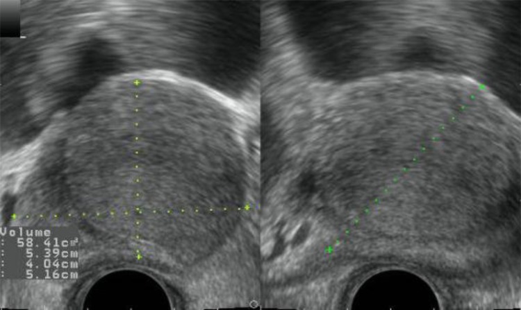
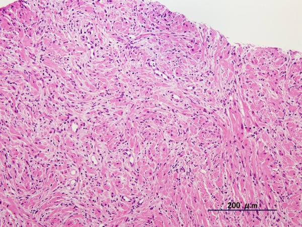
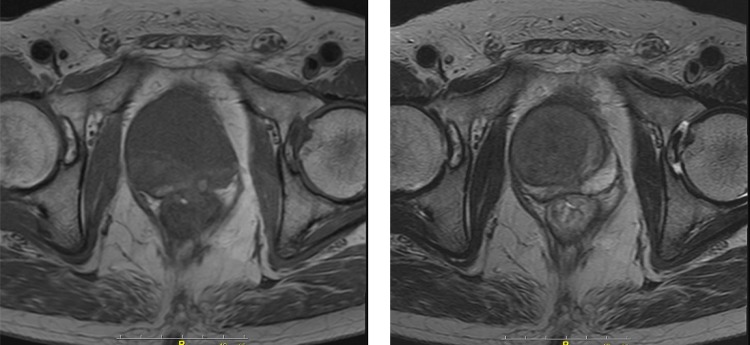
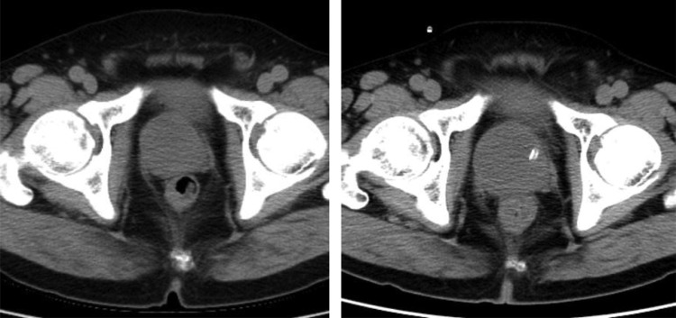
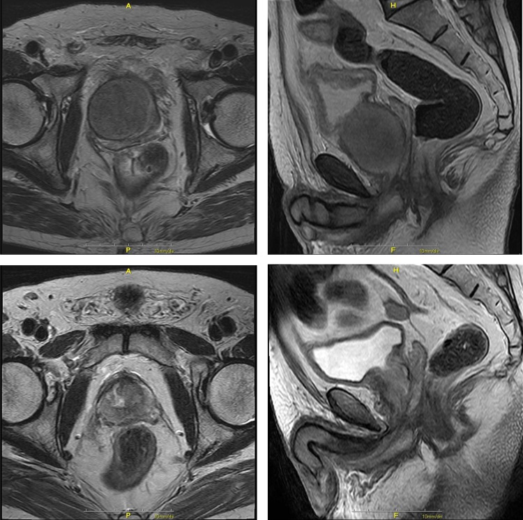
Leiomyoma of prostate are rare benign neoplasms that are usually diagnosed incidentally through postoperative pathological examination. A 70-year-old man is presented with severe symptoms of lower urinary tract obstruction. Although a digital rectal examination and the prostate-specific antigen level did not suggest malignancy, transrectal ultrasonography showed an unusual homogenous echoic mass measuring 45×37 mm in size in the prostate. A needle biopsy was performed and pathological diagnosis was prostatic leiomyoma. Holmium laser enucleation of prostate (HoLEP) was chosen and performed to resect the tumour. Although the enucleation step presented no problems, the morcellation procedure using a conventional transurethral mechanical morcellator was very difficult owing to the firmness of the tissue. By using a bipolar erectrosurgical loop, fragmentation and removal of tissue was accomplished. This is the first case reported of these rare neoplasms in which HoLEP was offered as a less invasive surgical approach. We emphasise the firmness of the leiomyomatous tissue, which would lead to morcellation failure.
Figures
Similar articles
-
Feasibility of holmium laser enucleation of the prostate (HoLEP) for recurrent/residual benign prostatic hyperplasia (BPH).BJU Int. 2012 Dec;110(11 Pt C):E845-50. doi: 10.1111/j.1464-410X.2012.11290.x. Epub 2012 Jun 15. BJU Int. 2012. PMID: 22702908
-
The diagnostic value of prostate cancer between holmium laser enucleation of the prostate and transurethral resection of the prostate for benign prostatic hyperplasia: A retrospective comparative study.Int J Surg. 2020 Jul;79:217-221. doi: 10.1016/j.ijsu.2020.05.025. Epub 2020 May 21. Int J Surg. 2020. PMID: 32447004
-
One-year Surgical Outcomes of Complete or Incomplete Enucleation of Prostate by Monopolar Electrocoagulation, Photoselective Vapoenucleation of 120-W GreenLight Laser, and Holmium Laser.Urology. 2017 Oct;108:142-148. doi: 10.1016/j.urology.2017.07.012. Epub 2017 Jul 20. Urology. 2017. PMID: 28735019
-
Current status of holmium laser enucleation of the prostate.Int J Urol. 2018 Mar;25(3):206-211. doi: 10.1111/iju.13507. Epub 2017 Dec 3. Int J Urol. 2018. PMID: 29205507 Review.
-
Holmium laser enucleation of the prostate and holmium laser ablation of the prostate: indications and outcome.Curr Opin Urol. 2009 Jan;19(1):38-43. doi: 10.1097/MOU.0b013e32831a7008. Curr Opin Urol. 2009. PMID: 19057214 Review.
Cited by
-
Device Malfunctions and Complications Associated with Benign Prostatic Hyperplasia Surgery: Review of the Manufacturer and User Facility Device Experience Database.J Endourol. 2019 Jun;33(6):448-454. doi: 10.1089/end.2019.0067. Epub 2019 May 24. J Endourol. 2019. PMID: 30990073 Free PMC article.
-
Leiomyoma of the Prostate Treated via Holmium Laser Enucleation: A Case Report and Literature Review.Cureus. 2023 Sep 14;15(9):e45273. doi: 10.7759/cureus.45273. eCollection 2023 Sep. Cureus. 2023. PMID: 37846242 Free PMC article.
-
Clinical effects of cluster technology optimization and innovations on laparoscopic splenectomy and azygoportal disconnection: a single-center retrospective study with 500 consecutive cases.Surg Endosc. 2022 Oct;36(10):7409-7418. doi: 10.1007/s00464-022-09159-0. Epub 2022 Mar 7. Surg Endosc. 2022. PMID: 35257212
References
-
- Hansel DE, Herawi M, Montgomery E, et al. Spindle cell lesions of the adult prostate. Mod Pathol 2007;2013:148–58 - PubMed
-
- Oderda M, Mondaini N, Bartoletti R, et al. Leiomyomata of the genitourinary tract: a case series from the “rare urological neoplasm” registry. Scand J Urol 2013;2013:158–62 - PubMed
-
- Nakamura K, Shiramizu M. [Pure leiomyoma of prostate presenting with rectal symptoms: a case report]. Hinyokika Kiyo 1992;2013:1067–9 - PubMed
-
- Hossain D, Meiers I, Qian J, et al. Prostatic leiomyoma with atypia: follow-up study of 10 cases. Ann Diagn Pathol 2008;2013:328–32 - PubMed
-
- Khalil KH, Rix GH, McBrien MP, et al. Bizarre leiomyoma of the prostate. Br J Urol 1997;2013:660. - PubMed
Publication types
MeSH terms
LinkOut - more resources
Full Text Sources
Other Literature Sources
Medical
Molecular Biology Databases