. 2013 Oct;24(20):3187-204.
doi: 10.1091/mbc.E13-06-0301. Epub 2013 Aug 21.

Cooperation between Rho-GEF Gef2 and its binding partner Nod1 in the regulation of fission yeast cytokinesis

Affiliations

Cooperation between Rho-GEF Gef2 and its binding partner Nod1 in the regulation of fission yeast cytokinesis

Yi-Hua Zhu et al. Mol Biol Cell. 2013 Oct.

Abstract

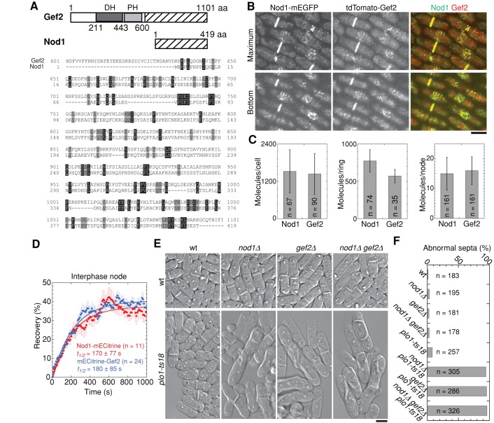
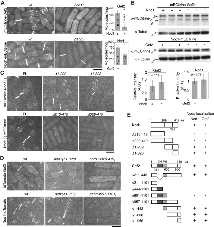
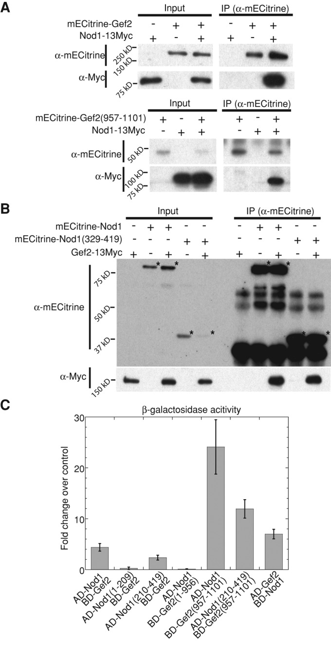
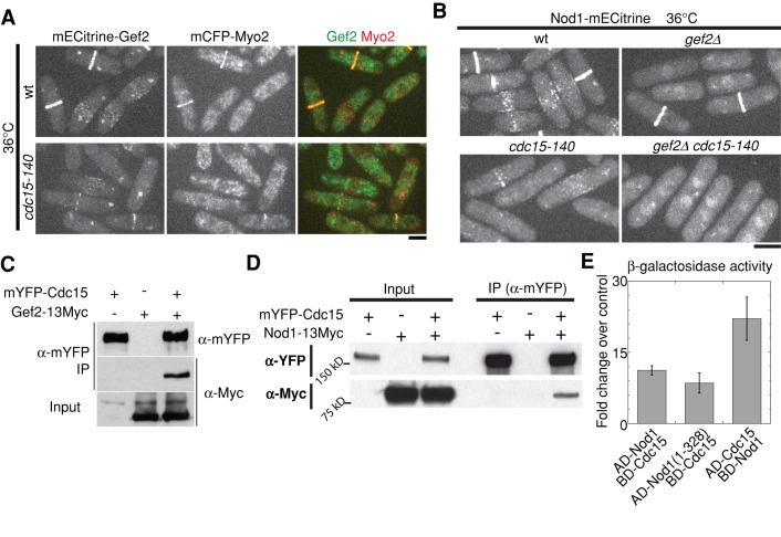
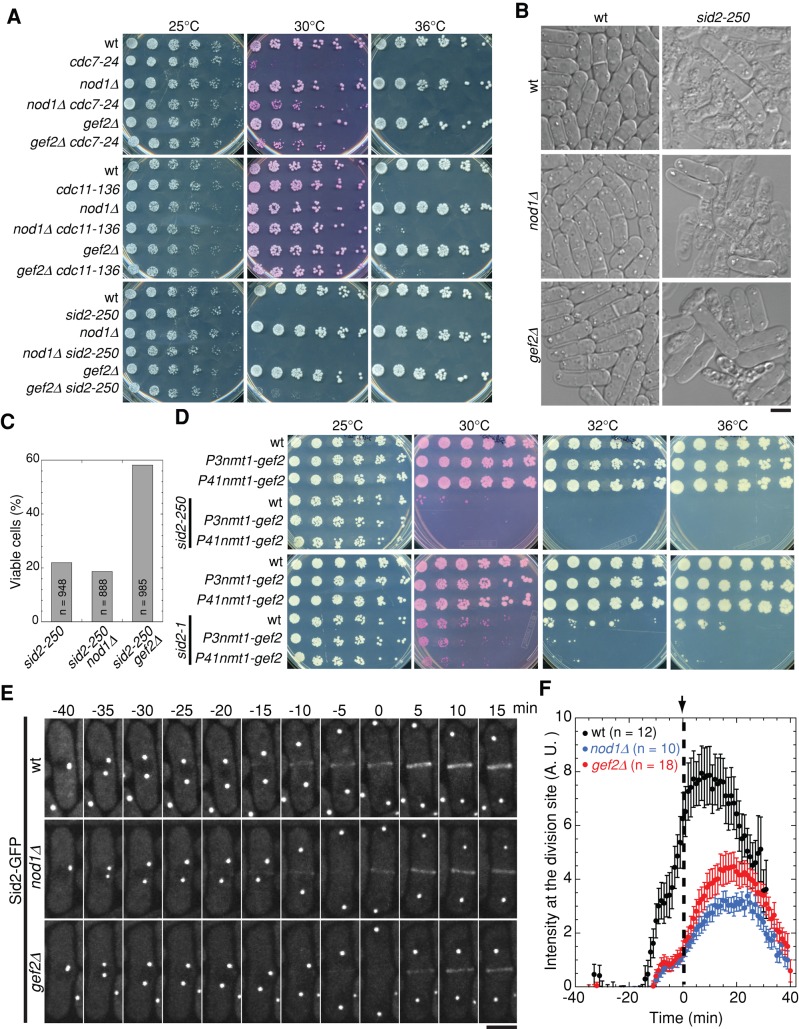
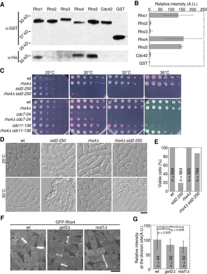
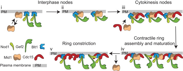
Cytokinesis is the last step of the cell-division cycle, which requires precise spatial and temporal regulation to ensure genetic stability. Rho guanine nucleotide exchange factors (Rho GEFs) and Rho GTPases are among the key regulators of cytokinesis. We previously found that putative Rho-GEF Gef2 coordinates with Polo kinase Plo1 to control the medial cortical localization of anillin-like protein Mid1 in fission yeast. Here we show that an adaptor protein, Nod1, colocalizes with Gef2 in the contractile ring and its precursor cortical nodes. Like gef2, nod1 has strong genetic interactions with various cytokinesis mutants involved in division-site positioning, suggesting a role of Nod1 in early cytokinesis. We find that Nod1 and Gef2 interact through the C-termini, which is important for their localization. The contractile-ring localization of Nod1 and Gef2 also depends on the interaction between Nod1 and the F-BAR protein Cdc15, where the Nod1/Gef2 complex plays a role in contractile-ring maintenance and affects the septation initiation network. Moreover, Gef2 binds to purified GTPases Rho1, Rho4, and Rho5 in vitro. Taken together, our data indicate that Nod1 and Gef2 function cooperatively in a protein complex to regulate fission yeast cytokinesis.


Figures

FIGURE 1:
FIGURE 1:
Nod1 colocalizes with Gef2 in cortical nodes and the contractile ring and shares similar function with Gef2 in division-site selection. (A) Nod1 shares similarity with Gef2 C-terminus. Top, schematics of Gef2 and Nod1 domains or regions. The similar regions between Nod1 and Gef2 are marked with the same pattern. Bottom, sequence alignment between Gef2 aa 601–1101 (top row) and FL Nod1 (bottom row) using Vector NTI program. Identical and similar (D/E, I/L/V, K/R, N/Q, and S/T) aa are shaded in black and gray, respectively. (B–F) Cells were grown and imaged at 25°C. (B) Colocalization of Nod1 with Gef2 in cortical nodes and the contractile ring (strain JW4457). Top, maximum intensity projection. Bottom, single slice at cell bottom. (C) Molecule numbers of mECitrine-Gef2 (JW4912) and Nod1-mECitrine (JW4008) globally in whole cells and locally in the contractile ring and interphase nodes. (D) FRAP analysis of Nod1 (JW4008) and Gef2 (JW3825). Cells were bleached at time zero. Mean ± SEM. (E, F) Nod1 and Gef2 have similar function in division-site positioning. (E) Differential interference contrast (DIC) images and (F) quantification of the division-site positioning. The abnormal septa are defined as septa not placed within the central 20% of the cell or not within 80–100° angle to the long axis of the cell. Strains used: wt (JW81), nod1∆ (JW3773), gef2∆ (JW1826), nod1∆ gef2∆ (JW3814), plo1-ts18 (IH1600), nod1∆ plo1-ts18 (JW3815), gef2∆ plo1-ts18 (JW3078), and nod1∆ gef2∆ plo1-ts18 (JW3873). Bars, 5 μm.
FIGURE 2:
FIGURE 2:
Nod1 and Gef2 are interdependent on their C-termini for cortical-node localization and partially interdependent for contractile-ring localization. (A) Micrographs of Nod1 and Gef2 localization in wt and deletion mutants (left). Molecules in the contractile ring were counted (right). Cells expressing mECitrine-Gef2 (JW3825 and JW4014) and Nod1-mECitrine (JW4008 and JW4038) were used. (B) Nod1 and Gef2 protein levels in wt (+) and the deletion (−) mutants. Cells extracts from the strains used in A were loaded in triplicate in Western blotting (top). Tubulin was used as the loading control. The graphs show the quantification of the protein levels (bottom). (C) Micrographs of Nod1 localization in cells expressing mECitrine-tagged FL Nod1 (JW4750 and JW4008) or Nod1 truncations (JW5065, JW4856, JW4325, and JW4326). (D) Micrographs of localization of Nod1 and Gef2 (strains JW4226, JW5107, JW4359, JW4010, JW4256, and JW4355). (E) Summary of Nod1 and Gef2 localization to cortical nodes in different truncation mutants. +, localized to cortical nodes; −, not localized to cortical nodes. Bars, 5 μm.
FIGURE 3:
FIGURE 3:
Nod1 and Gef2 physically interact through their C-termini. (A, B) Antibodies against mECitrine were used in IP. Monoclonal antibodies against mECitrine and Myc were used in Western blotting. (A) Nod1 co-IP with Gef2 C-terminus. IPs were carried out from cell extracts of nod1-13Myc (JW4013), mECitrine-gef2 (JW3825), mECitrine-gef2 nod1-13Myc (JW4330), mECitrine-gef2(957-1101) (JW3826), nod1-13Myc (JW4013), and mECitrine-gef2(957-1101) nod1-13Myc (JW4331). (B) Gef2 co-IP with Nod1 C-terminus. Strains JW3622, JW4453, JW5093, JW4455, and JW5095 were used. Asterisks mark the expected bands. (C) Nod1 and Gef2 interact via their C-termini in yeast two-hybrid assays. β‑Galactosidase activities (mean ± SD, n = 2) are shown as fold changes over the highest negative control.
FIGURE 4:
FIGURE 4:
The F-BAR protein Cdc15 recruits or stabilizes Gef2 and Nod1 localization to the contractile ring by interaction with the Nod1 N-terminus. (A, B) Involvement of Cdc15 in Gef2 and Nod1 localization at the contractile ring. Cells expressing mECitrine-Gef2 (A) and Nod1-mECitrine (B) were cultured at 25°C and shifted to 36°C for 2 h before imaging at 36°C. Myo2 was used to mark the contractile ring in A. Strains used were JW4008, JW4038, JW5027, JW5028, JW5582, and JW5583. (C, D) Cdc15 interacts with Gef2 and Nod1 in co-IPs (similar to Figure 3A). Strains used were JW1063, JW5120, JW4013, JW3325, and JW3204. (E) Cdc15 interacts with Nod1 N-terminus in yeast two-hybrid assays. β‑Galactosidase activities (mean ± SD, n = 2) as fold changes over the highest negative control are shown. Bars, 5 μm.
FIGURE 5:
FIGURE 5:
Nod1 and Gef2 affect contractile-ring stability. (A) nod1∆ and gef2∆ display synthetic interaction with cdc15-140. Serial dilutions (3×) of indicated strains (JW81, JW1743, JW4259, JW4016, JW2854, and JW2937) on YE5S plates at 25, 30, and 36°C, respectively. (B–D) nod1∆ cdc15-140 and gef2∆ cdc15-140 cells display typical cytokinesis defects with elongated and multinucleated cells. Relevant strains used in A were cultured in YE5S liquid at 25° (top) or 30°C (bottom) for 6 h before imaging. (B) Before imaging at 30°C, cells were stained with Hoechst for 10 min at 30°C to visualize DNA (green). DIC in gray. (C) Cell length and (D) number of nuclei in cells grown at 30°C for 6 h. (E–G) Nod1 and Gef2 affect contractile-ring stability during cytokinesis at 30°C. Rlc1 and Bgs1 were used to monitor the contractile ring and septum formation. Cells were grown at 30°C for 6 h before imaging at 30°C. Strains used: JW5357, JW5329, and JW5330. (E) Time courses of selected images from a movie with 1-min delay. The entire series can be viewed in Supplemental Videos S1 and S2. (F) Quantification of cells that fail to maintain the contractile ring (CR) after ring assembly. (G) Mean intensity of Rlc1-tdTomato at CR. Rlc1 intensity is significantly reduced in nod1∆ cdc15-140 (p < 0.001) and gef2∆ cdc15-140 (p < 0.001) cells vs. cdc15-140 cells. Bars, 5 μm.
FIGURE 6:
FIGURE 6:
nod1∆ and gef2∆ suppress SIN mutants by reducing cell lysis. (A) Serial dilutions (3×) of indicated strains on YE5S or YE5S + phloxin B (red dye accumulated in dead cells) plates at 25, 30, and 36°C. Strains used: wt (JW81), cdc7-24 (TP34), nod1∆ (JW4259), nod1∆ cdc7-24 (JW4304), gef2∆ (JW2854), gef2∆ cdc7-24 (JW3021), cdc11-136 (TP47), nod1∆ cdc11-136 (JW4306), gef2∆ cdc11-136 (JW2972), sid2-250 (YDM429), nod1∆ sid2-250 (JW4294), and gef2∆ sid2-250 (JW3009). (B, C) gef2∆ but not nod1∆ partially rescued cell lysis in sid2-250. Cells were grown in liquid culture at 25°C and then shifted 30°C for 6 h. (B) DIC images of sid2 mutant strains used in A. (C) Percentage of viable cells. Dead or lysed cells were identified as those that failed to maintain their cytoplasm. (D) Overexpression of Gef2 is synthetic lethal with sid2 mutants. Serial dilutions (3×) of indicated strains on YE5S or YE5S + phloxin B plates at 25, 30, and 36°C. Strains used: JW81, JW3561, JW3562, YDM429, JW5360, JW5361, VS2367, JW5405, and JW5406. (E, F) Sid2 localization at the division site is compromised in nod1∆ and gef2∆. Time 0 marks the end of anaphase B. (E) Time courses of representative cells expressing Sid2-GFP in wt (YDM415), gef2∆ (JW5580), and nod1∆ (JW5581). (F) Quantification of the intensity (mean ± SEM) of Sid2-GFP at the division site for strains in E. Black arrow and dashed line mark time 0. Bars, 5 μm.
FIGURE 7:
FIGURE 7:
Gef2 GEF domain binds to GTPases Rho1, Rho4, and Rho5 in vitro. (A, B) Purified GST-Rho GTPases and GST control were bound to the beads and then incubated with purified His‑GEF domain (aa 211–600) of Gef2. The amount of pulled down Gef2 was detected by Western blotting (A) and quantified (B). The intensities of His-Gef2(GEF) bands were measured, background subtracted, corrected for Rho GTPase amount, and normalized by setting the intensity of His-Gef2(GEF) in GST control as 1. The experiment was repeated, and mean ± SD is shown in B. (C–E) rho4∆ suppresses SIN mutants. Strains used: JW81, JW3041, YDM429, JW5505, TP34, JW5503, TP47, and JW5504. (C) Serial dilutions (3×) of indicated strains on YE5S or YE5S + phloxin B plates at 25, 30, 32, and 36°C for 3 d. (D, E) rho4∆ rescues the cell-lysis phenotype of sid2-250. (D) DIC images of cells grown in liquid culture at 25°C or after 6 h at 30°C. (E) Quantification of viable (not lysed or dead) cells after 6 h at 30°C. (F, G) Gef2 and Nod1 play a role in Rho4 localization. (F) Micrographs of GFP-Rho4 in wt (PPG1580) and the deletion mutants (JW4909 and JW4910). (G) Quantification of Rho4 intensity at the division site for strains in (F). Bars, 5 μm.
FIGURE 8:
FIGURE 8:
Model of Nod1 and Gef2 localization and interactions with other proteins on the cytoplasmic side of the plasma membrane during the cell cycle. i) During interphase, Nod1 and Gef2 localize to interphase nodes via Blt1 or other interphase-node proteins, ii) where they help to recruit and stabilize anillin-related protein Mid1. iii) The nodes mature into cytokinesis nodes and coalesce into the contractile ring as more Mid1 and other cytokinesis proteins like F-BAR protein Cdc15 arrive at the division site. iv) Cdc15 continuously recruits or stabilize the Nod1/Gef2 complex during ring maturation, which helps to maintain the contractile-ring integrity and stability. v) Mid1 disappears from the ring at the onset of its constriction. For clarity, the potential interactions between Gef2 and Rho GTPases are not shown.

Similar articles

Cited by

References

    1. Almonacid M, Celton-Morizur S, Jakubowski JL, Dingli F, Loew D, Mayeux A, Chen JS, Gould KL, Clifford DM, Paoletti A. Temporal control of contractile ring assembly by Plo1 regulation of myosin II recruitment by Mid1/anillin. Curr Biol. 2011;21:473–479. - PMC - PubMed
    1. Almonacid M, Moseley JB, Janvore J, Mayeux A, Fraisier V, Nurse P, Paoletti A. Spatial control of cytokinesis by Cdr2 kinase and Mid1/anillin nuclear export. Curr Biol. 2009;19:961–966. - PubMed
    1. Arasada R, Pollard TD. Distinct roles for F-BAR proteins Cdc15p and Bzz1p in actin polymerization at sites of endocytosis in fission yeast. Curr Biol. 2011;21:1450–1459. - PMC - PubMed
    1. Arellano M, Duran A, Pérez P. Localisation of the Schizosaccharomyces pombe rho1p GTPase and its involvement in the organisation of the actin cytoskeleton. J Cell Sci. 1997;110:2547–2555. - PubMed
    1. Bähler J, Steever AB, Wheatley S, Wang Y-L, Pringle JR, Gould KL, McCollum D. Role of polo kinase and Mid1p in determining the site of cell division in fission yeast. J Cell Biol. 1998a;143:1603–1616. - PMC - PubMed

Publication types

MeSH terms

LinkOut - more resources