. 2013 Nov;110(10):2350-7.
doi: 10.1152/jn.00218.2013. Epub 2013 Aug 21.

Short- and long-term plasticity in CA1 neurons from mice lacking h-channel auxiliary subunit TRIP8b

Affiliations

Short- and long-term plasticity in CA1 neurons from mice lacking h-channel auxiliary subunit TRIP8b

Darrin H Brager et al. J Neurophysiol. 2013 Nov.

Abstract

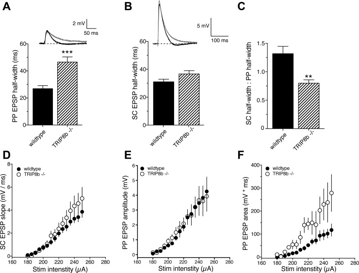
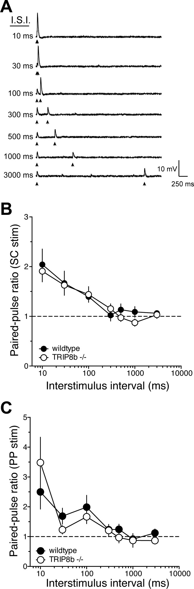
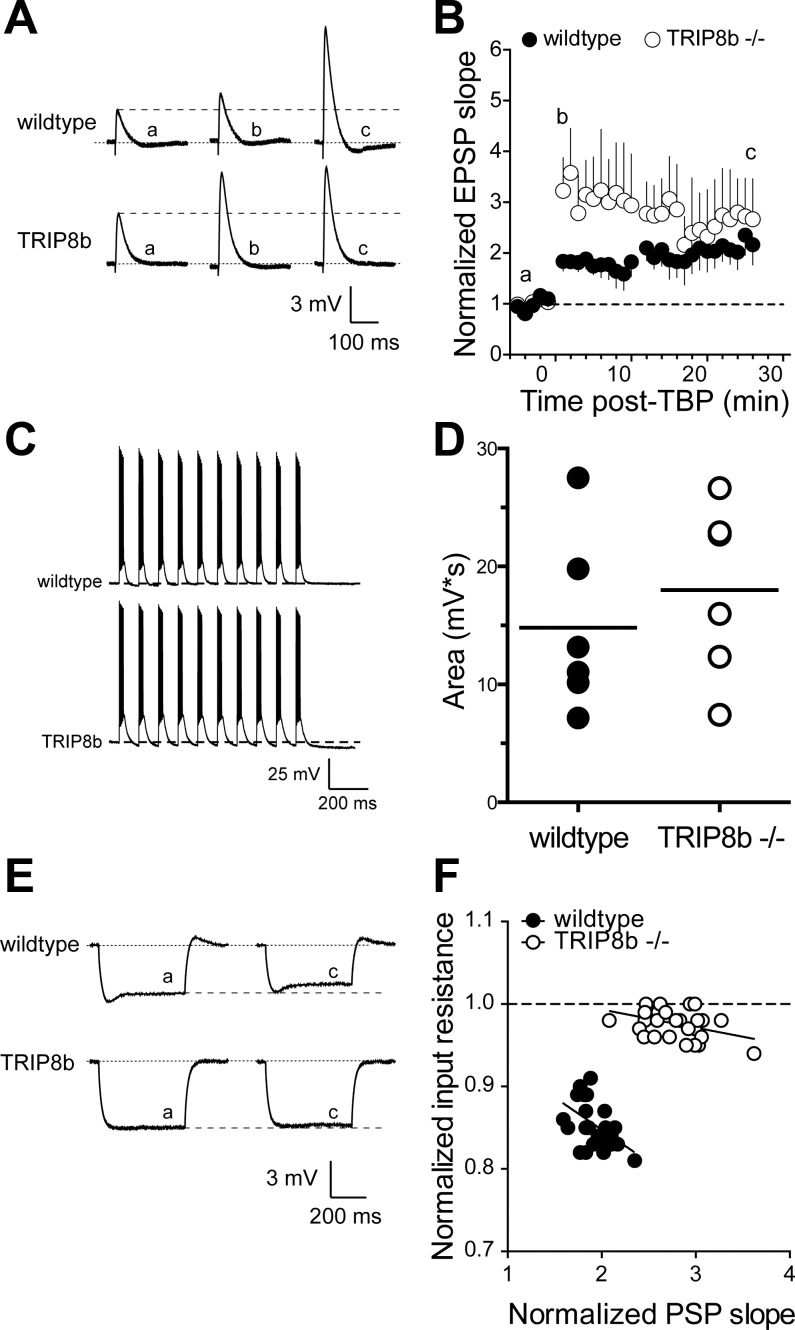
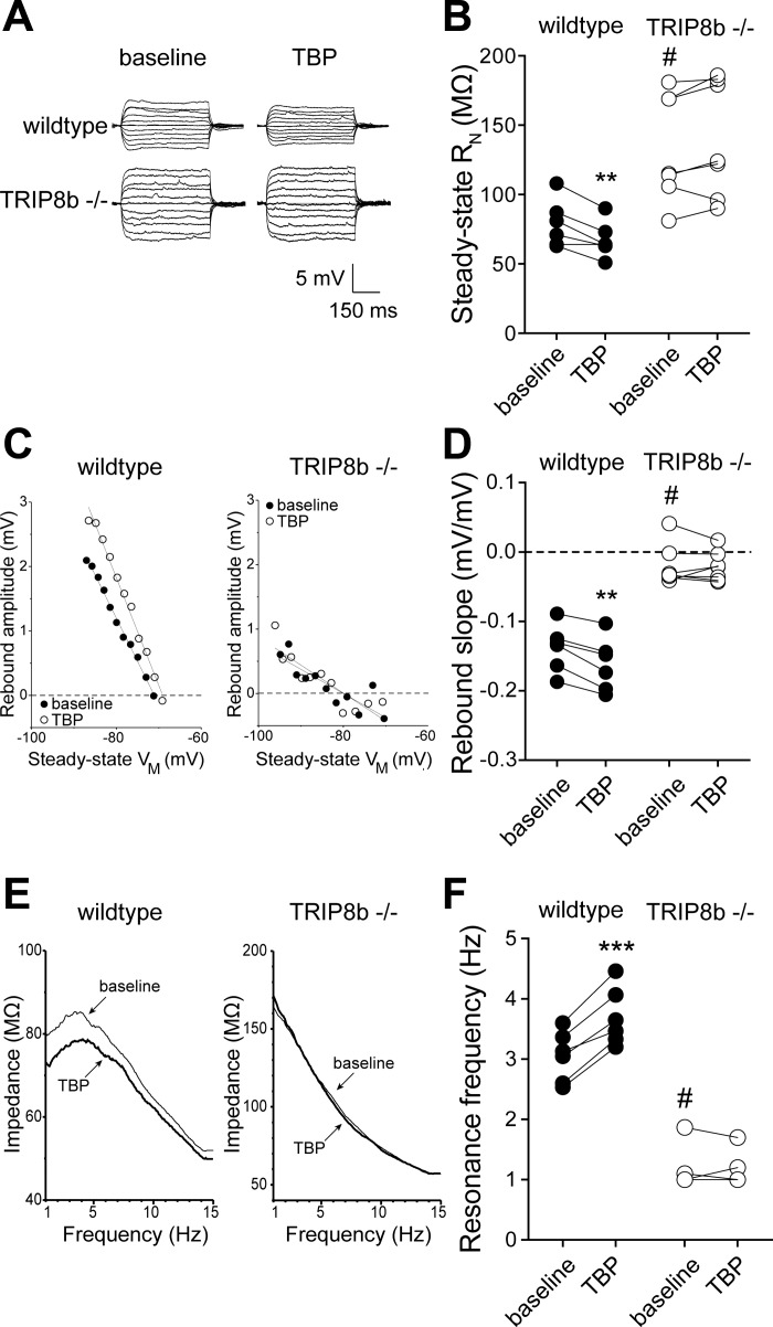
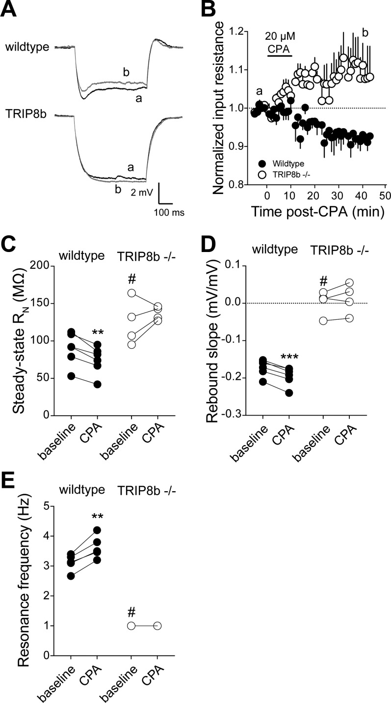
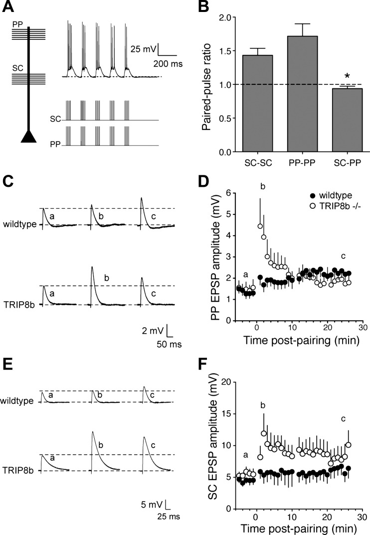
Hyperpolarization-activated cyclic nucleotide-gated nonselective cation channels (HCN or h-channels) are important regulators of neuronal physiology contributing to passive membrane properties, such as resting membrane potential and input resistance (R(N)), and to intrinsic oscillatory activity and synaptic integration. The correct membrane targeting of h-channels is regulated in part by the auxiliary h-channel protein TRIP8b. The genetic deletion of TRIP8b results in a loss of functional h-channels, which affects the postsynaptic integrative properties of neurons. We investigated the impact of TRIP8b deletion on long-term potentiation (LTP) at the two major excitatory inputs to CA1 pyramidal neurons: Schaffer collateral (SC) and perforant path (PP). We found that SC LTP was not significantly different between neurons from wild-type and TRIP8b-knockout mice. There was, however, significantly more short-term potentiation in knockout neurons. We also found that the persistent increase in h-current (I(h)) that normally occurs after LTP induction was absent in knockout neurons. The lack of I(h) plasticity was not restricted to activity-dependent induction, because the depletion of intracellular calcium stores also failed to produce the expected increase in I(h). Interestingly, pairing of SC and PP inputs resulted in a form of LTP in knockout neurons that did not occur in wild-type neurons. These results suggest that the physiological impact of TRIP8b deletion is not restricted to the integrative properties of neurons but also includes both synaptic and intrinsic plasticity.

Keywords: Ih; Schaffer collateral; hippocampus; intrinsic plasticity; perforant path.


Figures

Fig. 1.
Fig. 1.
TRIP8b deletion preferentially affects perforant path (PP) excitatory postsynaptic potentials (EPSPs). A: PP EPSP half-width was significantly greater in TRIP8b−/− neurons compared with wild-type neurons. B: Schaffer collateral (SC) EPSP half-width was not significantly different between wild-type and TRIP8b−/− neurons. C: the ratio of SC to PP EPSP half-width was significantly smaller in TRIP8b−/− neurons. D: relationship between SC EPSP slope and stimulus intensity for wild-type and TRIP8b−/− neurons. E and F: amplitude (E) and area (F) of PP EPSPs as a function of stimulus intensity. **P < 0.01 vs. wild type; ***P < 0.005 vs. wild type.
Fig. 2.
Fig. 2.
TRIP8b deletion does not affect paired-pulse ratio at either SC or PP synapses. A: representative traces showing the effect of paired stimulation [interstimulus interval (ISI) 10–3,000 ms] used to determine paired-pulse ratio. B and C: paired-pulse ratio as a function of ISI for SC (B) and PP (C) EPSPs.
Fig. 3.
Fig. 3.
SC EPSPs in TRIP8b-knockout mice show greater short-term potentiation but normal long-term potentiation after theta burst pairing (TBP). A: representative EPSPs in response to SC stimulation before, 1–2 min after, and 25 min after TBP. Note that a, b, c refer to the indicated time points in B. B: time course of EPSP potentiation after TBP. There was significantly more short-term potentiation, indicated by b, in TRIP8b-knockout neurons compared with control. C: representative traces from wild-type and TRIP8b-knockout neurons showing the voltage response during the TBP protocol. D: total depolarizing area under the voltage waveform during LTP induction for all TBP experiments. E: representative voltage traces indicating that input resistance decreased after TBP in wild-type but not TRIP8b-knockout neurons. Note that a and c refer to the indicated times in B. F: relationship between the change in input resistance and the change in EPSP slope. Data represent the time course of the change in input resistance and EPSP slope after TBP. Note the significantly reduced slope in TRIP8b-knockout neurons compared with wild-type neurons.
Fig. 4.
Fig. 4.
TBP-dependent intrinsic plasticity is absent in neurons from TRIP8b-knockout mice. A: representative voltage traces used to measure input resistance before and after TBP in wild-type and TRIP8b-knockout neurons. B: summary graph showing a significant decrease in input resistance (RN) after TBP in wild-type but not TRIP8b-knockout neurons. Note that baseline input resistance in TRIP8b-knockout neurons was significantly higher than wild type. C: representative plots of rebound amplitude vs. steady-state voltage (VM) from wild-type and TRIP8b-knockout neurons used to calculate rebound slope. Note both the reduced slope and lack of change after TBP in TRIP8b-knockout neurons. D: summary graph showing a significant increase in rebound slope after TBP in wild-type but not TRIP8b-knockout neurons. Note that baseline rebound slope in TRIP8b-knockout neurons was significantly lower than wild type. E: representative impedance amplitude profiles for wild-type and TRIP8b-knockout neurons before and after TBP. Resonance frequency (fR) is the frequency at which the impedance is maximal. F: summary graph showing a significant increase in fR after TBP in wild-type but not TRIP8b-knockout neurons. Note that baseline fR in TRIP8b-knockout neurons was significantly lower than wild type. **P < 0.01, ***P < 0.005 vs. TBP; #P < 0.05 vs. wild type.
Fig. 5.
Fig. 5.
Intracellular calcium store depletion-dependent intrinsic plasticity is absent in neurons from TRIP8b-knockout mice. A: representative voltage traces indicating that input resistance decreased after a 10-min application of 20 μM cyclopiazonic acid (CPA) in wild-type but not TRIP8b-knockout neurons. Note that a and b represent the indicated time points in B. B: summary graph showing the time course of the change in input resistance after CPA application. C: summary graph showing a significant decrease in input resistance after CPA application in wild-type but not TRIP8b-knockout neurons. D: summary graph showing a significant increase in rebound slope after CPA application in wild-type but not TRIP8b-knockout neurons. E: summary graph showing a significant increase in fR after CPA application in wild-type but not TRIP8b-knockout neurons. **P < 0.01, ***P < 0.005 vs. TBP; #P < 0.05 vs. wild type.
Fig. 6.
Fig. 6.
Deletion of TRIP8b results in greater short-term potentiation of PP EPSPs and heterosynaptic potentiation of SC EPSPs. A: representative voltage traces showing the response to paired activation of PP and SC inputs onto wild-type and TRIP8b-knockout CA1 neurons. B: lack of paired-pulse facilitation during paired SC-PP stimulation (ISI = 100 ms), in contrast to SC-SC or PP-PP stimulation. C: representative EPSPs in response to PP stimulation before, 1–2 min after, and 25 min after TBP. Note that a, b, and c refer to the indicated time points in D. D: time course of PP EPSP potentiation after PP-SC pairing. There was significantly more short-term potentiation in TRIP8b-knockout neurons compared with control. E: representative EPSPs in response to SC stimulation before, 1–2 min after, and 25 min after TBP. Note that a, b, and c refer to the indicated time points in F. F: time course of SC EPSP potentiation after PP-SC pairing. Note that LTP was produced in the SC pathway in TRIP8b-knockout neurons but not in wild-type neurons. *P < 0.05.

References

    1. Amaral DG, Witter MP. The three-dimensional organization of the hippocampal formation: a review of anatomical data. Neuroscience 31: 571–591, 1989 - PubMed
    1. Bender RA, Kirschstein T, Kretz O, Brewster AL, Richichi C, Rüschenschmidt C, Shigemoto R, Beck H, Frotscher M, Baram TZ. Localization of HCN1 channels to presynaptic compartments: novel plasticity that may contribute to hippocampal maturation. J Neurosci 27: 4697–4706, 2007 - PMC - PubMed
    1. Bliss TV, Collingridge GL. A synaptic model of memory: long-term potentiation in the hippocampus. Nature 361: 31–39, 1993 - PubMed
    1. Brager DH, Akhavan AR, Johnston D. Impaired dendritic expression and plasticity of h-channels in the fmr1(-/y) mouse model of fragile X syndrome. Cell Rep 1: 225–233, 2012 - PMC - PubMed
    1. Brager DH, Johnston D. Plasticity of intrinsic excitability during long-term depression is mediated through mGluR-dependent changes in Ih in hippocampal CA1 pyramidal neurons. J Neurosci 27: 13926–13937, 2007 - PMC - PubMed

Publication types

MeSH terms

LinkOut - more resources