. 2013 Aug 21;33(34):13882-7.
doi: 10.1523/JNEUROSCI.2524-13.2013.

Perivascular fibroblasts form the fibrotic scar after contusive spinal cord injury

Affiliations

Perivascular fibroblasts form the fibrotic scar after contusive spinal cord injury

Cynthia Soderblom et al. J Neurosci. .

Abstract

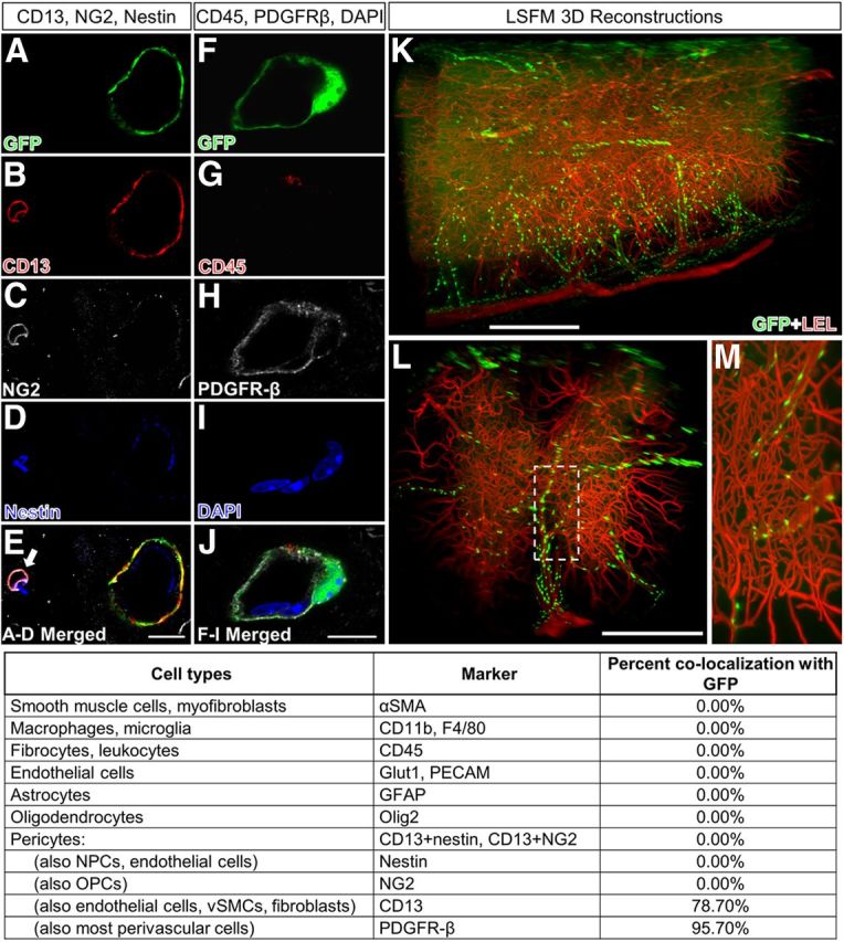
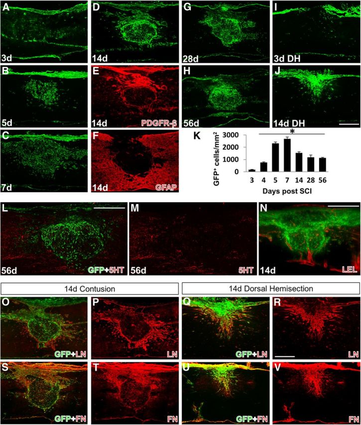
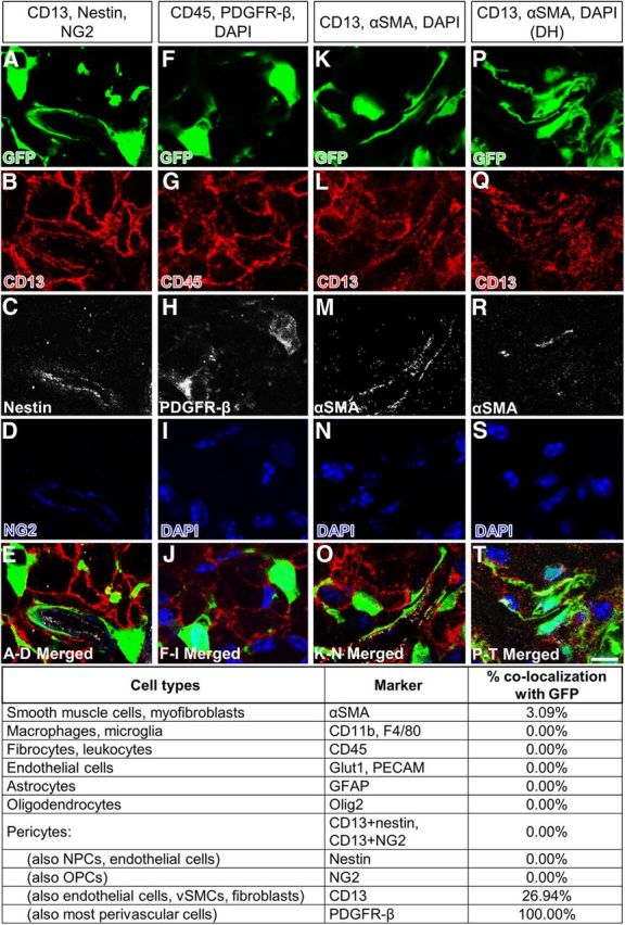
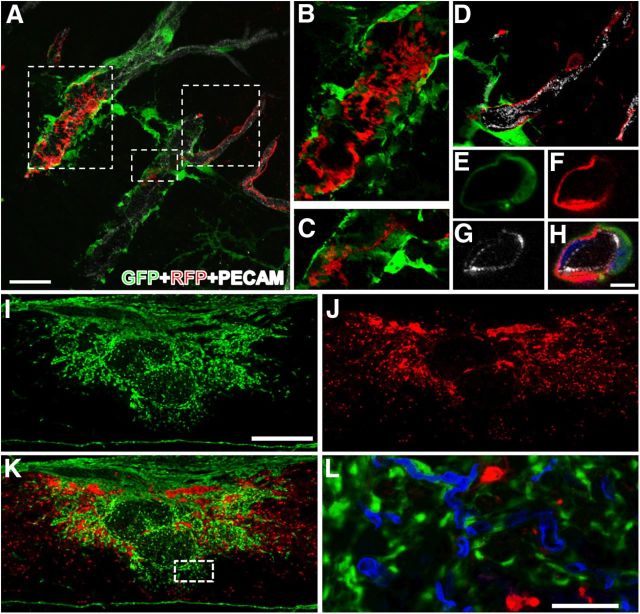
Injury to the CNS leads to formation of scar tissue, which is important in sealing the lesion and inhibiting axon regeneration. The fibrotic scar that comprises a dense extracellular matrix is thought to originate from meningeal cells surrounding the CNS. However, using transgenic mice, we demonstrate that perivascular collagen1α1 cells are the main source of the cellular composition of the fibrotic scar after contusive spinal cord injury in which the dura remains intact. Using genetic lineage tracing, light sheet fluorescent microscopy, and antigenic profiling, we identify collagen1α1 cells as perivascular fibroblasts that are distinct from pericytes. Our results identify collagen1α1 cells as a novel source of the fibrotic scar after spinal cord injury and shift the focus from the meninges to the vasculature during scar formation.


Figures

Figure 1.
Figure 1.
Col1α1-GFP+ cells are preferentially associated with larger blood vessels and express CD13 and PDGFR-β in uninjured spinal cord. Col1α1-GFP+ cells (A,F) express CD13 (B) and PDGFR-β (H), but not NG2 (C), nestin (D), CD45 (G), or markers for fibrocytes, neural cells, or immune cells (pictures not shown). Table lists percentage of Col1α1-GFP+ cells that express each cell type-specific antigen. LSFM reconstructions of spinal vasculature (LEL, red) in Col1α1-GFP mouse spinal cords show Col1α1+ cells (GFP, green) along large spinal vessels but not microvessels (K–M). E, Arrow refers to pericyte. Scale bars: A–J, 10 μm: K, L, 500 μm.
Figure 2.
Figure 2.
Spatiotemporal distribution of Col1α1-GFP+ cells after SCI. Col1α1-GFP+ cells enter the injury site between 3 and 5 dpi (A,B) and persist chronically (C–H). The cell density is highest at 7 dpi (C,K) and by 14 dpi (D) condense into a fibrous scar that apposes to the GFAP+ astrocytic scar (F) and are the predominant PDGFR-β+ cells in the scar (E). Serotonin (5HT) axons fail to penetrate the Col1α1-GFP+ region (L). M, Red channel in L. After dorsal hemisection, Col1α1+ cells also do not enter the injury site until after 3 dpi (I) and fill the injury epicenter by 14 dpi (J). LEL-labeled vasculature (red) encloses the fibrotic scar along the dense peripheral rim of Col1α1-GFP+ cells. N, a single LSFM section. O–V, Representative images of laminin (LN) and fibronectin (FN) expression relative to Col1α1+ cells in the scar. *p < 0.0001 (one-way ANOVA). Scale bars: A–J, L–V, 500 μm.
Figure 3.
Figure 3.
Antigenic profile of Col1α1-GFP+ cells at 14 dpi. Col1α1-GFP+ cells (A,F,K) continue to express PDGFR-β (H) after contusive SCI (A–O). A smaller percentage of cells continue to express CD13 (B,L). Most Col1α1-GFP+ cells expressed αSMA at 5 dpi (data not shown) but decrease to ∼3% by 14 dpi (M). Similar αSMA expression was observed after dorsal hemisection (DH) (P–T). Table lists percentage of Col1α1-GFP+ cells that express each cell type-specific antigen after contusive SCI. Scale bar, 10 μm.
Figure 4.
Figure 4.
Col1α1+ cells are distinct from NG2+ pericytes before and after injury. A, D, Col1α1+ cells surround larger-diameter arterioles with NG2+ pericytes (red) along smaller microvessels. A–C, NG2+ cells are also present as stellate cells. NG2+ cells are always encircled by Col1α1+ cells (E–H; blue represents DAPI) when present in overlapping regions. At 14 dpi (n = 3, I–L), Col1α1+ and NG2+ cells that have migrated to the injury site occupy distinct regions, with Col1α1+ cells occupying injury epicenter and NG2+ cells in the surrounding region. Col1α1+ cells appear to be coming off of blood vessels (Glut1, blue, L) at the outer edges of the fibrotic scar. L represents dashed region in K. Scale bars: A, 30 μm; E–H, 5 μm; I–K, 500 μm; L, 50 μm.

References

    1. Alliot F, Rutin J, Leenen PJ, Pessac B. Pericytes and periendothelial cells of brain parenchyma vessels co-express aminopeptidase N, aminopeptidase A, and nestin. J Neurosci Res. 1999;58:367–378. doi: 10.1002/(SICI)1097-4547(19991101)58:3<367::AID-JNR2>3.0.CO%3B2-T. - DOI - PubMed
    1. Armulik A, Genové G, Betsholtz C. Pericytes: developmental, physiological, and pathological perspectives, problems, and promises. Dev Cell. 2011;21:193–215. doi: 10.1016/j.devcel.2011.07.001. - DOI - PubMed
    1. Bundesen LQ, Scheel TA, Bregman BS, Kromer LF. Ephrin-B2 and EphB2 regulation of astrocyte-meningeal fibroblast interactions in response to spinal cord lesions in adult rats. J Neurosci. 2003;23:7789–7800. - PMC - PubMed
    1. Donnelly DJ, Popovich PG. Inflammation and its role in neuroprotection, axonal regeneration and functional recovery after spinal cord injury. Exp Neurol. 2008;209:378–388. doi: 10.1016/j.expneurol.2007.06.009. - DOI - PMC - PubMed
    1. Göritz C, Dias DO, Tomilin N, Barbacid M, Shupliakov O, Frisén J. A pericyte origin of spinal cord scar tissue. Science. 2011;333:238–242. doi: 10.1126/science.1203165. - DOI - PubMed

Publication types

MeSH terms