Review
. 2013 Aug 8:7:729-45.
doi: 10.2147/DDDT.S15467. eCollection 2013.

Developing therapeutic approaches for metachromatic leukodystrophy

Affiliations
Review

Developing therapeutic approaches for metachromatic leukodystrophy

Shilpa A Patil et al. Drug Des Devel Ther. .

Abstract

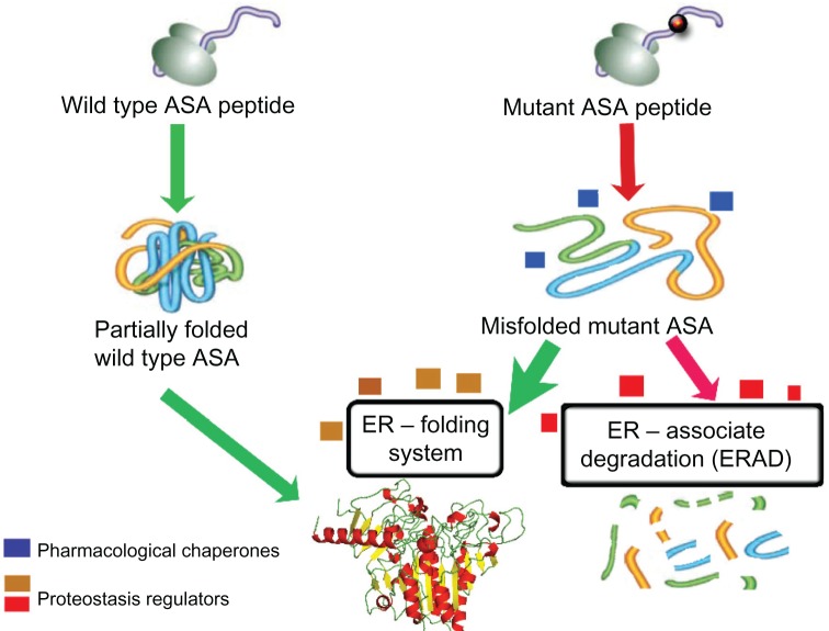
Metachromatic leukodystrophy (MLD) is an autosomal recessive lysosomal disorder caused by the deficiency of arylsulfatase A (ASA), resulting in impaired degradation of sulfatide, an essential sphingolipid of myelin. The clinical manifestations of MLD are characterized by progressive demyelination and subsequent neurological symptoms resulting in severe debilitation. The availability of therapeutic options for treating MLD is limited but expanding with a number of early stage clinical trials already in progress. In the development of therapeutic approaches for MLD, scientists have been facing a number of challenges including blood-brain barrier (BBB) penetration, safety issues concerning therapies targeting the central nervous system, uncertainty regarding the ideal timing for intervention in the disease course, and the lack of more in-depth understanding of the molecular pathogenesis of MLD. Here, we discuss the current status of the different approaches to developing therapies for MLD. Hematopoietic stem cell transplantation has been used to treat MLD patients, utilizing both umbilical cord blood and bone marrow sources. Intrathecal enzyme replacement therapy and gene therapies, administered locally into the brain or by generating genetically modified hematopoietic stem cells, are emerging as novel strategies. In pre-clinical studies, different cell delivery systems including microencapsulated cells or selectively neural cells have shown encouraging results. Small molecules that are more likely to cross the BBB can be used as enzyme enhancers of diverse ASA mutants, either as pharmacological chaperones, or proteostasis regulators. Specific small molecules may also be used to reduce the biosynthesis of sulfatides, or target different affected downstream pathways secondary to the primary ASA deficiency. Given the progressive neurodegenerative aspects of MLD, also seen in other lysosomal diseases, current and future therapeutic strategies will be complementary, whether used in combination or separately at specific stages of the disease course, to produce better outcomes for patients afflicted with this devastating inherited disorder.

Keywords: arylsulfatase A; enzyme enhancement therapy; enzyme replacement therapy; gene therapy; metachromatic leukodystrophy; small molecules.


Figures

Figure 1
Figure 1
Sulfatide metabolic pathway. Notes: The figure shows the biosynthetic pathways of sulfatide, the sphingolipid that accumulates during metachromatic leukodystrophy MLD. The specific enzymes described in each step of the pathway are in italics. The rounded boxes depict the products or substrates of the reactions. The green shaded square shapes show different therapeutic approaches for MLD treatment. Two current substrate reduction therapies for another lysosomal disease (Gaucher disease) are also shown: N-Butyldeoxynojirimycin (NB-DNJ) miglustat (Zavesca®, Actelion Pharmaceuticals Ltd, Allschwil, Switzerland), and eliglustat tartrate. Both of these small molecules competitively inhibit ceramide glucosyltransferase. Abbreviations: ASA, arylsulfatase A; EET, enzyme enhancement therapy; ERT, enzyme replacement therapy; Gal, galactose; HSCT, hematopoietic stem cell transplantation; Glc, glucose; SGal-Glc-cer, sulfo-galactose-glucosylceramide; MLD, metachromatic leukodystrophy; SRT, substrate reduction therapy.
Figure 2
Figure 2
Schematic representation of the mechanism of action of enzyme enhancement agents including pharmacological chaperones (PCs) and proteostasis regulators (PRs). Notes: ASA is synthesized in the endoplasmic reticulum (ER) as several other proteins target to different intracellular organelles, plasma membrane, and those destined to be secreted to the extracellular environment. Concomitant to the translation process, nascent ASA peptide interacts with several components of the ER folding system. Once folded properly, ASA is targeted to the ER exit pathway and ultimately reaches the lysosomal compartment, where it exerts its physiological function. Missense mutations in the ARSA gene can affect physicochemical interactions between amino acids, resulting in impairment of the folding process of the mutant ASA. PCs (blue boxes) are small molecules that physically interact with the target protein, in this case ASA, and assist its folding, preventing it from being targeted to the ERAD pathway and ultimately degraded by the ubiquitin-proteasomal system. Thus, increased levels of mutant ASA reach the lysosomes to degrade the natural substrate (sulfatides). eventually, other small molecules function as PRs (beige/brown boxes), which can improve the cellular folding capacity by interacting with ER folding components. These molecules can also promote physiological cellular responses, such as unfolded protein response, which can be advantageous to enhance folding of specific misfolded ASA mutants. Other PRs (red boxes) can also potentially inhibit components of ERAD and favor the interactions of misfolded ASA mutants with the ER folding elements, increasing the probability of reaching a folding-like conformation. Abbreviations: ASA, arylsulfatase A; ER, endoplasmic reticulum; ERAD, endoplasmic reticulum associate degradation; PC, pharmacological chaperones; PR, proteostasis regulators.
Figure 3
Figure 3
Cell-based high-throughput screening (HTS) assay. Notes: Here, a cell-based HTS assay for galactocerebrosidase (GALC), which is the lysosomal enzyme deficient in Krabbe disease or globoid cell leukodystrophy (GLD), is depicted for (A and B). The 1,536-well plate contains GLD patient cells that are cultured and treated with a small molecule collection for a specific period of time. After the treatment period, the specific enzyme GALC can be measured using a fluorescent substrate, 6-hexadecanoylamino-4-methylumbelliferyl-β-D-galactoside (HMU) for the enzyme. In each 1,536-well plate, control cells (columns 2 and 3) are seeded and later assayed. These control cells have normal Gal-ase enzymatic activity and are not treated with the library. The columns 1 and 2 contain only culture medium and no cells, and are used as backgrounds for the assays. Columns 5–48 contain cultured patients cells (B)– in this case, fibroblasts. Each well (yellow square) corresponds to a specific small molecule from the library, which is tested in different concentrations. (C) Two-plate view diagrams are shown with the fluorescent signals generated from the HMU against the different rows of a 1,536-well plate. Substantial assay window is observed between GALC enzymatic activity of controls (triangles) and GLD patient cells (circles). The small molecule candidates are selected based on the enhancement of the residual GALC enzyme activity. (D) The quantitative nature of the HTS assay, where seven different concentrations of the target library were tested, allows the generation of concentration-response curves (CRCs) given by the HMU fluorescence signal increase. Based on the characteristics of the CRCs, prioritization of “hits” can be done during the validation process with secondary and tertiary assays.

References

    1. Austin J, McAfee D, Armstrong D, O’Rourke M, Shearer L, Bachhawat B. Low sulfatase activities in metachromatic leukodystrophy (MLD). A controlled study of enzymes in 9 living and 4 autopsied patients with MLD. Trans Am Neurol Assoc. 1964;89:147–150. - PubMed
    1. Bosio A, Binczek E, Stoffel W. Functional breakdown of the lipid bilayer of the myelin membrane in central and peripheral nervous system by disrupted galactocerebroside synthesis. Proc Natl Acad Sci U S A. 1996;1293(23):13280–13285. - PMC - PubMed
    1. Aggarwal S, Yurlova L, Simons M. Central nervous system myelin: structure, synthesis and assembly. Trends Cell Biol. 2011;21(10):585–593. - PubMed
    1. Faust PL, Kaye EM, Powers JM. Myelin lesions associated with lysosomal and peroxisomal disorders. Expert Rev Neurother. 2010;10(9):1449–1466. - PubMed
    1. Gieselmann V. Metachromatic leukodystrophy: genetics, pathogenesis and therapeutic options. Acta Paediatr Suppl. 2008;97(457):15–21. - PubMed

Publication types

MeSH terms