. 2013;11(8):e1001624.
doi: 10.1371/journal.pbio.1001624. Epub 2013 Aug 13.

A Bacteriophage tailspike domain promotes self-cleavage of a human membrane-bound transcription factor, the myelin regulatory factor MYRF

Affiliations

A Bacteriophage tailspike domain promotes self-cleavage of a human membrane-bound transcription factor, the myelin regulatory factor MYRF

Zhihua Li et al. PLoS Biol. 2013.

Abstract

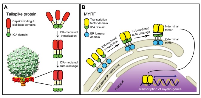
Myelination of the central nervous system (CNS) is critical to vertebrate nervous systems for efficient neural signaling. CNS myelination occurs as oligodendrocytes terminally differentiate, a process regulated in part by the myelin regulatory factor, MYRF. Using bioinformatics and extensive biochemical and functional assays, we find that MYRF is generated as an integral membrane protein that must be processed to release its transcription factor domain from the membrane. In contrast to most membrane-bound transcription factors, MYRF proteolysis seems constitutive and independent of cell- and tissue-type, as we demonstrate by reconstitution in E. coli and yeast. The apparent absence of physiological cues raises the question as to how and why MYRF is processed. By using computational methods capable of recognizing extremely divergent sequence homology, we identified a MYRF protein domain distantly related to bacteriophage tailspike proteins. Although occurring in otherwise unrelated proteins, the phage domains are known to chaperone the tailspike proteins' trimerization and auto-cleavage, raising the hypothesis that the MYRF domain might contribute to a novel activation method for a membrane-bound transcription factor. We find that the MYRF domain indeed serves as an intramolecular chaperone that facilitates MYRF trimerization and proteolysis. Functional assays confirm that the chaperone domain-mediated auto-proteolysis is essential both for MYRF's transcriptional activity and its ability to promote oligodendrocyte maturation. This work thus reveals a previously unknown key step in CNS myelination. These data also reconcile conflicting observations of this protein family, different members of which have been identified as transmembrane or nuclear proteins. Finally, our data illustrate a remarkable evolutionary repurposing between bacteriophages and eukaryotes, with a chaperone domain capable of catalyzing trimerization-dependent auto-proteolysis in two entirely distinct protein and cellular contexts, in one case participating in bacteriophage tailspike maturation and in the other activating a key transcription factor for CNS myelination.


Conflict of interest statement

The authors have declared that no competing interests exist.

Figures

Figure 1
Figure 1. Full-length MYRF is generated as a membrane protein.
(A) Predicted sequence features of MYRF and sequence diagrams of various MYRF constructs used for IF microscopy. Stars in blue indicate predicted NLSs at K245KRK248 and K482KGK485. (B) IF images of GFP-MYRF, MYRF-GFP, MYRFΔTM-GFP, and MYRF-1:756-GFP in HeLa cells. (C) IF image of 3F-MYRF-GFP in HeLa cells. Scale bar, 10 µm.
Figure 2
Figure 2. Full-length MYRF is a type-II membrane protein.
(A) Predicted sequence features of MYRF and sequence diagrams of various MYRF constructs used for experiments. (B) Western blot of HeLa cells transfected with pcDNA3 and 3F-MYRF. (C) The top band of HeLa cells that were transfected with 5M-MYRF-3F has the same electrophoretic mobility as full-length protein products for the same construct that were obtained with an in vitro translation system. (D) Full-length forms of MYRF consist of two closely spaced bands that represent glycosylated and unglycosylated full-length MYRF, respectively (indicated by the two arrows). (E) HeLa cells transfected with 3F-MYRF were disrupted using a Dounce-type homogenizer, and then centrifuged at 200× g for 5 min to obtain a supernatant fraction. It was mixed with 0.1 volume of each of the following chemicals: 5 M NaCl, 1 M Na2CO3 (pH 11), and 10% SDS. After incubation for 20 min at room temperature, mixtures were centrifuged at 20,000× g for 15 min at 4°C to separate supernatant (S) from pellet (P). Calnexin, a known integral membrane protein, served as a control. (F) Membrane topology of GFP-MYRF-3F and 3F-MYRF-L690A-GFP in HeLa cells. When cell membranes were selectively permeated by digitonin, FLAG IF signals of GFP-MYRF-3F could not be detected, indicating that the C-terminus of MYRF is located within the ER lumen. In contrast, FLAG IF signals of 3F-MYRF-L690A-GFP were robustly detected even when cell membranes were selectively permeated by digitonin, indicating that the N-terminus of full-length MYRF is located on the cytoplasmic side of ER membranes. Scale bar, 10 µm.
Figure 3
Figure 3. The ICA domain autonomously mediates the proteolytic processing of MYRF.
(A) Multiple sequence alignment of the ICA domains from eukaryotes, a bacterium, and a phage, generated with ClustalW . Strictly conserved residues are shown in red. The numbering system is based on MYRF. (B) The auto-processing mechanism for MYRF postulated based on the ICA domain and its known properties. (C) Western blots of HeLa cells transfected with various MYRF constructs, showing the effects of mutations in the ICA domain on the proteolytic processing of MYRF. (D) IF image of 3F-MYRF-S578A and 3F-MYRF-K583A in HeLa cells. (E) The amino acid sequence of MYRF (residues N567-R692) was mapped onto the crystal structure of an ICA domain (PDB ID: 3GW6) using the alignment shown in Figure S3A. In the zoomed active site are shown two key catalytic residues (S578 and K583, both belonging to the same subunit) in stick model and two strictly conserved residues (V670 of one subunit and G626 of a different subunit) in space filling model. Shown below are L683, I687, and L690 that were predicted to form a leucine zipper. For visual clarity, clipped images were generated when deemed necessary. (F) (Left) Western blot showing that the proteolytic processing of MYRF is independent of its membrane insertion. MYRF-1:756 is a mutant truncated before the TM domain at L756. (Middle) Western blot showing the proteolytic processing of MYRF-319:708 in HeLa cells and E. coli. (Right) Western blot showing the normal processing of full-length MYRF in budding yeast. Scale bar, 10 µm.
Figure 4
Figure 4. The N-terminal trimer is formed by the ICA domain and enters the nucleus.
(A) Predicted sequence features of MYRF and sequence diagrams of various MYRF constructs used for experiments. (B) Western blots showing co-immunoprecipitation results for the MYRF constructs. “Input” was incubated with FLAG antibody-coated beads and then spun down to separate “Sup” from “Bead” fractions. The failure of MYRF-1:577 to homo-oligomerize demonstrated the importance of the ICA domain for the N-terminal trimer formation. (C) When the NLSs (NLS1 and NLS2) were deleted, the nuclear translocation of the N-terminal trimer was partially blocked. Scale bar, 20 µm.
Figure 5
Figure 5. Auto-processing is essential for the functions of MYRF.
(A) The transcriptional activity of various MYRF constructs was estimated by their ability to activate the transcription of Edn2 in HeLa cells. Values are means ± SEM. (B) Examples of transfected CG4 cells that matured to express MBP or O1. (C) Quantification of the proportion of transfected CG4 cells expressing MBP or O1. Values are means ± SEM. *p<0.05, **p<0.01, and ***p<0.001. Scale bar, 10 µm.
Figure 6
Figure 6. The ICA domain catalyzes trimerization-dependent auto-proteolysis in entirely distinct protein and cellular contexts.
(A) In K1F bacteriophage, the C-terminal ICA domain within each tailspike endosialidase auto-catalytically removes itself following tailspike trimerization, guiding maturation of the six tailspikes surrounding the phage tail (shown for phage K1E, adapted from [55]). (B) Once generated as a type-II membrane protein, the ICA domain is thought to induce the trimerization of MYRF, upon which it cleaves itself, generating two independent trimers. The N-terminal trimer translocates to the nucleus and activates the transcription of myelin genes by direct DNA binding. The transcriptional role of the N-terminal trimer serves to promote the terminal differentiation of OLs, likely aided by an as-yet-unknown function of the C-terminal trimer that remains in the ER.

Comment in

  • Myelination borrows a trick from phage.
    Roberts RG. Roberts RG. PLoS Biol. 2013;11(8):e1001626. doi: 10.1371/journal.pbio.1001626. Epub 2013 Aug 13. PLoS Biol. 2013. PMID: 23966834 Free PMC article. No abstract available.

References

    1. Hoppe T, Matuschewski K, Rape M, Schlenker S, Ulrich HD, et al. (2000) Activation of a membrane-bound transcription factor by regulated ubiquitin/proteasome-dependent processing. Cell 102: 577–586. - PubMed
    1. Stewart EV, Nwosu CC, Tong Z, Roguev A, Cummins TD, et al. (2011) Yeast SREBP cleavage activation requires the Golgi Dsc E3 ligase complex. Mol Cell 42: 160–171. - PMC - PubMed
    1. Wang X, Sato R, Brown MS, Hua X, Goldstein JL (1994) SREBP-1, a membrane-bound transcription factor released by sterol-regulated proteolysis. Cell 77: 53–62. - PubMed
    1. Goldstein JL, DeBose-Boyd RA, Brown MS (2006) Protein sensors for membrane sterols. Cell 124: 35–46. - PubMed
    1. Shen J, Chen X, Hendershot L, Prywes R (2002) ER stress regulation of ATF6 localization by dissociation of BiP/GRP78 binding and unmasking of Golgi localization signals. Dev Cell 3: 99–111. - PubMed

Publication types

MeSH terms