. 2013;11(8):e1001625.
doi: 10.1371/journal.pbio.1001625. Epub 2013 Aug 13.

MYRF is a membrane-associated transcription factor that autoproteolytically cleaves to directly activate myelin genes

Affiliations

MYRF is a membrane-associated transcription factor that autoproteolytically cleaves to directly activate myelin genes

Helena Bujalka et al. PLoS Biol. 2013.

Abstract

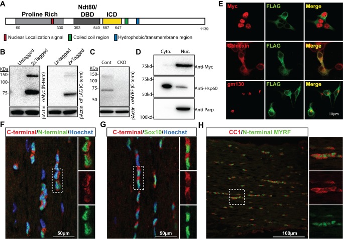
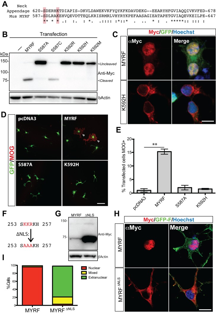
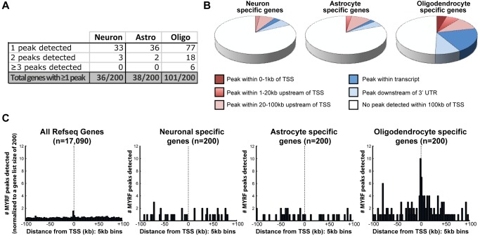
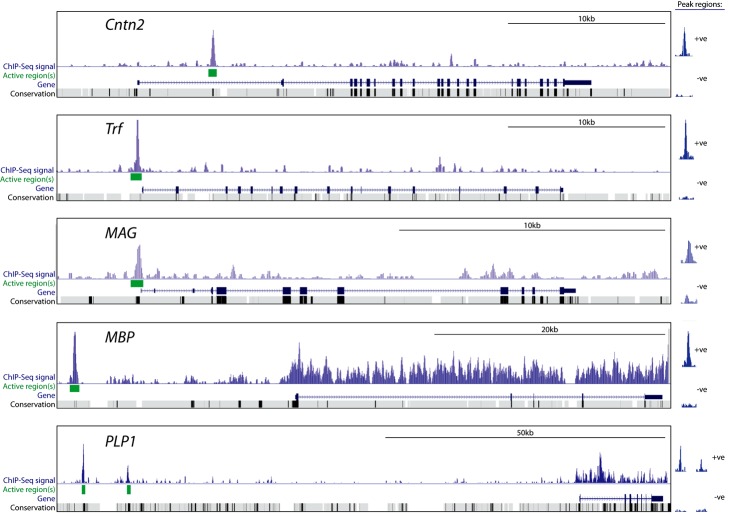
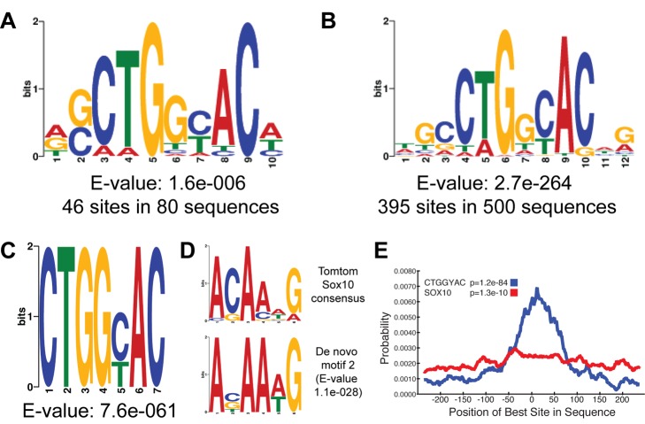
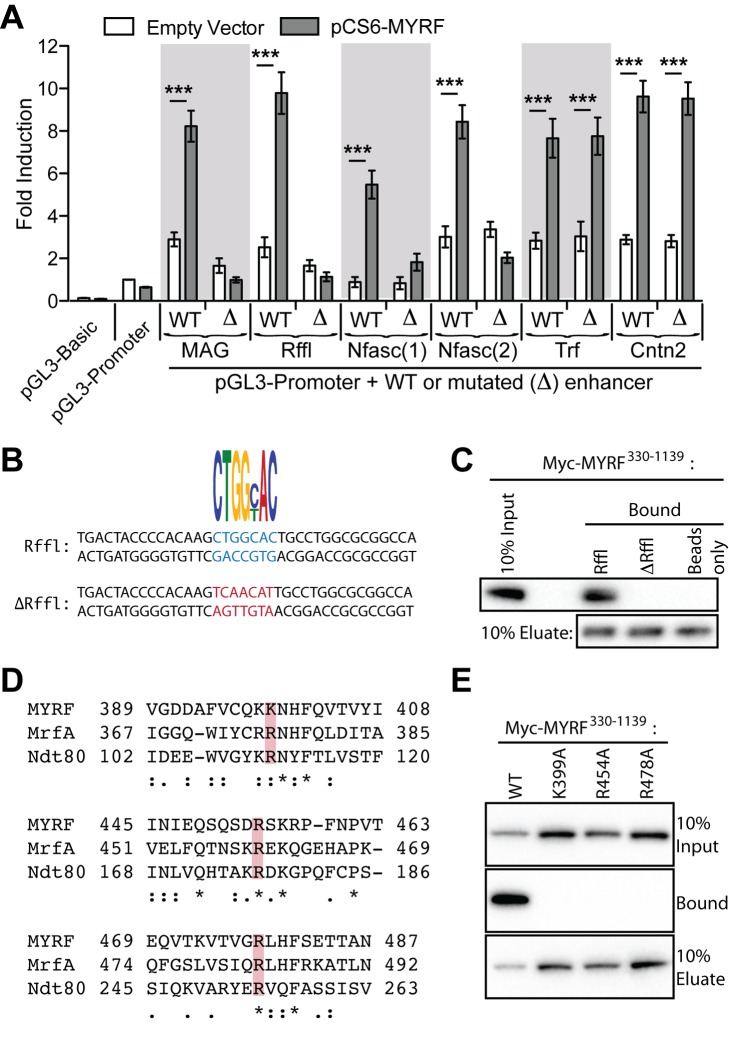
The myelination of axons is a crucial step during vertebrate central nervous system (CNS) development, allowing for rapid and energy efficient saltatory conduction of nerve impulses. Accordingly, the differentiation of oligodendrocytes, the myelinating cells of the CNS, and their expression of myelin genes are under tight transcriptional control. We previously identified a putative transcription factor, Myelin Regulatory Factor (Myrf), as being vital for CNS myelination. Myrf is required for the generation of CNS myelination during development and also for its maintenance in the adult. It has been controversial, however, whether Myrf directly regulates transcription, with reports of a transmembrane domain and lack of nuclear localization. Here we show that Myrf is a membrane-associated transcription factor that undergoes an activating proteolytic cleavage to separate its transmembrane domain-containing C-terminal region from a nuclear-targeted N-terminal region. Unexpectedly, this cleavage event occurs via a protein domain related to the autoproteolytic intramolecular chaperone domain of the bacteriophage tail spike proteins, the first time this domain has been found to play a role in eukaryotic proteins. Using ChIP-Seq we show that the N-terminal cleavage product directly binds the enhancer regions of oligodendrocyte-specific and myelin genes. This binding occurs via a defined DNA-binding consensus sequence and strongly promotes the expression of target genes. These findings identify Myrf as a novel example of a membrane-associated transcription factor and provide a direct molecular mechanism for its regulation of oligodendrocyte differentiation and CNS myelination.


Conflict of interest statement

The authors have declared that no competing interests exist.

Figures

Figure 1
Figure 1. The MYRF protein is subject to posttranslational cleavage.
(A) Schematic of the MYRF protein showing positions of the predicted NLS, Ndt80-like DBD, ICD, coiled coil region, and transmembrane region. (B) Western blot analysis of a double tagged (N-terminal Myc, C-terminal FLAG tagged) or untagged MYRF expression construct in 293T cells. Probing with anti-Myc or anti-FLAG reveals the presence of a ∼140 kDa full-length fragment and truncated cleavage products. (C) Western blot of control and Myrf CKO cultured mouse oligodendrocyte lysates with anti-C-terminal MYRF mAb. (D) Cell fractionation experiment showing that the majority of Myc-tagged N-terminal cleaved MYRF product is present in the nucleus of 293T cells (Anti-Hsp60 and anti-Parp used for cytoplasmic and nuclear controls, respectively). (E) CG-4 cells transfected with the Myc-MYRF-FLAG construct were co-stained for anti-FLAG and either anti-Myc, anti-Calnexin, or anti-golgi matrix protein 130 (gm130). (F–H) Adult mouse optic nerve stained with the anti-N-terminal-MYRF and anti-C-terminal-MYRF-mab antibodies (F), anti-C-terminal-MYRF-mab and anti-Sox10 (G), or anti-N-terminal-MYRF-mab and CC1 (H).
Figure 2
Figure 2. Autoproteolytic processing via the ICD and a NLS are required for nuclear localization of MYRF.
(A) ClustalW2 alignment of the peptide sequence of MYRF and the ICD domain of the bacteriophage GA-1 Neck appendage protein. The serine/lysine dyad residues subjected to mutagenesis are highlighted. (B) Western blot analysis of un-mutated or ICD mutant (K592R, K592H, K592M) Myc-tagged MYRF constructs in 293T cells. (C) Immunofluorescence for the N-terminal Myc tagged MYRF and the K587H mutant in CG-4 cells. Prevention of the cleavage is associated with a loss of nuclear localization of the N-terminus. (D) Primary rat OPC cultures co-transfected with GFP and either empty vector (pcDNA3) or pcDNA3 containing MYRF or the S587A and K592H mutant constructs, stained for MOG. (E) Quantification of the percentage of transfected (GFP+) cells expressing MOG 48 h posttransfection in each condition. **p<0.01 by t test. (F) Predicted NLS within the proline-rich region of MYRF showing the KKR to AAA mutation in the Myc-MYRFΔNLS construct. (G) Western analysis of the Myc-MYRF and Myc-MYRFΔNLS construct; mutation of the NLS has no effects on the cleavage of MYRF, though it routinely led to an increase in protein levels. (H) Representative images of immunostaining for the N-terminal Myc tag showing shift from nuclear to extranuclear staining in the Myc-MYRFΔNLS construct. (I) Quantification of the proportion of predominantly nuclear, mixed, or predominantly extranuclear staining seen with each construct (100 cells assessed/condition). Scale bars, (C and H) 10 µm and (D) 50 µm.
Figure 3
Figure 3. MYRF binds to regions of the genome surrounding oligodendrocyte enriched/myelin genes.
(A) Number of the neuron-, astrocyte-, and oligodendrocyte-specific genes (from , 200 genes per list) with 1, 2, or ≥3 MYRF peaks detected within 100 kb of their TSS. (B) Pie charts showing the proportion of oligodendrocyte-, astrocyte-, or neuron-specific genes that have MYRF binding sites detected within 1 kb, 20 kb, or 100 kb upstream of the TSS, within the gene or downstream of the 3′ UTR (but still within 100 kb of the TSS). (C) Histograms showing the incidence of MYRF peaks relative to the TSS of either an unbiased list of rat Refseq genes (17,090 genes) or the neuron-, astrocyte-, or oligodendrocyte-specific genes. A modest increase in the incidence of MYRF binding is detectable proximal to the TSS of the unbiased gene list; a more pronounced increase in MYRF binding is observed around the TSS of oligodendrocyte-specific genes.
Figure 4
Figure 4. Examples of MYRF peaks proximal to oligodendrocyte-enriched genes.
Signal track shows MYRF occupancy for the Cntn2, Trf, Mag, Mbp, and Plp1 genes. Statistically significant peaks/active regions (green boxes) were identified in regions corresponding to conserved intronic regions (Cntn2), promoter regions (Trf and Mag), and upstream conserved regions (Mbp and Plp1). Inserts to the right show the Myc-MYRF signal and untagged MYRF control signal for each of the peaks/active regions identified. Note high background signal within the Plp1 and Mbp genes (see text for further details).
Figure 5
Figure 5. MYRF binding identifies enhancers of myelin gene expression.
(A) DNA sequences (from 400–700 bp) encompassing the MYRF peaks proximal to the Cntn2, Trf, Mag, Mbp, and Plp1 genes (Figure 4) were cloned into pGL3 upstream of the SV40 promoter and luciferase gene and co-transfected into the CG-4 cell line with either empty (pCS6) or MYRF overexpression (pCS6-MYRF) vectors. Co-expression of MYRF strongly induces luciferase expression in the presence of these enhancers, but has no effect on luciferase activity in their absence. (B) The MYRF-bound regions identified upstream of the Mbp and Plp1 genes strongly promote luciferase expression in control oligodendrocytes relative to Myrf CKO oligodendrocytes, mirroring the loss of expression of MBP protein in the Myrf CKO cells (C). Fold inductions for all conditions are expressed relative to the pGL3-Promoter condition in control cells (pCS6 transfected in A, Myrf Wt/Fl in B). Data are shown as means and SEMs from 4–5 independent experiments. *p<0.05, **p = 0.01, **p<0.001 based on two-way ANOVA with Bonferroni posttest.
Figure 6
Figure 6. De novo identification of consensus sequences from MYRF peaks.
(A) De novo sequence analysis using MEME from 80 peaks identified within 100 kb of oligodendrocyte-enriched genes identifies the sequence CTGGYAC, where Y = C or T. In separate analyses, essentially the same motif was identified using the 100 bp sequences surrounding the 500 strongest peaks using MEME (B) or 500 bp sequences of all 2,085 peaks using DREME (C). The second strongest motif identified in the DREME analysis from (C) was ACAA(A/T)G (D), a strong match for the known consensus sequence for Sox10. (E) Central enrichment analysis of the CTGGYAC and ANAA(A/T)G (Sox10) motifs in the 2,085 500 bp input sequences.
Figure 7
Figure 7. MYRF binds DNA via the CTGGYAC motif.
(A) Mutational analysis of the CTGGYAC motif in six luciferase reporters containing MYRF-bound DNA regions from near the Trf, Mag, Cntn2, Rffl, and Nfasc genes in CG-4 cells. In four out of the six sequences, mutation of a single CTGGYAC sequence (Δ) was sufficient to abolish the effect of MYRF. Fold inductions for all conditions are expressed relative to the pGL3-Promoter and pCS6 co-transfected control cells. Data are shown as means and SEMs from three independent experiments. ***p<0.001. (B) DNA pulldowns using double-stranded oligonucleotides corresponding to the predicted MYRF binding site in the Rffl intronic enhancer, or equivalent oligonucleotides with the seven base pair motif mutated, conjugated to magnetic beads. The wild-type Rffl sequence efficiently captured Myc-MYRF330–1139 from cell lysates, whereas no interaction was detected for the mutated sequence or beads without DNA (C). (D) Sequence alignment between MYRF, Dictyostelium MrfA (Uniprot Q54PT9), and S. cerevisiae Ndt80 (Uniprot P38830) showing conservation of basic amino acids required for DNA binding by Ndt80 (highlighted). (E) DNA pulldown assay measuring interaction between the DNA sequence from the Rffl enhancer and the DNA binding domain of wild-type or mutant MYRFs. All detections performed with anti-Myc.

Comment in

  • Myelination borrows a trick from phage.
    Roberts RG. Roberts RG. PLoS Biol. 2013;11(8):e1001626. doi: 10.1371/journal.pbio.1001626. Epub 2013 Aug 13. PLoS Biol. 2013. PMID: 23966834 Free PMC article. No abstract available.

References

    1. Lu QR, Sun T, Zhu Z, Ma N, Garcia M, et al. (2002) Common developmental requirement for Olig function indicates a motor neuron/oligodendrocyte connection. Cell 109: 75–86. - PubMed
    1. Zhou Q, Anderson DJ (2002) The bHLH transcription factors OLIG2 and OLIG1 couple neuronal and glial subtype specification. Cell 109: 61–73. - PubMed
    1. Yu Y, Chen Y, Kim B, Wang H, Zhao C, et al. (2013) Olig2 targets chromatin remodelers to enhancers to initiate oligodendrocyte differentiation. Cell 152: 248–261. - PMC - PubMed
    1. Xin M, Yue T, Ma Z, Wu FF, Gow A, et al. (2005) Myelinogenesis and axonal recognition by oligodendrocytes in brain are uncoupled in Olig1-null mice. J Neurosci 25: 1354–1365. - PMC - PubMed
    1. Qi Y, Cai J, Wu Y, Wu R, Lee J, et al. (2001) Control of oligodendrocyte differentiation by the Nkx2.2 homeodomain transcription factor. Development 128: 2723–2733. - PubMed

Publication types