. 2013;9(8):e1003173.
doi: 10.1371/journal.pcbi.1003173. Epub 2013 Aug 15.

Simulating cortical development as a self constructing process: a novel multi-scale approach combining molecular and physical aspects

Affiliations

Simulating cortical development as a self constructing process: a novel multi-scale approach combining molecular and physical aspects

Frederic Zubler et al. PLoS Comput Biol. 2013.

Erratum in

Abstract

Current models of embryological development focus on intracellular processes such as gene expression and protein networks, rather than on the complex relationship between subcellular processes and the collective cellular organization these processes support. We have explored this collective behavior in the context of neocortical development, by modeling the expansion of a small number of progenitor cells into a laminated cortex with layer and cell type specific projections. The developmental process is steered by a formal language analogous to genomic instructions, and takes place in a physically realistic three-dimensional environment. A common genome inserted into individual cells control their individual behaviors, and thereby gives rise to collective developmental sequences in a biologically plausible manner. The simulation begins with a single progenitor cell containing the artificial genome. This progenitor then gives rise through a lineage of offspring to distinct populations of neuronal precursors that migrate to form the cortical laminae. The precursors differentiate by extending dendrites and axons, which reproduce the experimentally determined branching patterns of a number of different neuronal cell types observed in the cat visual cortex. This result is the first comprehensive demonstration of the principles of self-construction whereby the cortical architecture develops. In addition, our model makes several testable predictions concerning cell migration and branching mechanisms.


Conflict of interest statement

The authors have declared that no competing interests exist.

Figures

Figure 1
Figure 1. Regulating mechanisms for generating the cell lineage tree during preplate and cortical plate formation.
Preplate formation: The initial progenitor in the ventricular zone (VZ) contains precise amounts of the intracellular substances X and Y. Through symmetric and later asymmetric divisions with differential partition of the substances in the daughter cells, the VZ progenitor pool increases, and the future layer 1 cells (L1) as well as the the subplate (SP) are formed from cells that have left the proliferative state. Cortical plate formation: After the preplate formation, the VZ cells which are still in a proliferative stage express a gene regulatory network (GRN) for the sequential formation of neuron precursors. The GRN consists of pairs of self enhancing, mutually inhibiting genes, sequentially activated at each branch point in the lineage tree, and deciding between further division (in the VZ and later in the subventricular zone – SVZ) or exit from the cell cycle and differentiation into a cortical neuron. (X, Y: intracellular substances; T1, T2: thresholds on intracellular substances; a6, b6, a5, b5, a4, b4, a23, b23: genes. See text and Table 1 for details).
Figure 2
Figure 2. Simulation of the preplate formation.
Projection view exported from the CX3D simulator. (A) Symmetric divisions of the initial precursor have formed a layer of progenitors in the ventricular zone (VZ). (B) The VZ cells (dark grey) undergo asymmetric divisions, forming first the marginal zone, i.e. the future L1 cells (yellow), and then the subplate (light grey). (C). View after completion of the preplate formation.
Figure 3
Figure 3. Simulation of the cortical plate formation.
Slice view exported from the CX3D simulator; for clarity the subplate cells have been removed. (A) After formation of the preplate, the cells in the ventricular zone (VZ) either form radial glial cells (black cells) extending a radial glial process (RGP), or stay in proliferative mode, increasing the precursor pool of the VZ (turquoise). (B) When the first neuron precursors (blue) are formed in the VZ, they migrate by climbing along the RGP toward their final position, forming the future layer 6. Once the neuron precursors detect a contact with either the top-most L1 cells (yellow) or with cells of their own kind, they stop their migration by detaching from the radial fibers. (C) When L5 cells (magenta) are formed they migrate through the L6 cells (some of which are still being produced) and stop just below L1; at the same time the second pool of precursors is formed in the subventricular zone (grey). (D,E) L4 (red) and then L23 cells (green) are produced in the SVZ, and migrate toward L1. L1 cells are physically pushed upward by the neuron precursors, which displaces the stopping signal. (F) In the final laminated structure, there is no VZ or SVZ anymore. The somata start to secrete a diffusible, cell type specific guidance cue in order to guide the axonal and dendritic outgrowth.
Figure 4
Figure 4. Layer specificity of five different cell-types.
(A) Real cortical neurons reconstructed from the cat visual cortex; from left to right: a pyramidal cell of layer 2/3, a spiny stellate cell of layer 4, a pyramidal cell of layer 5, a pyramidal cell of layer 6 (axons drawn in black, dendrites in blue) and a basket cell of layer 2/3. Scale bar: formula image
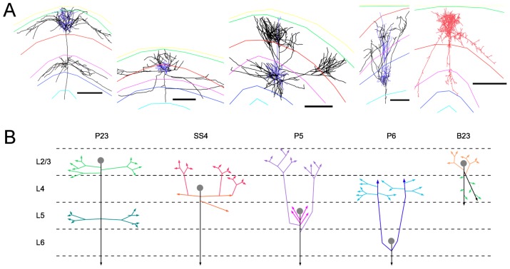
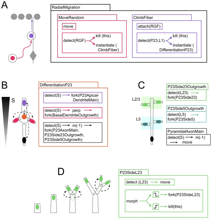
. (Reconstructed cells are a courtesy of Kevan Martin and colleagues, Institute of Neuroinformatics, Zurich). (B) Schematic representation of the axonal branching pattern for encoding in the instruction language G-code. Each color corresponds to a segment of the axonal arbor encoded by a specific G-code machine.
Figure 5
Figure 5. Qualitative analysis of reconstructed cells' morphologies.
To gain a better understanding of its structure, we visualize selectively subparts of each cell and describe them qualitatively. (A) Orientation of the neurites of a P23 cell; the axon (black) leaves the soma toward the white matter, the apical dendrite (blue) toward the pia, the basal dendrites (red, pink, yellow, green) in a perpendicular direction. The lengths of all basal dendrites (path lengths from the tip of the branches to the soma) seem to be constant. The apical dendrite ramifies before entering layer 1. (B) Similar neurite orientation for a P6 cell; the basal dendrites make less bifurcation than in the P23 cell, but here too the path length seems to be constant. Here the apical dendrite does not ramify (no apical ‘tuft’). (C) Large scale axonal structure of a P23 cell. The main trunk of the axon goes down (initially toward the white matter), and makes side branches in layer 2/3 (green, blue) and in layer 5 (pink, red). These side branches usually extend before forming a patch in the same layer (several side branches have been removed for clarity; the complete axon is displayed in E). (D) Large scale axonal structure of a P6 cell (for clarity several side branches have been removed). (E) One quantitative measure which helps to differentiate between the ‘backbone’ (forming the large scale shape of a neuron) and the ‘patches’ is the bouton density, illustrated here with a P23 cell: the segments with more than 0.2 boutons per formula image 
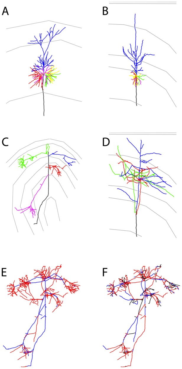
are in red, the segments with fewer boutons are in blue. (F) The differentiation between backbone and patches based on the branching angles is less obvious. Segments displayed in black make an angle formula image degrees to the parent branch (corresponding in principle to side branches); for segments in red the angle with the parent branch is 20–80 degrees (corresponding in principle to bifurcation); the segments in blue are continuations of the mother branch (angle formula image degrees; continuation of a major shaft). Reconstructed cells are courtesy of Kevan Martin and colleagues, Institute of Neuroinformatics, Zurich.
Figure 6
Figure 6. Simulation of axonal and dendritic outgrowth.
Projection view exported from the CX3D simulator. The figure shows the axonal (black) and dendritic (grey) branching pattern for five different cells (from left to right: P23, SS4, P5, P6, B23) in the layered cortical environment grown in Figure 3. The layer specificity reflects the layer specificity observed from the model cells (see also Figure 7). The grey somata are cells that have migrated in the wrong layer, and have been ‘deactivated’ (see text).
Figure 7
Figure 7. Influence of the physical environment on neural growth.
(A,B) Branching pattern of the P23 and the P6 cell after differentiation in a cortical environment. The tortuosity of the branches is due to the mechanical interactions with obstacles (other cells) and to the fluctuating concentrations of signaling molecules. P23: From the down-going main axonal trunk, side branches are formed in layer 2/3 and at the border of layer 5, which tend to extend and ramify in the middle of their respective layers. When branches leave the simulated column, the concentration gradients they are following point in the opposite direction (toward the column), resulting in a turning of the branches. P6: From the down-going main axonal trunk, side branches move up toward layer 4, where they ramify. (C) Similar cells (containing the same G-code machines) in an an environment without any obstacles, and with analytically defined noiseless gradients of guiding cues. Note the smoothness of the branches.
Figure 8
Figure 8. G-code machines used for the growth of a P23 cell.
(A) After it has exited the cell cycle, the P23 precursor cell expresses ‘RadialMigration’, a machine which consists of two sequentially activated internal machines. The first internal machine (‘MoveRandom’) performs a random walk (with an instance of the G-code primitive ‘move’ without directional input) until the cell touches a radial glial fiber (RGF); this contact (recognized with the primitive ‘detect’) triggers the removal (primitive ‘kill’) of the internal machine, and the activation (primitive ‘instantiate’) of the second internal machine (‘ClimbFiber’) for migration along the RGF, until the cell enters layer 1 or touches other P23 cells. When this happens, the migration stops and the differentiation machine is launched. (B) The machine ‘DifferentiationP23’ is expressed when a P23 cell stops its migration. It extends (primitive ‘fork’) an axon, an apical dendrite and several basal dendrite and instantiates into them specific growth cone machines. The direction of sprouting is determined by the gradient of the extracellular substance S (Semaphorin). (C) The axon growth cone is composed of three sub-machines: the first one moves the branch tip down the gradient of Semaphorin; the two others produce side branches when crossing a region with high concentration of the P23 or P5 signaling molecule. (D) The growth cone machine used in the terminal branches forms a recurrent branching process, inserting copies of itself in each daughter branch after bifurcation. During elongation the direction is chosen along the gradient of the layer specific signaling cue. The diameter decreases during elongation and at bifurcation, and is constantly assessed (with the primitive ‘morph’): as long as the diameter is large enough branching occurs further; when the diameter falls under a threshold, the growth cone removes itself and the elongation stops.

References

    1. Kitano H (2002) Systems biology: a brief overview. Science 295: 1662–1664. - PubMed
    1. De Schutter E (2008) Why are computational neuroscience and systems biology so separate? PLoS Comput Biol 4: e1000078. - PMC - PubMed
    1. Alon U (2006) An Introduction to Systems Biology: Design Principles of Biological Circuits. Chapman & Hall/CRC.
    1. Joyner MJ, Pedersen BK (2011) Ten questions about systems biology. J Physiol 589: 1017–1030. - PMC - PubMed
    1. Marée AFM, Hogeweg P (2001) How amoeboids self-organize into a fruiting body: multicellular coordination in Dictyostelium discoideum. Proc Natl Acad Sci U S A 98: 3879–3883. - PMC - PubMed

Publication types

LinkOut - more resources