. 2013 Aug;9(8):e1003525.
doi: 10.1371/journal.ppat.1003525. Epub 2013 Aug 15.

The smallest capsid protein mediates binding of the essential tegument protein pp150 to stabilize DNA-containing capsids in human cytomegalovirus

Affiliations

The smallest capsid protein mediates binding of the essential tegument protein pp150 to stabilize DNA-containing capsids in human cytomegalovirus

Xinghong Dai et al. PLoS Pathog. 2013 Aug.

Abstract

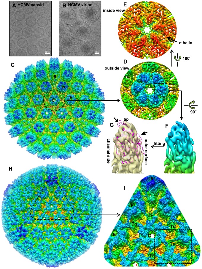
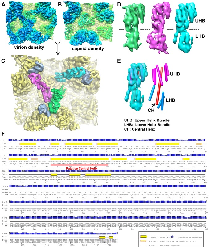
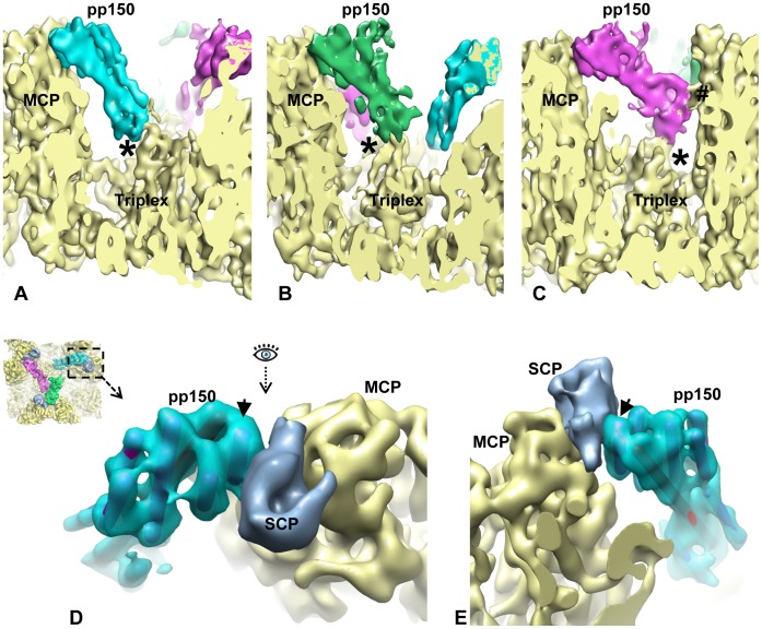
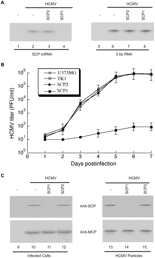
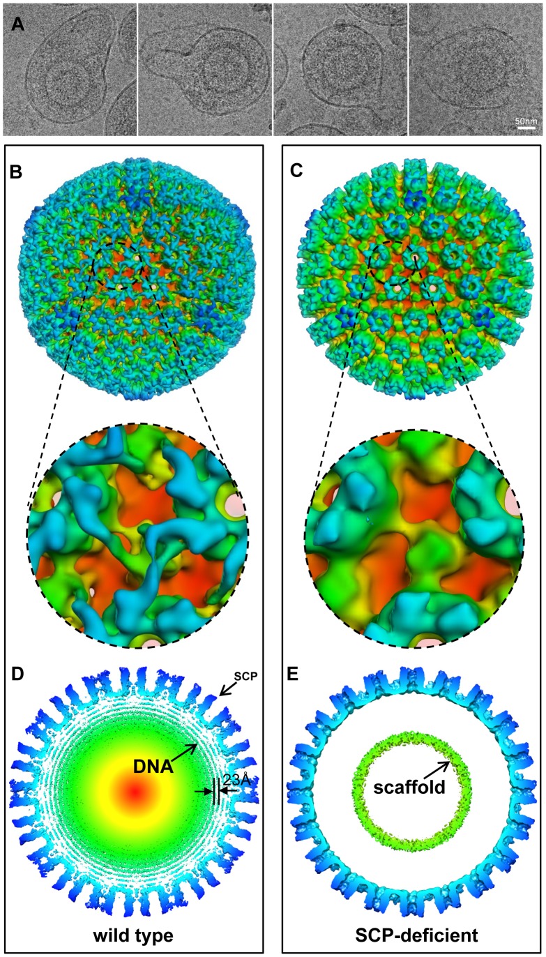
Human cytomegalovirus (HCMV) is a ubiquitous herpesvirus that causes birth defects in newborns and life-threatening complications in immunocompromised individuals. Among all human herpesviruses, HCMV contains a much larger dsDNA genome within a similarly-sized capsid compared to the others, and it was proposed to require pp150, a tegument protein only found in cytomegaloviruses, to stabilize its genome-containing capsid. However, little is known about how pp150 interacts with the underlying capsid. Moreover, the smallest capsid protein (SCP), while dispensable in herpes simplex virus type 1, was shown to play essential, yet undefined, role in HCMV infection. Here, by cryo electron microscopy (cryoEM), we determine three-dimensional structures of HCMV capsid (no pp150) and virion (with pp150) at sub-nanometer resolution. Comparison of these two structures reveals that each pp150 tegument density is composed of two helix bundles connected by a long central helix. Correlation between the resolved helices and sequence-based secondary structure prediction maps the tegument density to the N-terminal half of pp150. The structures also show that SCP mediates interactions between the capsid and pp150 at the upper helix bundle of pp150. Consistent with this structural observation, ribozyme inhibition of SCP expression in HCMV-infected cells impairs the formation of DNA-containing viral particles and reduces viral yield by 10,000 fold. By cryoEM reconstruction of the resulting "SCP-deficient" viral particles, we further demonstrate that SCP is required for pp150 functionally binding to the capsid. Together, our structural and biochemical results point to a mechanism whereby SCP recruits pp150 to stabilize genome-containing capsid for the production of infectious HCMV virion.


Conflict of interest statement

The authors have declared that no competing interests exist.

Figures

Figure 1
Figure 1. Comparison of 3D reconstructions of the HCMV capsid and virion.
(A, B) CryoEM images of HCMV capsid (A) and virion (B). (C) Radially colored surface representation of the 3D reconstruction of the capsid at 6 Å resolution viewed along a 3-fold axis. Capsomers in an asymmetric unit, including a penton and three hexons, are labeled as “5”, “C”, “P” and “E”, respectively, as in the nomenclature of . (D, E) Close-up views of the C hexon demarked in the capsid reconstruction, viewed from outside (D) or inside (E) of the capsid. An α-helix with typical sausage shape is denoted in (E). (F, G) MCPud. The density map of the MCPud denoted by the square in (D) was extracted and radially colored (F). In (G), the same MCPud is shown in semi-transparent yellow and superimposed with the HSV-1 MCPud atomic model (magenta ribbon). Note all helices match in the two structures but the loops at the tip (arrow) and at the outer surface (arrowhead) of HSV-1 MCPud do not fit the cryoEM density map of HCMV MCPud, suggesting possible structural differences. (H) Radially colored surface representation of the 3D reconstruction of the HCMV virion viewed along a 3-fold axis. (I) Zoom-in view of the area denoted in (H). Structural components in an asymmetric unit are labeled, including a penton (“5”), three hexons (“C”, “P” and “E”), and six triplexes (“Ta”, “Tb”, “Tc”, “Td”, “Te” and “Tf”). Dashed square demarcates a region encompassing Te that is segmented out for averaging with similar regions around Tb and Td (see text and Figure 2).
Figure 2
Figure 2. Structure of the capsid-interacting pp150 tegument protein.
(A, B) Averaged density of the regions surrounding triplexes Tb, Td and Te (as marked by the dashed square in Figure 1I) from the virion (A) or capsid (B) reconstructions. The two density maps are colored the same as in Figure 1H or 1C. (C) SCP (light blue) and tegument (green, magenta and cyan) densities segmented from the virion densities of (A) superimposed on the capsid densities (yellow) of (B). (D) Views of the three tegument densities of (C) after alignment to each other. The most prominent feature is the sausage-shaped densities due to helices. The dashed line denotes the boundary of the upper helix bundle (UHB) and the lower helix bundle (LHB). (E) The cyan tegument density in (D) is shown semi-transparently and superimposed with cylinders representing helices. Helices in the upper helix bundle (magenta) and lower helix bundle (cyan) are connected by a 67 Å-long central helix (CH, red). (F) Secondary structure prediction of pp150 based on its amino acid sequence. The putative location for the long central helix identified in (E) is indicated by the red line.
Figure 3
Figure 3. SCP mediates pp150 binding to the capsid.
(A–C) Density slices showing that pp150 tegument protein binds to the capsid triplex with its LHB (lower helix bundle). The binding sites on the triplex are labeled with “*”. The LHB of one molecule in the group-of-three tegument densities also has contact with MCP. It is labeled with “#”. (D) A close-up, top view of the region demarcated by the dashed square in the inset, revealing the interactions between pp150 (cyan), SCP (light blue) and MCPud (yellow). (E) Same as in (D) but viewed from the direction indicated by the eye symbol in (D). Arrow in both (D) and (E) points to the α-helix in pp150 UHB (upper helix bundle) that interacts with SCP.
Figure 4
Figure 4. Ribozyme-mediated inhibition of HCMV SCP expression and viral growth.
(A) Northern analysis of HCMV mRNAs in infected cells. RNA samples were isolated from parental U373MG cells (lanes 1, 2, 5 and 6) or M1GS-expressing cells (lanes 3, 4, 7 and 8) that were either mock-infected (lanes 1 and 5) or infected with HCMV (MOI = 0.5–1; all other lanes) for 48 h, separated by denaturing gels, and transferred to membranes. Membranes were hybridized with radiolabled probes containing the sequence of HCMV SCP mRNA (lanes 1–4) or IE 5 kb RNA (lanes 5–8). SCP1, ribozyme targeting HCMV SCP mRNA for degradation; SCP2, control ribozyme that binds but cannot degrade HCMV SCP mRNA. (B) Growth of HCMV in U373MG cells and cell lines expressing M1GS RNA. Cells (5×10⁵) were infected with HCMV at MOI = 3. Values are means derived from triplicate experiments. Standard deviation is indicated by error bars. TK1, control ribozyme targeting HSV-1 thymidine kinase mRNA. (C) Western analysis of HCMV SCP and MCP proteins. Protein samples were either isolated from the parental U373MG cells or ribozyme-expressing cells (Infected cells, lanes 9–12) or from viral particle preparations purified from these cells (HCMV particles, lanes 13–15), separated in SDS-polyacrylamide gels, transferred to membranes, and reacted with antibodies against HCMV SCP (anti-SCP) and MCP (anti-MCP) .
Figure 5
Figure 5. Confirmation of the role of SCP by structural comparison of SCP-deficient and wild-type viral particles.
(A) Representative cryoEM images of SCP-deficient viral particles showing enveloped particles without the dsDNA genome. (B, C) Radially colored surface representations of 3D reconstructions of wild type (B) and SCP-deficient (C) HCMV viral particles at 20 Å resolution. Lower panels are zoom-in views of the region containing a triplex, revealing that pp150 is present in the wild-type structure but absent in the SCP-deficient viral particles. (D, E) 15 Å-thick central slices extracted from reconstructions of wild type (D) and SCP-deficient (E) particles respectively. Concentric shells of density inside the capsid in (D) are attributable to the viral dsDNA genome, and they are uniformly spaced (23 Å). A ring of scaffold densities are identified in (E), but there is no DNA density. Small bulge on tip of MCP in (D) corresponds to the density of SCP. There is no such bulge at the corresponding position in the SCP-deficient reconstruction.

References

    1. Mocarski ES, Shenk T, Pass RF (2007) Cytomegalovirus. In: Knipe DM, Howley PM, Griffin DE, Martin MA, Lamb RA et al.., editors. Fields Virology. Philadelphia, PA: Lippincott/Williams & Wilkins. pp. 2701–2771.
    1. Britt WJ, Boppana S (2004) Human cytomegalovirus virion proteins. Hum Immunol 65: 395–402. - PubMed
    1. Chee M, Rudolph SA, Plachter B, Barrell B, Jahn G (1989) Identification of the major capsid protein gene of human cytomegalovirus. J Virol 63: 1345–1353. - PMC - PubMed
    1. Gibson W, Baxter MK, Clopper KS (1996) Cytomegalovirus “missing” capsid protein identified as heat-aggregable product of human cytomegalovirus UL46. J Virol 70: 7454–7461. - PMC - PubMed
    1. Gibson W, Clopper KS, Britt WJ, Baxter MK (1996) Human cytomegalovirus (HCMV) smallest capsid protein identified as product of short open reading frame located between HCMV UL48 and UL49. J Virol 70: 5680–5683. - PMC - PubMed

Publication types

MeSH terms