

2-methoxyestradiol induces mitotic arrest, apoptosis, and synergistic cytotoxicity with arsenic trioxide in human urothelial carcinoma cells
- PMID: 23967052
- PMCID: PMC3742604
- DOI: 10.1371/journal.pone.0068703
2-methoxyestradiol induces mitotic arrest, apoptosis, and synergistic cytotoxicity with arsenic trioxide in human urothelial carcinoma cells
Abstract
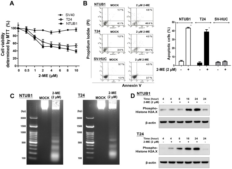
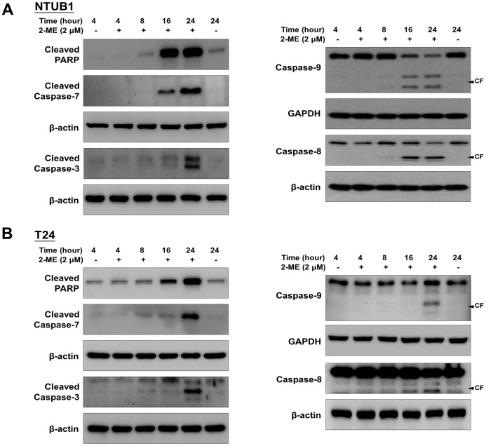
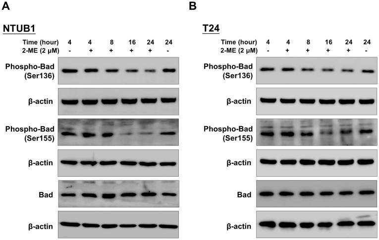
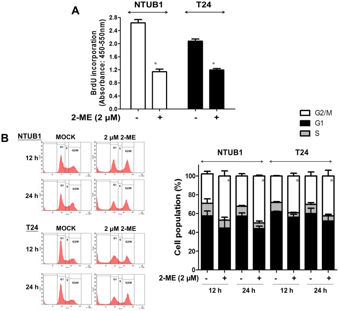
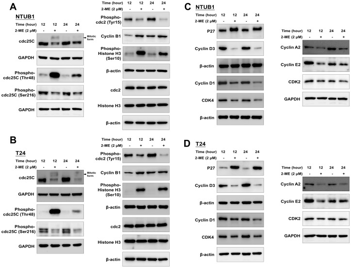
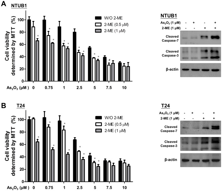
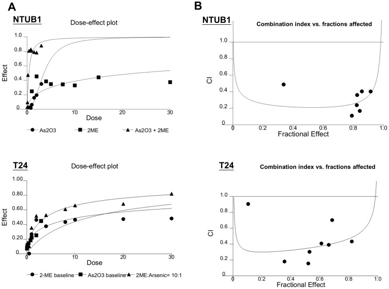
2-Methoxyestradiol (2-ME), an endogenous derivative of 17β-estradiol, has been reported to elicit antiproliferative responses in various tumors. In this study, we investigated the effects of 2-ME on cell viability, proliferation, cell cycle, and apoptosis in human urothelial carcinoma (UC) cell lines. We used two high-grade human bladder UC cell lines (NTUB1 and T24). After treatment with 2-ME, the cell viability and apoptosis were measured by MTT assay and flow cytometry (fluorescence-activated cell sorting), with annexin V-FITC staining and propidium iodide (PI) labeling. DNA fragmentation was analyzed by agarose gel electrophoresis. Flow cytometry with PI labeling was used for the cell cycle analyses. The protein levels of caspase activations, poly (ADP-ribose) polymerase (PARP) cleavage, phospho-histone H2A.X, phospho-Bad, and cell cycle regulatory molecules were measured by Western blot. The effects of the drug combinations were analyzed using the computer software, CalcuSyn. We demonstrated that 2-ME effectively induces dose-dependent cytotoxicity and apoptosis in human UC cells after 24 h exposure. DNA fragmentation, PARP cleavage, and caspase-3, 7, 8, 9 activations can be observed with 2-ME-induced apoptosis. The decreased phospho-Bad (Ser136 and Ser155) and mitotic arrest of the cell cycle in the process of apoptosis after 2-ME treatment was remarkable. In response to mitotic arrest, the mitotic forms of cdc25C, phospho-cdc2, cyclin B1, and phospho-histone H3 (Ser10) were activated. In combination with arsenic trioxide (As2O3), 2-ME elicited synergistic cytotoxicity (combination index <1) in UC cells. We concluded that 2-ME significantly induces apoptosis through decreased phospho-Bad and arrests bladder UC cells at the mitotic phase. The synergistic antitumor effect with As2O3 provides a novel implication in clinical treatment of UC.
Conflict of interest statement
Figures
Similar articles
-
MLN4924, a novel protein neddylation inhibitor, suppresses proliferation and migration of human urothelial carcinoma: In vitro and in vivo studies.Cancer Lett. 2015 Jul 28;363(2):127-36. doi: 10.1016/j.canlet.2015.01.015. Epub 2015 Jan 20. Cancer Lett. 2015. PMID: 25615422
-
Arsenic trioxide induces apoptosis in pancreatic cancer cells via changes in cell cycle, caspase activation, and GADD expression.Pancreas. 2003 Aug;27(2):174-9. doi: 10.1097/00006676-200308000-00011. Pancreas. 2003. PMID: 12883267
-
Arsenic trioxide causes redistribution of cell cycle, caspase activation, and GADD expression in human colonic, breast, and pancreatic cancer cells.Cancer Invest. 2004;22(3):389-400. doi: 10.1081/cnv-200029068. Cancer Invest. 2004. PMID: 15493360
-
Arsenic trioxide preferentially induces nonapoptotic cell deaths as well as actin cytoskeleton rearrangement in the CHO AA8 cell line.Postepy Hig Med Dosw (Online). 2014 Dec 21;68:1492-500. doi: 10.5604/17322693.1133098. Postepy Hig Med Dosw (Online). 2014. PMID: 25531713 Review.
-
Therapeutic promises of 2-methoxyestradiol and its drug disposition challenges.Mol Pharm. 2010 Dec 6;7(6):2030-9. doi: 10.1021/mp100190f. Epub 2010 Oct 21. Mol Pharm. 2010. PMID: 20831190 Free PMC article. Review.
Cited by
-
The pro-apoptotic actions of 2-methoxyestradiol against ovarian cancer involve catalytic activation of PKCδ signaling.Oncotarget. 2020 Oct 6;11(40):3646-3659. doi: 10.18632/oncotarget.27760. eCollection 2020 Oct 6. Oncotarget. 2020. PMID: 33088425 Free PMC article.
-
2-methoxyestradiol and disorders of female reproductive tissues.Horm Cancer. 2014 Oct;5(5):274-83. doi: 10.1007/s12672-014-0181-2. Epub 2014 Apr 25. Horm Cancer. 2014. PMID: 24764201 Free PMC article. Review.
-
A molecular understanding of D-homoestrone-induced G2/M cell cycle arrest in HeLa human cervical carcinoma cells.J Cell Mol Med. 2015 Oct;19(10):2365-74. doi: 10.1111/jcmm.12587. Epub 2015 Jul 31. J Cell Mol Med. 2015. PMID: 26228523 Free PMC article.
-
Selenium-enriched Spirulina protects INS-1E pancreatic beta cells from human islet amyloid polypeptide-induced apoptosis through suppression of ROS-mediated mitochondrial dysfunction and PI3/AKT pathway.Eur J Nutr. 2015 Jun;54(4):509-22. doi: 10.1007/s00394-014-0732-x. Epub 2014 Aug 12. Eur J Nutr. 2015. PMID: 25112514
-
cJun-N-terminal kinase activity and mitosis perturbation drive the 2-methoxyestradiol-mediated enhancement of viral oncolysis by Epizootic Hemorrhagic Disease Virus-Tel Aviv University.Sci Rep. 2025 Jul 9;15(1):24624. doi: 10.1038/s41598-025-08687-8. Sci Rep. 2025. PMID: 40634455 Free PMC article.
References
-
- Jemal A, Siegel R, Xu J, Ward E (2010) Cancer statistics, 2010. CA Cancer JClin 60: 277–300. - PubMed
-
- Cohen MH, Rothmann M (2001) Gemcitabine and cisplatin for advanced, metastatic bladder cancer. JClinOncol 19: 1229–1231. - PubMed
-
- von der MH, Hansen SW, Roberts JT, Dogliotti L, Oliver T, et al. (2000) Gemcitabine and cisplatin versus methotrexate, vinblastine, doxorubicin, and cisplatin in advanced or metastatic bladder cancer: results of a large, randomized, multinational, multicenter, phase III study. JClinOncol 18: 3068–3077. - PubMed
-
- Latini DM, Lerner SP, Wade SW, Lee DW, Quale DZ (2010) Bladder cancer detection, treatment and outcomes: opportunities and challenges. Urology 75: 334–339. - PubMed
-
- Lakhani NJ, Sarkar MA, Venitz J, Figg WD (2003) 2-Methoxyestradiol, a promising anticancer agent. Pharmacotherapy 23: 165–172. - PubMed
Publication types
MeSH terms
Substances
LinkOut - more resources
Full Text Sources
Other Literature Sources
Molecular Biology Databases
Research Materials
Miscellaneous
