

Aging, estrogen loss and epoxyeicosatrienoic acids (EETs)
- PMID: 23967089
- PMCID: PMC3742755
- DOI: 10.1371/journal.pone.0070719
Aging, estrogen loss and epoxyeicosatrienoic acids (EETs)
Abstract
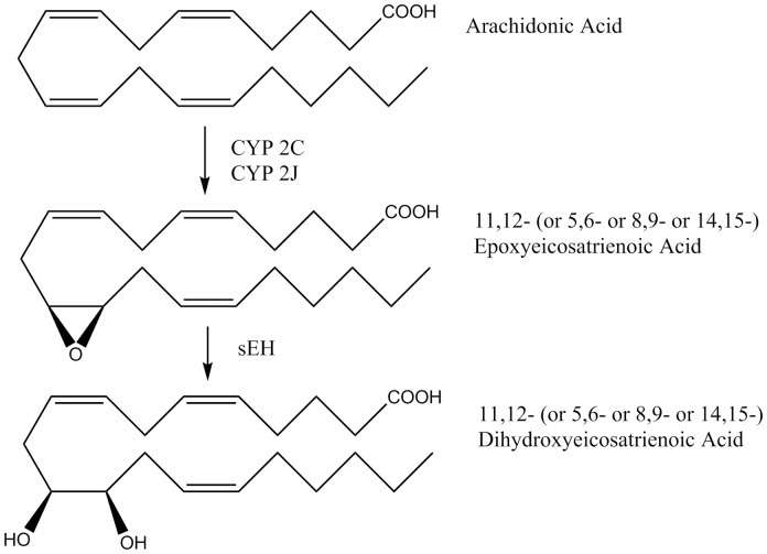
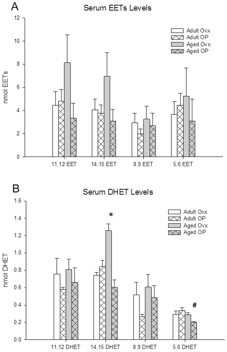
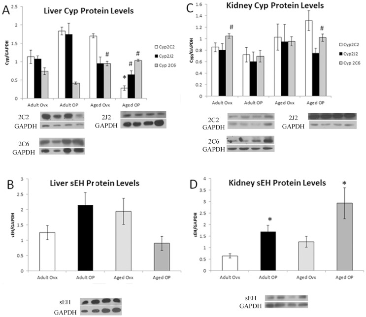
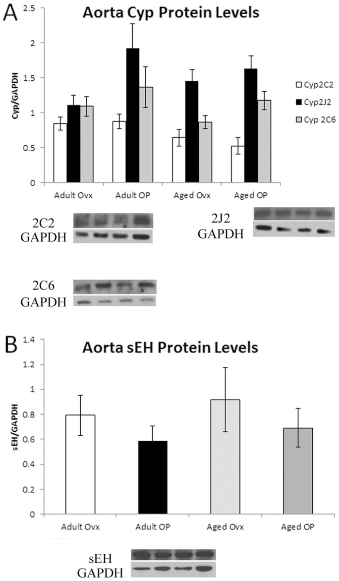
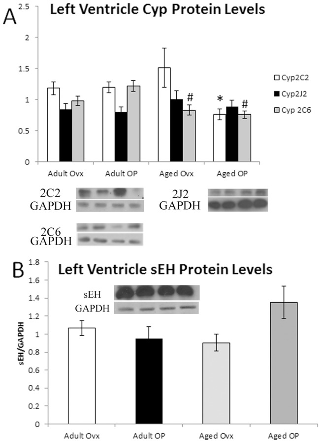
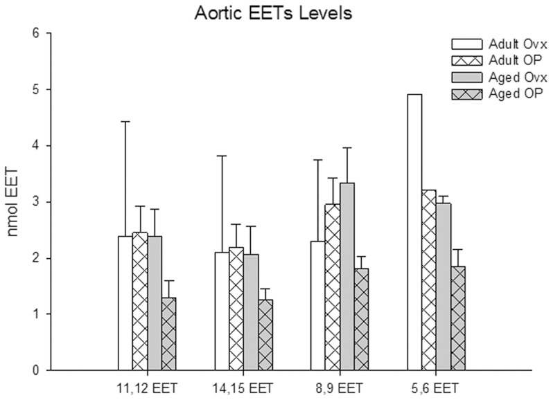
Inflammation is a key element in many cardiovascular diseases. Both estrogen loss, caused by menopause, and aging have inflammatory consequences. Epoxyeicosatrienoic acids (EETs) are anti-inflammatory molecules synthesized by various cytochrome P450 (Cyp) enzymes from arachidonic acid. EETs are in the third (Cytochrome P450) pathway of arachindonic acid metabolism, others being cyclooxygenases and lipoxygenases. We hypothesized that aging and estrogen loss would reduce levels of anti-inflammatory EETs. Adult (6 mo) and aged (22 mo) ovariectomized rats with (OP) and without (Ovx) 17-∃-estradiol replacement were used in this study. Mass spectrometry was used to measure levels of EETs and their metabolites, dihydroxyeicosatrienoic acids (DHETs). Levels of Cyp2C2, Cyp2C6, and Cyp2J2, the principal Cyps responsible for EETs synthesis, as well as soluble epoxide hydrolase (sEH), which metabolizes EETS to DHETs, were determined via western blot. Overall Cyp levels decreased with age, though Cyp2C6 increased in the liver. sEH was increased in the kidney with estrogen replacement. Despite protein changes, no differences were measured in plasma or aortic tissue levels of EETs. However, plasma 14,15 DHET was increased in aged Ovx, and 5,6 DHET in adult OP. In conclusion neither aging nor estrogen loss decreased the anti-inflammatory EETs in the cardiovascular system.
Conflict of interest statement
Figures
Similar articles
-
11,12-Epoxyecosatrienoic acids mitigate endothelial dysfunction associated with estrogen loss and aging: Role of membrane depolarization.J Mol Cell Cardiol. 2016 May;94:180-188. doi: 10.1016/j.yjmcc.2016.03.019. Epub 2016 Apr 11. J Mol Cell Cardiol. 2016. PMID: 27079253 Free PMC article.
-
Epoxyeicosatrienoic and dihydroxyeicosatrienoic acids dilate human coronary arterioles via BK(Ca) channels: implications for soluble epoxide hydrolase inhibition.Am J Physiol Heart Circ Physiol. 2006 Feb;290(2):H491-9. doi: 10.1152/ajpheart.00927.2005. Epub 2005 Oct 28. Am J Physiol Heart Circ Physiol. 2006. PMID: 16258029 Free PMC article.
-
Increased epoxyeicosatrienoic acids and reduced soluble epoxide hydrolase expression in the preeclamptic placenta.J Hypertens. 2016 Jul;34(7):1364-70. doi: 10.1097/HJH.0000000000000942. J Hypertens. 2016. PMID: 27115337 Free PMC article.
-
CYP450 Epoxygenase Metabolites, Epoxyeicosatrienoic Acids, as Novel Anti-Inflammatory Mediators.Molecules. 2022 Jun 16;27(12):3873. doi: 10.3390/molecules27123873. Molecules. 2022. PMID: 35744996 Free PMC article. Review.
-
Arachidonic acid cytochrome P450 epoxygenase pathway.J Lipid Res. 2009 Apr;50 Suppl(Suppl):S52-6. doi: 10.1194/jlr.R800038-JLR200. Epub 2008 Oct 23. J Lipid Res. 2009. PMID: 18952572 Free PMC article. Review.
Cited by
-
11,12-Epoxyecosatrienoic acids mitigate endothelial dysfunction associated with estrogen loss and aging: Role of membrane depolarization.J Mol Cell Cardiol. 2016 May;94:180-188. doi: 10.1016/j.yjmcc.2016.03.019. Epub 2016 Apr 11. J Mol Cell Cardiol. 2016. PMID: 27079253 Free PMC article.
-
TLR4 mutation and HSP60-induced cell death in adult mouse cardiac myocytes.Cell Stress Chaperones. 2015 May;20(3):527-35. doi: 10.1007/s12192-015-0577-0. Epub 2015 Feb 27. Cell Stress Chaperones. 2015. PMID: 25716072 Free PMC article.
-
Soluble Epoxide Hydrolase Is Associated with Postprandial Anxiety Decrease in Healthy Adult Women.Int J Mol Sci. 2022 Oct 5;23(19):11798. doi: 10.3390/ijms231911798. Int J Mol Sci. 2022. PMID: 36233100 Free PMC article.
-
Age‑related differences for expression of the nerve‑specific proteins after peripheral nerve injury.Exp Ther Med. 2022 Sep 21;24(5):682. doi: 10.3892/etm.2022.11618. eCollection 2022 Nov. Exp Ther Med. 2022. PMID: 36185767 Free PMC article.
-
Indirubin-3'-oxime as a dual-action agent: mitigating heat-induced male infertility in Drosophila melanogaster and inhibiting soluble epoxide hydrolase.J Enzyme Inhib Med Chem. 2025 Dec;40(1):2447719. doi: 10.1080/14756366.2024.2447719. Epub 2025 Jan 22. J Enzyme Inhib Med Chem. 2025. PMID: 39840826 Free PMC article.
References
-
- Oparil S, Levine RL, Chen SJ, Durand J, Chen YF (1997) Sexually Dimorphic Response of the Balloon-Injured Rat Carotid Artery to Hormone Treatment. Circulation 95: 1301–1307. - PubMed
-
- Walsh BA, Busch BL, Mullick AE, Reiser KM, Rutledge JC (1999) 17β-Estradiol Reduces Glycoxidative Damage in the Artery Wall. Arterioscler Thromb Vasc Biol 19: 840–846. - PubMed
-
- Hulley S, Grady D, Bush T, Furberg C, Herrington D, et al. (1998) Randomized trial of estrogen plus progestin for secondary prevention of coronary heart disease in postmenopausal women. Heart and Estrogen/progestin Replacement Study (HERS) Research Group. JAMA 280: 605–613. - PubMed
-
- Women's Health Initiative Steering Committee (2004) Effects of conjugated equine estrogen in postmenopausal women with hysterectomy: The women's health initiative randomized controlled trial. JAMA 291: 1701–1712. - PubMed
Publication types
MeSH terms
Substances
Grants and funding
LinkOut - more resources
Full Text Sources
Other Literature Sources
Medical
Miscellaneous
