. 2013 Aug 15;8(8):e70843.
doi: 10.1371/journal.pone.0070843. eCollection 2013.

Identification of potential Plk1 targets in a cell-cycle specific proteome through structural dynamics of kinase and Polo box-mediated interactions

Affiliations

Identification of potential Plk1 targets in a cell-cycle specific proteome through structural dynamics of kinase and Polo box-mediated interactions

Nousheen Bibi et al. PLoS One. .

Abstract

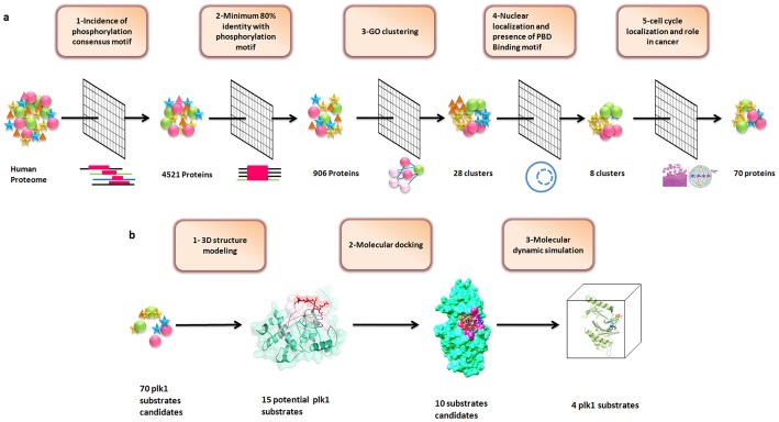
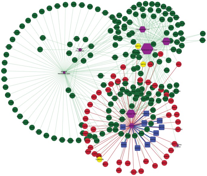
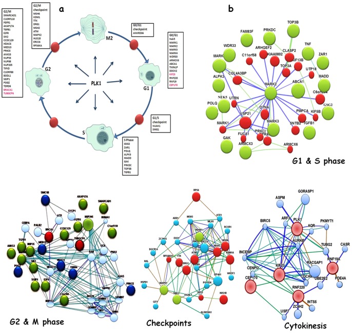
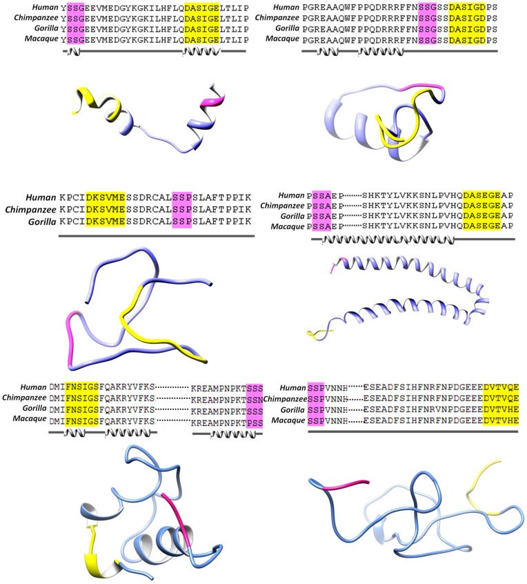
Polo like kinase 1 (Plk1) is a key player in orchestrating the wide variety of cell-cycle events ranging from centrosome maturation, mitotic entry, checkpoint recovery, transcriptional control, spindle assembly, mitotic progression, cytokinesis and DNA damage checkpoints recovery. Due to its versatile nature, Plk1 is considered an imperative regulator to tightly control the diverse aspects of the cell cycle network. Interactions among Plk1 polo box domain (PBD) and its putative binding proteins are crucial for the activation of Plk1 kinase domain (KD). To date, only a few substrate candidates have been characterized through the inclusion of both polo box and kinase domain-mediated interactions. Thus it became compelling to explore precise and specific Plk1 substrates through reassessment and extension of the structure-function paradigm. To narrow this apparently wide gap in knowledge, here we employed a thorough sequence search of Plk1 phosphorylation signature containing proteins and explored their structure-based features like conceptual PBD-binding capabilities and subsequent recruitment of KD directed phosphorylation to dissect novel targets of Plk1. Collectively, we identified 4,521 phosphodependent proteins sharing similarity to the consensus phosphorylation and PBD recognition motifs. Subsequent application of filters including similarity index, Gene Ontology enrichment and protein localization resulted in stringent pre-filtering of irrelevant candidates and isolated unique targets with well-defined roles in cell-cycle machinery and carcinogenesis. These candidates were further refined structurally using molecular docking and dynamic simulation assays. Overall, our screening approach enables the identification of several undefined cell-cycle associated functions of Plk1 by uncovering novel phosphorylation targets.


Conflict of interest statement

Competing Interests: The authors have declared that no competing interests exist.

Figures

Figure 1
Figure 1. Schematic illustration of strategy.
(a) Plk1 candidates substrates were selected on the basis of series of filters applied to human proteome including, presence of Plk1 specific phosphorylation motif at sequence level, minimum 80% identity, clustering on the basis of biological process, cellular localization and molecular function, presence of PBD recognition motif, common cell-cycle localization and role in cancer. (b) Scheme depicting different structure-based techniques used for the validation of putative hits obtained from sequence-based approaches.
Figure 2
Figure 2. Gene network clusters enriched in cancer and cell-cycle related nodes.
Red nodes represent proteins involved in cancer and cell-cycle, yellow are known Plk1 substrates, while green nodes represent targets having no known contributions in cancer. The blue nodes are proteins selected for structural validation in this study. Green and red lines represent protein network edges.
Figure 3
Figure 3. Plk1-dependent substrates.
(a) Cell-cycle diagram depicting proteins sorted out through sequence based approach having Plk1 phosphorylation motif. Proteins are classified according to function and localization. Known Plk1 substrates are shown in red and novel putative substrates are shown in black. Red nodes indicate cell-cycle checkpoints. (b) Protein interaction analysis and interaction map in cell-cycle localized clusters (G1 and S, G2 and M, checkpoints, cytokinesis). Node color indicates the specific localization and other distinguishing features. In G1 and S phases, green nodes are G1 specific proteins and red nodes represent proteins involved in both G1 and S. In G2 and M phases, green nodes are both G2 and M specific and predicted as novel substrates in our study, red nodes are known Plk1 substrates and dark blue nodes are substrates shared by other kinesis. In checkpoints interaction map, red nodes represent G2/M specific checkpoints and green nodes are G1/S checkpoints. In cytokinesis interaction map, pink nodes are known Plk1 substrates. Blues lines delineate direct interactions, green lines represents co-expression, brown lines delineate shared protein domain.
Figure 4
Figure 4. Multiple sequence alignment (MSA), secondary and tertiary structures of four selected Plk1 specific substrates.
(a) SMARCAD1, (b) GSG2, (c) NUP35, (d) NEK5, (e) KIF23 and (f) CEP170. MSA illustrates the conservation pattern of predicted PBD recognition and kinase phosphorylation motifs across close homologues of selected proteins (a–d). Secondary structure for each substrate was predicted by JPred as shown below the alignments Tertiary structures of (a–f) substrates are shown in cornflower blue ribbon-form, phosphorylation motifs are shown in yellow, PBD recognition motifs are shown in pink.
Figure 5
Figure 5. Plk1 protein architecture and substrates placing in the binding pockets of PBD and KD.
(a) Schematic representation of Plk1 3D architecture: important residues lying at the KD and PBD (Polo Box domain involved in binding is highlighted by pink color); (b) Plk1 3D model, (i) KD, (ii) PBD with specific binding pocket (pink); selected Plk1 specific substrate candidates (putative novel and known) bind at the common position (with respect to motif signature) inside the binding pockets of KD and PBD. (c) Distinctive residues of Plk1 binding pocket and selected substrates; (i) KD anterior view and (ii) KD posterior view, (iii) PBD anterior view (iv) PBD posterior view.
Figure 6
Figure 6. Characterization of inhibitory molecular interaction between PBD and KD of Plk1.
Cyan blue meshy ribbon represents the KD with interacting residues in cyan stick form, while PBD is represented in pink meshy ribbon with interacting residues in pink stick. Hydrogen bonds are represented by black dotted lines with bond distances in angstrom.
Figure 7
Figure 7. PBD regulated Plk1 kinase activation and substrate phosphorylation.
(a) Hypothetical model for the activation of Plk1 proposed by Elia et al., 2003. PBDI and PBDII are shown in pink color, KD in sky blue and bound substrate by yellow balls. (b) Docking analysis data in support of Plk1 activation model. Polo box (pink ribbon) and kinase domains (sky blue ribbon) are tightly bound with each other in an inactive state (left), phosphopeptide (yellow ball and sticks) binding to PBD discharges the KD for phosphorylation (right).
Figure 8
Figure 8. Binding mode and molecular interaction analyses of putative substrates.
(I) localization of potential kinase phosphorylation and PBD recognition motif; (a) Plk1-SMARCAD1 complex, (b) Plk1-GSG2 complex, (c) Plk1-KIF23 complex and (d) Plk1-CEP170 complex. KD is shown in blue ribbon with interacting residues in pink sticks, while PBD is shown in pink ribbon and cyan sticks represent interacting residues. Substrate peptides are shown in yellow ball and stick mode and hydrogen bonding is shown by black dotted lines with calculated distances in angstrom.
Figure 9
Figure 9. Plots to investigate the stability and fluctuation of MD trajectories for all systems.
(a) RMSD plot computed through each system trajectory, (b) Rg plot, (c) RMSF plot (full protein), Plk1-NEK5 complex, (blue) Plk1-GSG2 complex (red), Plk1- SMARCAD1 complex (green), Plk1-NUP35 complex (black), (d) Significant Plk1 residues involved in substrates binding. Overall residues are stable with no more than 1Å fluctuation. Trp414, His538 and Lys540 are much stable in all systems.
Figure 10
Figure 10. Conformational switches of Plk1 structure upon substrate binding.
NEK5 (white), NUP35 (pink), GSG2 (blue) and SMARCAD1 (yellow). (I) Helical region is variable in NEK5, NUP35 and SMARCAD1. (II) Specific binding orientation of active site residues in different complexes. (III) Extension of β-strand at two regions, Ile553-Glu555 in Nup53, GSG2 and SMARCAD1 complexes and Leu435-Phe436 in NEK5, NUP35 complexes. (IV) Changes at linker region between two PBD domains of Plk1. (V) Three residues (Phe516-Arg518) extension of β strand in GSG2 complex.

References

    1. Barr FA, Sillje HH, Nigg EA (2004) Polo-like kinases and the orchestration of cell division. Nat Rev Mol Cell Biol 5: 429–440. - PubMed
    1. Glover DM (2005) Polo kinase and progression through M phase in Drosophila: a perspective from the spindle poles. Oncogene 24: 230–237. - PubMed
    1. van de Weerdt BC, Medema RH (2006) Polo-like kinases: a team in control of the division. Cell Cycle 5: 853–864. - PubMed
    1. Llamazares S, Moreira A, Tavares A, Girdham C, Spruce BA, et al. (1991) polo encodes a protein kinase homolog required for mitosis in Drosophila. Genes Dev 5: 2153–2165. - PubMed
    1. Sunkel CE, Glover DM (1988) Polo, a mitotic mutant of Drosophila displaying abnormal spindle poles. J Cell Sci 89: 25–38. - PubMed

Publication types

MeSH terms

LinkOut - more resources