. 2013 Aug 15;8(8):e70916.
doi: 10.1371/journal.pone.0070916. eCollection 2013.

Analysis of trafficking, stability and function of human connexin 26 gap junction channels with deafness-causing mutations in the fourth transmembrane helix

Affiliations

Analysis of trafficking, stability and function of human connexin 26 gap junction channels with deafness-causing mutations in the fourth transmembrane helix

Cinzia Ambrosi et al. PLoS One. .

Abstract

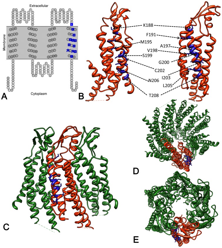
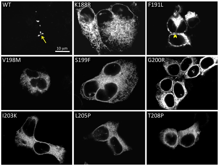
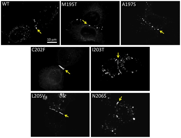
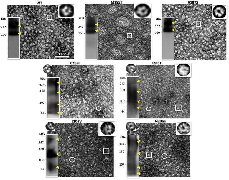
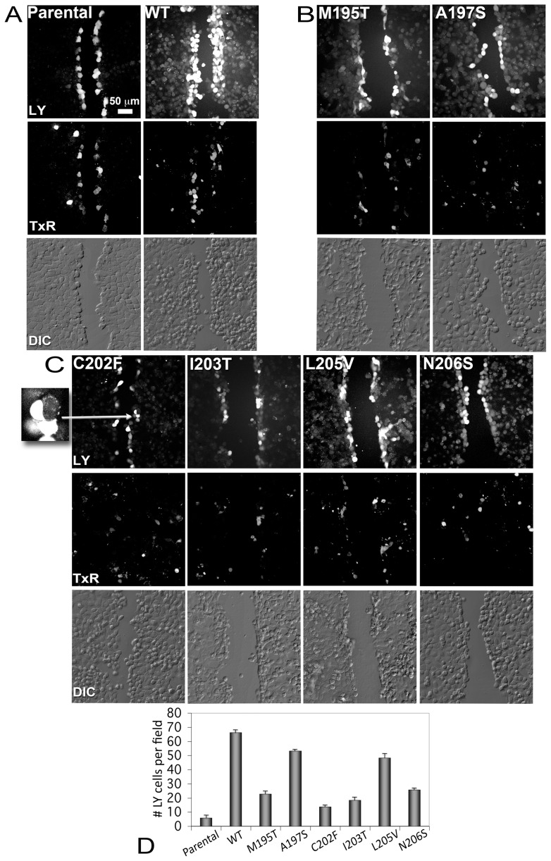
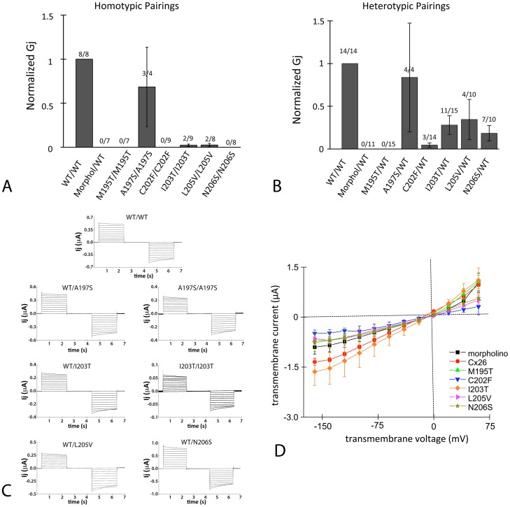
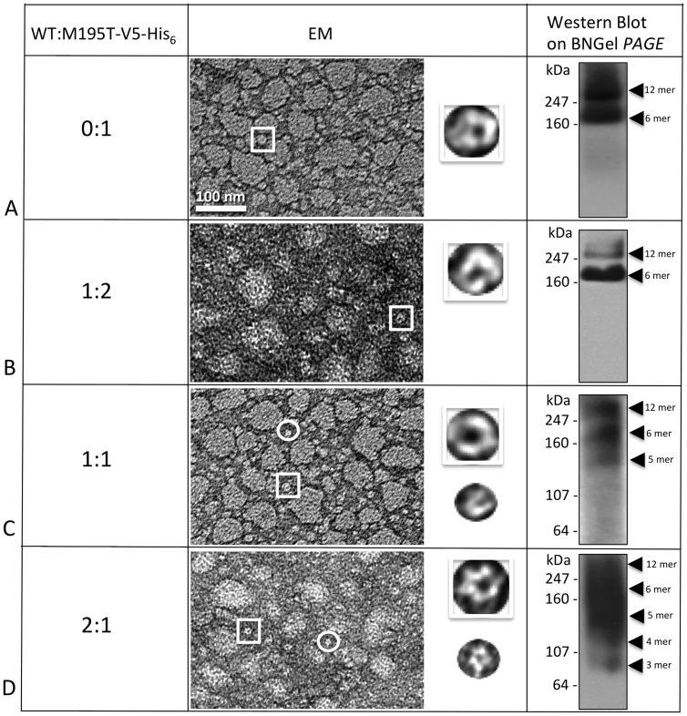
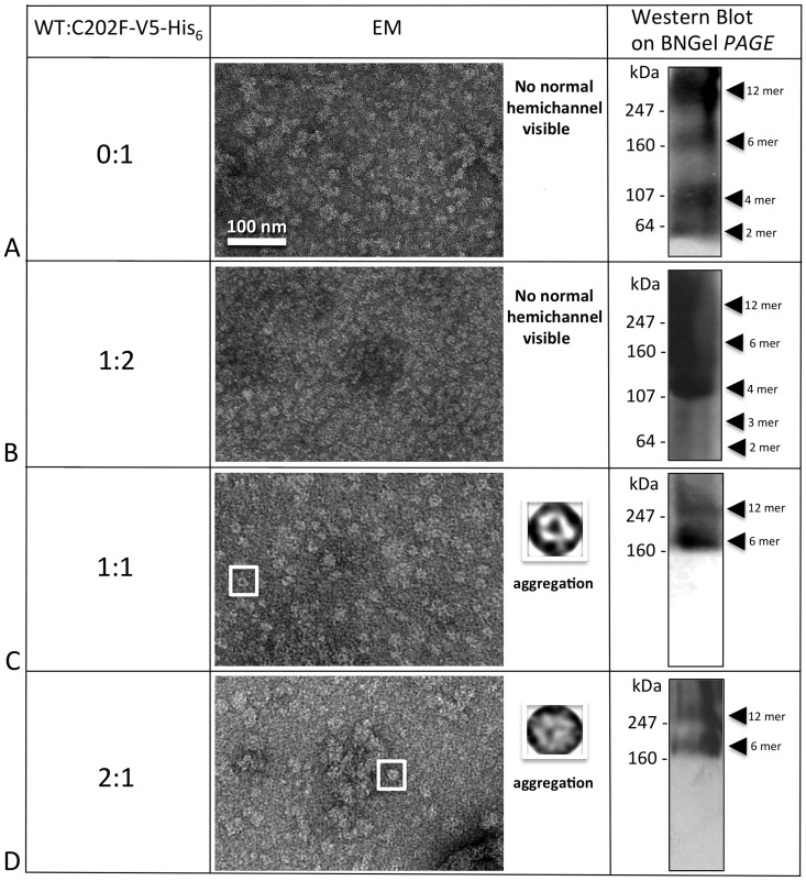
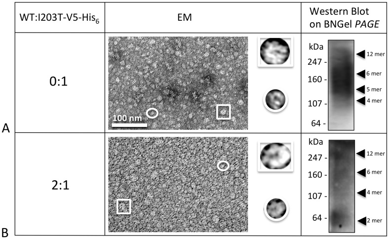
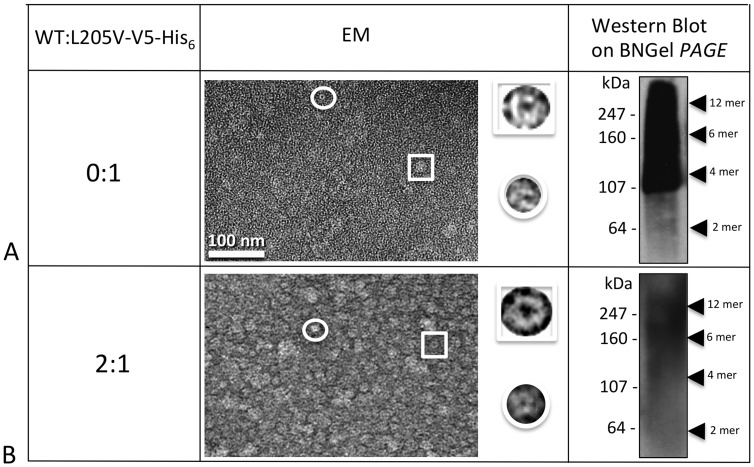
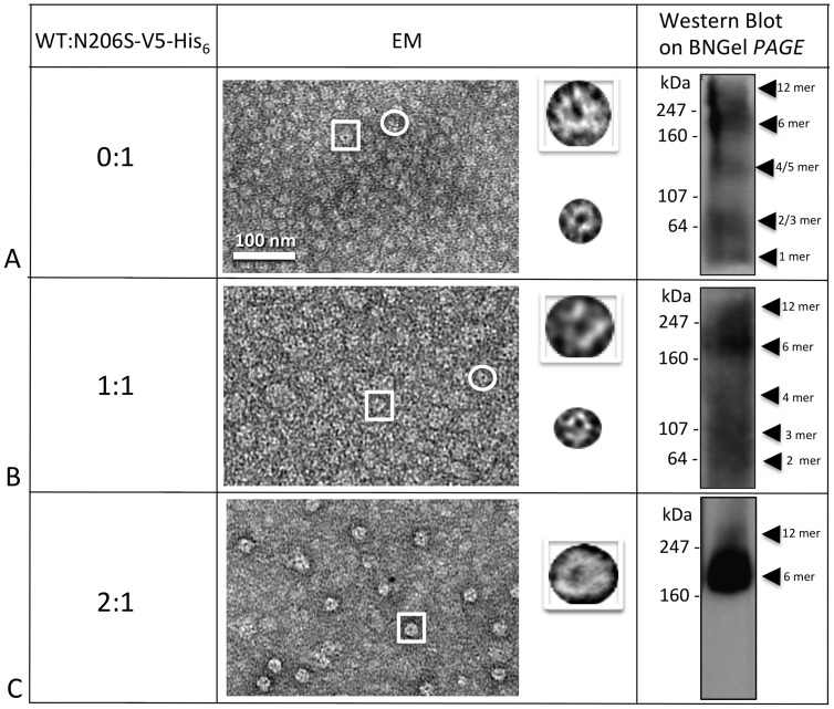
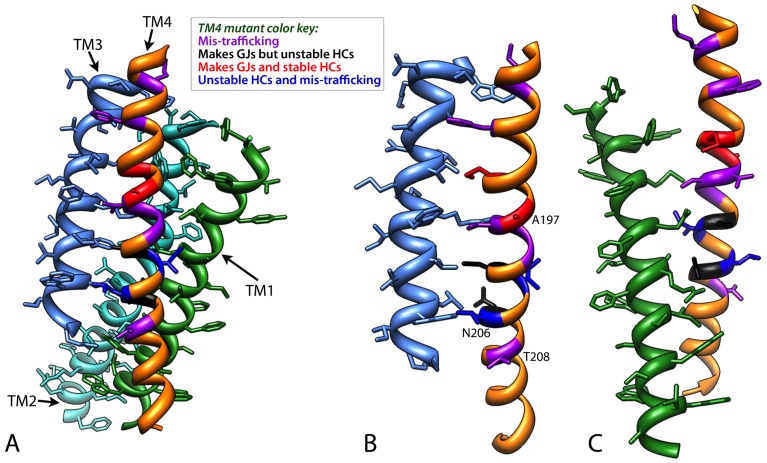
Human Connexin26 gene mutations cause hearing loss. These hereditary mutations are the leading cause of childhood deafness worldwide. Mutations in gap junction proteins (connexins) can impair intercellular communication by eliminating protein synthesis, mis-trafficking, or inducing channels that fail to dock or have aberrant function. We previously identified a new class of mutants that form non-functional gap junction channels and hemichannels (connexons) by disrupting packing and inter-helix interactions. Here we analyzed fourteen point mutations in the fourth transmembrane helix of connexin26 (Cx26) that cause non-syndromic hearing loss. Eight mutations caused mis-trafficking (K188R, F191L, V198M, S199F, G200R, I203K, L205P, T208P). Of the remaining six that formed gap junctions in mammalian cells, M195T and A197S formed stable hemichannels after isolation with a baculovirus/Sf9 protein purification system, while C202F, I203T, L205V and N206S formed hemichannels with varying degrees of instability. The function of all six gap junction-forming mutants was further assessed through measurement of dye coupling in mammalian cells and junctional conductance in paired Xenopus oocytes. Dye coupling between cell pairs was reduced by varying degrees for all six mutants. In homotypic oocyte pairings, only A197S induced measurable conductance. In heterotypic pairings with wild-type Cx26, five of the six mutants formed functional gap junction channels, albeit with reduced efficiency. None of the mutants displayed significant alterations in sensitivity to transjunctional voltage or induced conductive hemichannels in single oocytes. Intra-hemichannel interactions between mutant and wild-type proteins were assessed in rescue experiments using baculovirus expression in Sf9 insect cells. Of the four unstable mutations (C202F, I203T, L205V, N206S) only C202F and N206S formed stable hemichannels when co-expressed with wild-type Cx26. Stable M195T hemichannels displayed an increased tendency to aggregate. Thus, mutations in TM4 cause a range of phenotypes of dysfunctional gap junction channels that are discussed within the context of the X-ray crystallographic structure.


Conflict of interest statement

Competing Interests: Note that while Dr. Badger is affiliated with a commercial company, his authorship on this manuscript does not alter the authors' adherence to PLOS' policy on sharing of data and materials. His role on this manuscript was in the analysis stage; no data or materials were involved with his efforts. All authors have declared that no competing interests exist.

Figures

Figure 1
Figure 1. Spatial arrangement of 12 NSHL residues in TM4.
(A) Topology diagram of Cx26. Blue boxes indicate these 12 residues. (B–E) Positions of residues on current X-ray atomic model (PDB ID 2ZW3) marked in blue on (B) monomer (C–E) hexamer. There is an ∼54° angle along the y axis between the two monomer views in (B). (C) Side view of front three connexins. Views down cytoplasmic side (D) and extracellular side (E).
Figure 2
Figure 2. Eight TM4 deafness mutations cause mis-trafficking.
All 14 mutations were tested for their ability to make gap junctions in transiently transfected HeLa cells. Eight mutations (K188R, F191L, V198M, S199F, G200R, I203K, L205P, T208P) caused mis-trafficking, typically with intracellular aggregation. WT human Cx26 is shown at the top left for comparison, with an arrow pointing to a gap junction. The arrowhead in the F191L image points to a cell-cell apposition area with aggregate fluorescence.
Figure 3
Figure 3. Six Cx26 TM4 traffic properly in HeLa cells.
M195T, A197S, C202F, I203T, L205V and N206S each formed gap junctions between apposing cells when transiently expressed in mammalian cells. An arrow points to a gap junctions in each image.
Figure 4
Figure 4. Analysis of hemichannel stability for gap junction forming mutants.
Shown here are a representative electron micrograph and Blue Native westerns (bottom left hand inset) of purified hemichannels for each mutant purified from an Sf9/baculovirus expression system. The yellow arrowheads point to the recognizable bands. The insets on the top right are 6 fold enlargements of a normal hemichannel (the expected hexamer) indicated by the box in the micrograph, while the insets on the top left are 6 fold enlargements of the small oligomers visible only in unstable mutants and indicated by the circle in these micrographs.
Figure 5
Figure 5. Dye transfer assays for gap junction forming mutants.
Intercellular communication was assayed by scrape dye loading using Lucifer Yellow (LY) and Dextran Texas Red (TxR). LY will pass through gap junctions while Dextran TxR will not. (Panel A) Confluent gap-junction deficient parental HeLa cells (left column) and HeLa cells transiently transfected with Cx26 WT-GFP-4C (right column) serve as negative and positive controls, respectively. Top row: Images of LY fluorescence. Middle row: Images of TxR fluorescence Bottom row: Differential Interference Contrast (DIC) light micrograph of the same field of cells showing cell-cell contact. Dextran Texas Red acts as a reporter for dead or non-communicating cells and those along the scratch that also contained Lucifer Yellow were not counted in this analysis. (Panel B) Similar images as in (Panel A) for the two mutants, M195T and A197S, which made detergent stable hemichannels and channels. (Panel C) LY (top), TxR (middle) and DIC images (bottom) for mutants that made detergent unstable hemichannels and channels (left to right, C202F, I203T, L205V and N206S). The inset at the left is a 4.5x magnification of a LY non-transferring cell expressing C202F gap junctions. The arrow points to these cells in the C202F LY image. (D) Histogram of levels of communicating cells for each mutant. Error bars are standard errors of the mean (SEM).
Figure 6
Figure 6. Six mutant channels (M195T, A197S, C202F, I203T, L205V and N206S) were tested for their conductances in the paired Xenopus oocyte system.
(A) Mutant channels were tested in the homotypic configuration. Conductance measurements from two oocyte batches were pooled, each bar represents the mean ± SEM. The numbers above each bar represent the number of oocyte pairs in which coupling was observed as a fraction of the number tested. The negative-control (morpholino) involved oocytes injected only with the standard anti-XeCx38 antisense and intercellular conductance (Gj) was normalized to that of WT. Each bar represents a mean ± SEM and the numbers above each bar represent the number of oocyte pairs in which coupling was observed as a fraction of the number tested. (B) Mutants were paired heterotypically with WT and intercellular conductance (Gj) was normalized to that of WT. Conductance measurements from three oocyte batches were pooled, each bar represents the mean ± SEM, and the numbers above each bar represent the number of oocyte pairs in which coupling was observed as a fraction of the number tested. The negative-control (morpholino) involved oocytes injected only with the standard anti-XeCx38 antisense. (C) Characteristics of gap junctions induced by four of the six mutants after expression in Xenopus oocytes. Characteristic currents induced by WT are displayed on top Oocyte pairs were clamped at −20 mV and currents were recorded from a continuously clamped oocyte while its partner was pulsed in 10 mV increments to induce transjunctional voltages (Vj's) of up to ±100 mV. Only A197S and I203T formed distinguishable homotypic channels and these are observed on the right, adjacent to their corresponding heterotypic currents. (D) Transmembrane currents were measured in single oocytes to determine if mutants formed functionally conductive hemichannels. Each point represents the mean current (± SEM) for three oocytes from the same batch. The negative-control (morpholino) involved oocytes injected only with the standard anti-XeCx38 antisense.
Figure 7
Figure 7. Co-expression of WT with M195T–WT decreases the stability of heteromers.
(A–D) Hemichannels formed by M195T and the WT analyzed in different ratios showed increased instability from heteromeric interactions. EM images showed consistent aggregation of hemichannels for all four ratios analyzed, from 0:1 to 2:1. The Blue Native (BNGel PAGE) westerns confirmed stability with only two bands (hexamer and dodecamer) for the ratios 0 and 1:2 (A and B), but higher ratios of WT:M195T-V5-His6 are unstable with (C) three predominant bands for 1:1 and (D) a ladder of bands for the 2:1 ratio.
Figure 8
Figure 8. C202F hemichannel stability can be rescued by the WT.
(A) C202F forms unstable oligomers not easily recognizable by EM as hemichannels and a ladder of bands as detected by Blue Native western analysis. (B) The addition of WT protomers to the mutant oligomers did not result in stable hemichannels for the 0:1 ratio neither by EM or Blue Native western analysis. (C, D) The higher ratios (1:1 and 2:1) clearly showed only two bands on Blue Native westerns at the hexamer and dodecamer positions and more recognizable doughnut-like structures in EM images, but these heteromeric channels tended to aggregate.
Figure 9
Figure 9. I203T mutant stability cannot be rescued by the WT.
(A) Homomeric I203T hexamers and dodecamers are unstable. Both EM and Blue Native western analyses clearly showed no rescuing the I203T mutant hemichannels, even at the very high ratio of 2 (B), where the majority of the heteromer subunits should contain WT.
Figure 10
Figure 10. L205V mutant cannot be rescued by the WT.
(A) Homomeric oligomers of L205V are unstable. Similar to the I203T mutant shown in Fig. 9, (B) this mutant did not display stable hemichannels or channels when I203T monomers where mixed with the WT monomers in a 2:1 ratio of WT:L205V-V5-His6.
Figure 11
Figure 11. N206S forms stable hemichannels when expressed with WT.
(A–C) As visible by both EM and Blue Native Western analysis, stable hemichannels only resulted at 2:1 ratios of WT:N206S-V5-His6 (C). The EM image clearly showed distinct hemichannel structures and the hexamer band predominates in the Blue Native (BNGel PAGE) Western (C).
Figure 12
Figure 12. Mapping of the twelve TM4 residues on the current X-ray model for the four transmembrane helices.
Since the twelve amino acid positions analyzed fall only in the four α helical bundle domain of Cx26, here we show only (A) TM1 (medium blue), TM2 (cyan) TM3 (green) and TM4 (orange). The orientation of the Cx26 subunit is the same as in Fig. 1 with the extracellular loops at the top of the image. The mutant residues have been colored coded such that mutations that caused -trafficking are shown in purple, mutations that make gap junctions but unstable hemichannels are black and mutations that make stable gap junctions and hemichannels are indicated in red. Two positions, I203 and L205, have different phenotypes for the each of two mutations at these positions. These residues are dark blue. (B) Side chain interactions for these residues are between TM4 and TM1 and (C) TM4 and TM3. Note that the A197, N206S and T208 face the lipid bilayer. Note the mutant phenotypes are not mapped to any particular face of TM4.

References

    1. Sohl G, Willecke K (2004) Gap junctions and the connexin protein family. Cardiovasc Res 62: 228–232. - PubMed
    1. Harris A, Locke D (2009) Connexins : a guide. New York, N.Y.: Springer. xvi, 573 p.
    1. Saez JC, Berthoud VM, Branes MC, Martinez AD, Beyer EC (2003) Plasma membrane channels formed by connexins: their regulation and functions. Physiol Rev 83: 1359–1400. - PubMed
    1. Evans WH, Martin PE (2002) Gap junctions: structure and function (Review). Mol Membr Biol 19: 121–136. - PubMed
    1. Maeda S, Nakagawa S, Suga M, Yamashita E, Oshima A, et al. (2009) Structure of the connexin 26 gap junction channel at 3.5 A resolution. Nature 458: 597–602. - PubMed

Publication types

MeSH terms