

Comparison of magnetic resonance imaging in live vs. post mortem rat brains
- PMID: 23967148
- PMCID: PMC3742751
- DOI: 10.1371/journal.pone.0071027
Comparison of magnetic resonance imaging in live vs. post mortem rat brains
Abstract
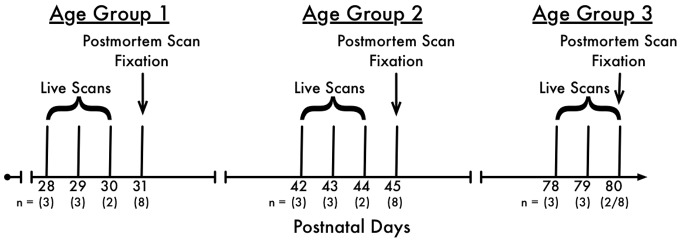
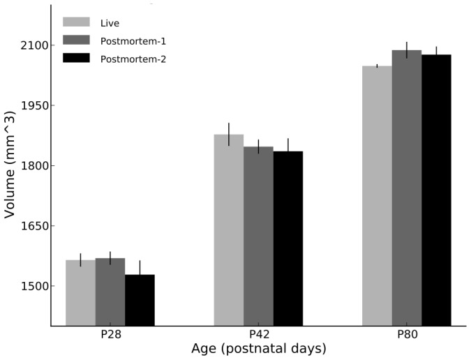
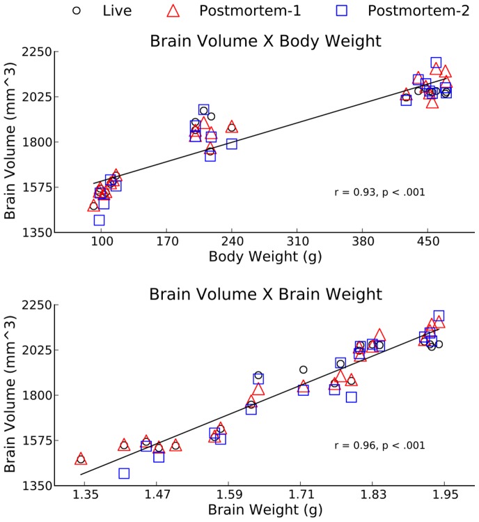
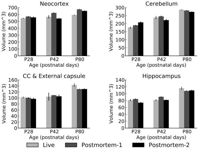
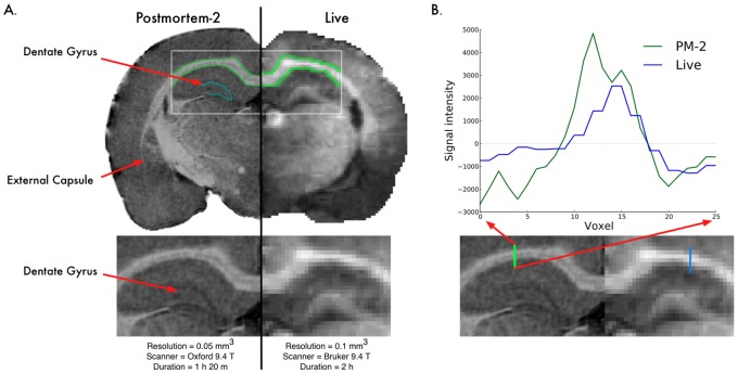
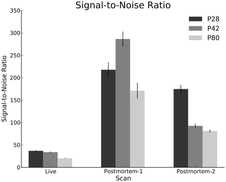
Magnetic Resonance Imaging (MRI) is an increasingly popular technique for examining neurobiology in rodents because it is both noninvasive and nondestructive. MRI scans can be acquired from either live or post mortem specimens. In vivo scans have a key advantage in that subjects can be scanned at multiple time-points in longitudinal studies. However, repeated exposure to anesthesia and stress may confound studies. In contrast, post mortem scans offer improved image quality and increased signal-to-noise ratio (SNR) due to several key advantages: First, the images are not disrupted by motion and pulsation artifacts. Second, they allow the brain tissue to be perfused with contrast agents, enhancing tissue contrast. Third, they allow longer image acquisition times, yielding higher resolution and/or improved SNR. Fourth, they allow assessment of groups of animals at the same age without scheduling complications. Despite these advantages, researchers are often skeptical of post mortem MRI scans because of uncertainty about whether the fixation process alters the MRI measurements. To address these concerns, we present a thorough comparative study of in vivo and post mortem MRI scans in healthy male Wistar rats at three age points throughout adolescence (postnatal days 28 through 80). For each subject, an in vivo scan was acquired, followed by perfusion and two post mortem scans at two different MRI facilities. The goal was to assess robustness of measurements, to detect any changes in volumetric measurements after fixation, and to investigate any differential bias that may exist between image acquisition techniques. We present this volumetric analysis for comparison of 22 anatomical structures between in vivo and post mortem scans. No significant changes in volumetric measurements were detected; however, as hypothesized, the image quality is dramatically improved in post mortem scans. These findings illustrate the validity and utility of using post mortem scans in volumetric neurobiological studies.
Conflict of interest statement
Figures

Similar articles
-
Post-mortem magnetic resonance microscopy (MRM) of the murine brain at 7 Tesla results in a gain of resolution as compared to in vivo MRM.Front Neuroanat. 2014 Jun 13;8:47. doi: 10.3389/fnana.2014.00047. eCollection 2014. Front Neuroanat. 2014. PMID: 24982617 Free PMC article.
-
The choice of embedding media affects image quality, tissue R2* , and susceptibility behaviors in post-mortem brain MR microscopy at 7.0T.Magn Reson Med. 2019 Apr;81(4):2688-2701. doi: 10.1002/mrm.27595. Epub 2018 Dec 2. Magn Reson Med. 2019. PMID: 30506939
-
Post mortem high resolution diffusion MRI for large specimen imaging at 11.7 T with 3D segmented echo-planar imaging.J Neurosci Methods. 2019 Jan 1;311:222-234. doi: 10.1016/j.jneumeth.2018.10.010. Epub 2018 Oct 12. J Neurosci Methods. 2019. PMID: 30321565
-
Perinatal post-mortem ultrasound (PMUS): radiological-pathological correlation.Insights Imaging. 2019 Aug 21;10(1):81. doi: 10.1186/s13244-019-0762-2. Insights Imaging. 2019. PMID: 31432284 Free PMC article. Review.
-
Overcoming hurdles: Enhancing post-mortem capabilities for neurological investigations in Africa.Aging Brain. 2023 Sep 28;4:100099. doi: 10.1016/j.nbas.2023.100099. eCollection 2023. Aging Brain. 2023. PMID: 37809277 Free PMC article. Review. No abstract available.
Cited by
-
Expression of Oligodendrocyte and Oligoprogenitor Cell Proteins in Frontal Cortical White and Gray Matter: Impact of Adolescent Development and Ethanol Exposure.Front Pharmacol. 2021 May 6;12:651418. doi: 10.3389/fphar.2021.651418. eCollection 2021. Front Pharmacol. 2021. PMID: 34025418 Free PMC article.
-
Sexual dimorphism of sulcal morphology of the ferret cerebrum revealed by MRI-based sulcal surface morphometry.Front Neuroanat. 2015 May 6;9:55. doi: 10.3389/fnana.2015.00055. eCollection 2015. Front Neuroanat. 2015. PMID: 25999821 Free PMC article.
-
Volumetric assessment and longitudinal changes of subcortical structures in formalinized Beagle brains.PLoS One. 2022 Oct 7;17(10):e0261484. doi: 10.1371/journal.pone.0261484. eCollection 2022. PLoS One. 2022. PMID: 36206292 Free PMC article.
-
Immune Cell Infiltration into the Brain After Ischemic Stroke in Humans Compared to Mice and Rats: a Systematic Review and Meta-Analysis.Transl Stroke Res. 2021 Dec;12(6):976-990. doi: 10.1007/s12975-021-00887-4. Epub 2021 Jan 26. Transl Stroke Res. 2021. PMID: 33496918 Free PMC article.
-
Adolescent Alcohol Exposure Persistently Impacts Adult Neurobiology and Behavior.Pharmacol Rev. 2016 Oct;68(4):1074-1109. doi: 10.1124/pr.115.012138. Pharmacol Rev. 2016. PMID: 27677720 Free PMC article. Review.
References
-
- Buynitsky T, Mostofsky DI (2009) Restraint stress in biobehavioral research: Recent developments. Neuroscience & Biobehavioral Reviews 33: 1089–1098. - PubMed
-
- Isgor C, Kabbaj M, Akil H, Watson SJ (2004) Delayed effects of chronic variable stress during peripubertal-juvenile period on hippocampal morphology and on cognitive and stress axis functions in rats. Hippocampus 14: 636–648. - PubMed
Publication types
MeSH terms
Grants and funding
- P41 EB015897/EB/NIBIB NIH HHS/United States
- P30-HD03110/HD/NICHD NIH HHS/United States
- P60 AA011605/AA/NIAAA NIH HHS/United States
- U24 AA020022/AA/NIAAA NIH HHS/United States
- R41-NS059095/NS/NINDS NIH HHS/United States
- R41 NS059095/NS/NINDS NIH HHS/United States
- 8P41EB015897-23/EB/NIBIB NIH HHS/United States
- RC1-AA019211/AA/NIAAA NIH HHS/United States
- P30 HD003110/HD/NICHD NIH HHS/United States
- U24 AA020024/AA/NIAAA NIH HHS/United States
- U01 AA020023/AA/NIAAA NIH HHS/United States
- RC1 AA019211/AA/NIAAA NIH HHS/United States
LinkOut - more resources
Full Text Sources
Other Literature Sources
Medical
