. 2013 Aug 13;8(8):e71092.
doi: 10.1371/journal.pone.0071092. eCollection 2013.

SNPs altering ammonium transport activity of human Rhesus factors characterized by a yeast-based functional assay

Affiliations

SNPs altering ammonium transport activity of human Rhesus factors characterized by a yeast-based functional assay

Aude Deschuyteneer et al. PLoS One. .

Abstract

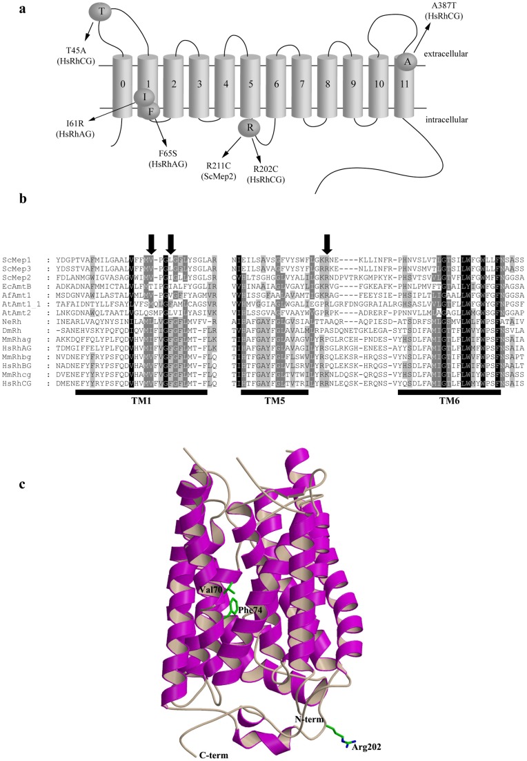
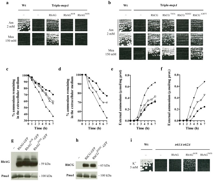
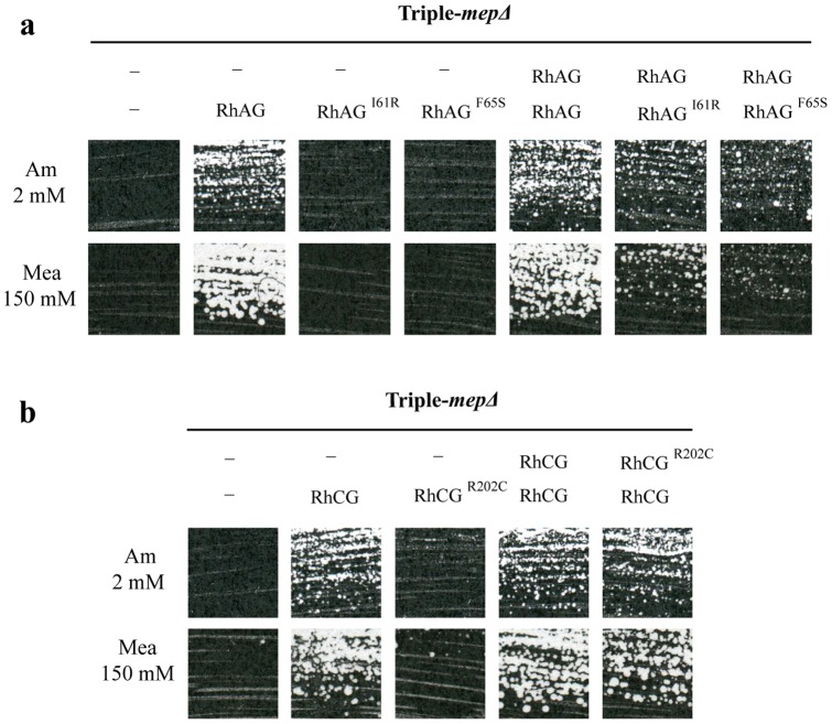
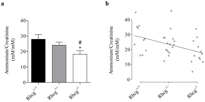
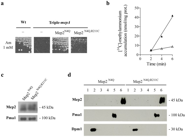
Proteins of the conserved Mep-Amt-Rh family, including mammalian Rhesus factors, mediate transmembrane ammonium transport. Ammonium is an important nitrogen source for the biosynthesis of amino acids but is also a metabolic waste product. Its disposal in urine plays a critical role in the regulation of the acid/base homeostasis, especially with an acid diet, a trait of Western countries. Ammonium accumulation above a certain concentration is however pathologic, the cytotoxicity causing fatal cerebral paralysis in acute cases. Alteration in ammonium transport via human Rh proteins could have clinical outcomes. We used a yeast-based expression assay to characterize human Rh variants resulting from non synonymous single nucleotide polymorphisms (nsSNPs) with known or unknown clinical phenotypes and assessed their ammonium transport efficiency, protein level, localization and potential trans-dominant impact. The HsRhAG variants (I61R, F65S) associated to overhydrated hereditary stomatocytosis (OHSt), a disease affecting erythrocytes, proved affected in intrinsic bidirectional ammonium transport. Moreover, this study reveals that the R202C variant of HsRhCG, the orthologue of mouse MmRhcg required for optimal urinary ammonium excretion and blood pH control, shows an impaired inherent ammonium transport activity. Urinary ammonium excretion was RHcg gene-dose dependent in mouse, highlighting MmRhcg as a limiting factor. HsRhCG(R202C) may confer susceptibility to disorders leading to metabolic acidosis for instance. Finally, the analogous R211C mutation in the yeast ScMep2 homologue also impaired intrinsic activity consistent with a conserved functional role of the preserved arginine residue. The yeast expression assay used here constitutes an inexpensive, fast and easy tool to screen nsSNPs reported by high throughput sequencing or individual cases for functional alterations in Rh factors revealing potential causal variants.


Conflict of interest statement

Competing Interests: The authors have declared that no competing interests exist.

Figures

Figure 1
Figure 1. Location of the considered amino acid substitutions in HsRhAG, HsRhCG, and ScMep2.
a Schematic representation of the predicted topology of Mep-Amt-Rh proteins. The first transmembrane segment (0) is absent in ScMep2 protein. b Partial primary sequence alignment of Mep-Amt-Rh proteins from different organisms. SWISS PROT or GeneBank accession numbers are referred hereafter. ScMep1, 2, 3: S. cerevisiae Mep1 (P40260), Mep2 (P41948), and Mep3 (P53390); EcAmtB: E. coli AmtB (P69681); AfAmt1: A. fulgidus Amt1 (O29285); AtAmt1_1, 2: A. thaliana Amt1;1 (P54144) and Amt2 (Q9M6N7); NeRh: N. europaea Rh50 (Q82X47); DmRh: D. melanogaster Rh (Q9V3T3); MmRhag, bg, cg: M. musculus Rhag (Q9QUT0), Rhbg (Q8BUX5) and Rhcg (Q9QXP0); HsRhAG, BG, CG: H. sapiens RhAG (Q02094), RhBG (AAL05978) and RhCG (Q9UBD6). The limits of transmembrane helices (TM) 1, 5 and 6 of human HsRhCG are deduced from the crystal structure of this protein (pdb code 3HD6) and are underlined. The location of Ile61 and Phe65 of HsRhAG and Arg202 of HsRhCG is indicated by an arrow. c Ribbon representation of the HsRhCG X-ray structure with labelled and depicted Val70, Phe74 and Arg202 residues.
Figure 2
Figure 2. OHSt-related HsRhAG variants and HsRhCGR202C are altered in inherent bidirectional ammonium transport.
a–b Growth tests on solid minimal medium containing as the sole nitrogen source 2 mM ammonium (Am 2 mM), or proline supplemented with a toxic concentration of methylammonium (Mea 150 mM). Wild-type cells (23344c) were transformed with the empty p426 vector (−), and triple-mepΔ cells (31019b) were transformed with the empty p426 vector (−), or with a multi-copy plasmid bearing the native (HsRhAG, HsRhCG) or the mutated HsRHAG and HsRHCG genes (HsRhAGI61R, HsRhAGF65S, HsRhCGT45A, HsRhCGR202C, HsRhCGA387T). Cells were incubated for 7 days (a), and for 7 days (Mea 150 mM, Am 2 mM upper line) or 14 days (Am 2 mM bottom line) (b), at 29°C. c-d Ammonium removal assays. Triple-mepΔ cells (31019b) were transformed with p426 (), p426-HsRhAG (), p426-HsRhAGI61R (Δ), p426-HsRhAGF65S (), p426-HsRhCG (▾) or p426-HsRhCGR202C (∇). E–f Ammonium excretion assays. Same cells used in (c) and (d). g–h Immunodetection of the HsRhAG-GFP and HsRhCG-GFP variants expressed in yeast. Triple-mepΔ cells (31019b) were transformed with the empty p426 vector (−), or with a multi-copy plasmid bearing GFP-tagged native (HsRhAG-GFP, HsRhCG-GFP) or mutated HsRHAG and HsRHCG genes (HsRhAGI61R-GFP, HsRhAGF65S-GFP, HsRhCGR202C-GFP). Membrane-enriched cell extracts were prepared from cells grown on glutamate minimal medium, separated by SDS-PAGE and immunoblotted with anti-GFP antibodies. ScPma1 was immunodetected as a loading control. i Growth tests of yeast strains on solid YNB medium with glutamine as the sole nitrogen source and supplemented with 5 mM KCl (K+ 5mM). Wild-type (S288c) and trk1Δ trk2Δ cells (CY162) were transformed as in (a) and incubated for 5 days at 29°C.
Figure 3
Figure 3. Analysis of trans-dominant effects of altered HsRhAG and HsRhCG variants.
Growth tests on solid minimal medium containing as the sole nitrogen source 2 mM ammonium (Am 2 mM) or, proline supplemented with a toxic concentration of methylammonium (Mea 150 mM). a Triple-mepΔ cells (31064a) were co-transformed with 2 multi-copy vectors: p424 (−) or p424-HsRhAG with p426 (−), p426-sRhAG, p426-HsRhAGI61R or p426-HsRhAGF65S. Cells were incubated for 7 days at 29°C. b Triple-mepΔ cells (31064a) were co-transformed with 2 multi-copy vectors: p424 (−), p424-HsRhCG or p424-HsRhCGR202C with p426 (−), p426-HsRhCG or p426-HsRhCGR202C. Cells were incubated for 7 (Am 2 mM) and 8 (Mea 150 mM) days at 29°C.
Figure 4
Figure 4. RHcg gene-dose effect on urinary ammonium content in mice.
Urinary ammonium and creatinine were assayed on urines of Rhcg+/+, Rhcg+/− and Rhcg−/− mice collected after a fasting of 16 hours. a Data expressed as means ± s.e.m.; 10≤n≤16; *, p<0.05 versus Rhcg+/+; #, p<0.05 versus Rhcg+/−. b Correlation of Spearman. The correlation between RHcg alleles number (0, 1 or 2) and ammonium urinary content is positive (rs  = 0.425) and the gradation observed between Rhcg−/−, Rhcg+/− and Rhcg+/+ is significant (p<0.05).
Figure 5
Figure 5. The R211C substitution alters inherent ammonium transport via yeast ScMep2.
a Growth tests of yeast strains on solid minimal medium containing 1 mM ammonium (Am 1mM) as the sole nitrogen source. Wild-type cells (23344c) were transformed with the empty pFL38 vector (−) and triple-mepΔ cells (31019b) were transformed with the empty pFL38 vector or with YCpMep2N4Q or YCpMep2N4Q,R211C. Cells were incubated for 4 days at 29°C. b Accumulation of [14C]-methylammonium (0.5 mM) was measured in proline-grown triple-mepΔ (31019b) cells transformed with YCpMep2N4Q (▴) or with YCpMep2N4Q,R211C (Δ) c Immunodetection of the ScMep2 variant. Same cells used in (b). Membrane-enriched cell extracts were separated by SDS-PAGE and immunoblotted with anti-Mep2 antibodies. ScPma1 was immunodetected as a loading control. d Subcellular localization of the ScMep2 variant. Same cells used in (b). Membrane-enriched yeast cell extracts were submitted to subcellular fractionation. The six different fractions were separated by SDS-PAGE and immunoblotted with anti-Mep2 antibodies. ScDpm1 and ScPma1 were immunodetected as markers for internal membranes (fractions 2, 3 and 4) and for plasma membrane (fractions 5 and 6), respectively.
Figure 6
Figure 6. The C-terminal extension of HsRhCG forms a network with intracellular loops.
a A cytoplasmic view of the trimeric HsRhCG crystal structure. Each monomer is depicted in coloured ribbon. To emphasize contacts between distant residues, the long intracellular loop between transmembrane helices TM5 and TM6 is in magenta, the short loop between TM3 and TM4 in blue and the C-terminus extension from Arg417 to Ser443 is coloured in red. The C-extension of one monomer is found oriented towards the adjacent monomer, in a circular manner. Note that the last C-terminal 36 residues of HsRhCG protein cannot be built due to lack of available structural data. In order to localize the substrate pore channel, side chains of the twin-histidines (His185 and His344) are showed and labelled in each monomer. The side chain of Arg202, as well as those of the two surrounding residues, Arg201 and Asn203, are also depicted and labelled in each monomer. The atomic colour scheme is carbon in green, nitrogen in blue, oxygen in red and sulphur in yellow. b Schematic 2D interacting plot of residues Arg201 to Asn203 in HsRhCG crystal structure. Hydrogen bonds are green dashed lines and van der Waals contacts are represented by red semi-circles with radiating spokes. Bonds of Arg202 and the two surrounding residues Arg201 and Asn203 are in purple and other bonds are in light brown. All atoms and residues are labelled. The atomic colour scheme is carbon in dark, nitrogen in blue, oxygen in red and sulphur in yellow. c Schematic 2D interacting plot of residues 422 to 443 of the HsRhCG C-terminus. See (b) for the legend.

References

    1. Auron A, Brophy PD (2012) Hyperammonemia in review: pathophysiology, diagnosis, and treatment. Pediatric nephrology 27: 207–222. - PubMed
    1. Weiner ID, Verlander JW (2011) Role of NH3 and NH4+ transporters in renal acid-base transport. American journal of physiology Renal physiology 300: F11–23. - PMC - PubMed
    1. Adeva MM, Souto G (2011) Diet-induced metabolic acidosis. Clinical nutrition 30: 416–421. - PubMed
    1. Cordain L, Eaton SB, Sebastian A, Mann N, Lindeberg S, et al. (2005) Origins and evolution of the Western diet: health implications for the 21st century. The American journal of clinical nutrition 81: 341–354. - PubMed
    1. Pitts RF, Lotspeich WD, Schiess WA, Ayer JL, Miner P (1948) The renal regulation of acid-base balance in man. I. The nature of the mechanism for acidifying the urine. The Journal of clinical investigation 27: 48–56. - PMC - PubMed

Publication types

MeSH terms

LinkOut - more resources