. 2013 Aug 13;8(8):e71641.
doi: 10.1371/journal.pone.0071641. eCollection 2013.

Fibroblast growth factor-4 enhances proliferation of mouse embryonic stem cells via activation of c-Jun signaling

Affiliations

Fibroblast growth factor-4 enhances proliferation of mouse embryonic stem cells via activation of c-Jun signaling

Sung-Ho Kook et al. PLoS One. .

Abstract

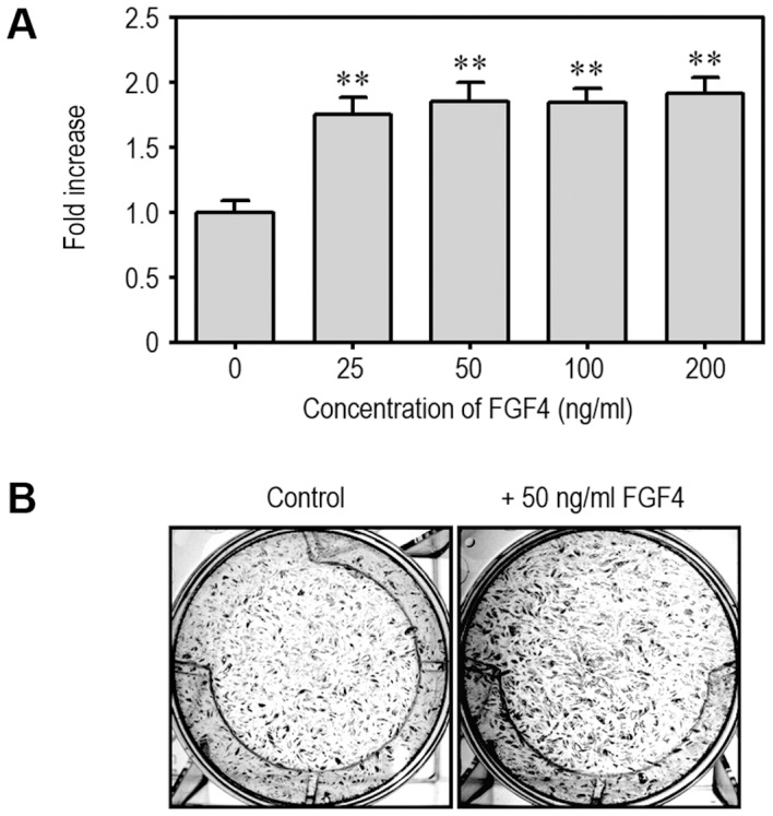
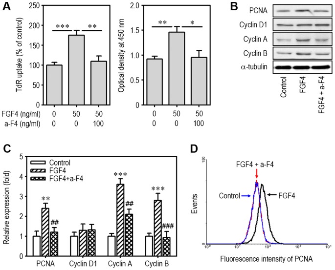
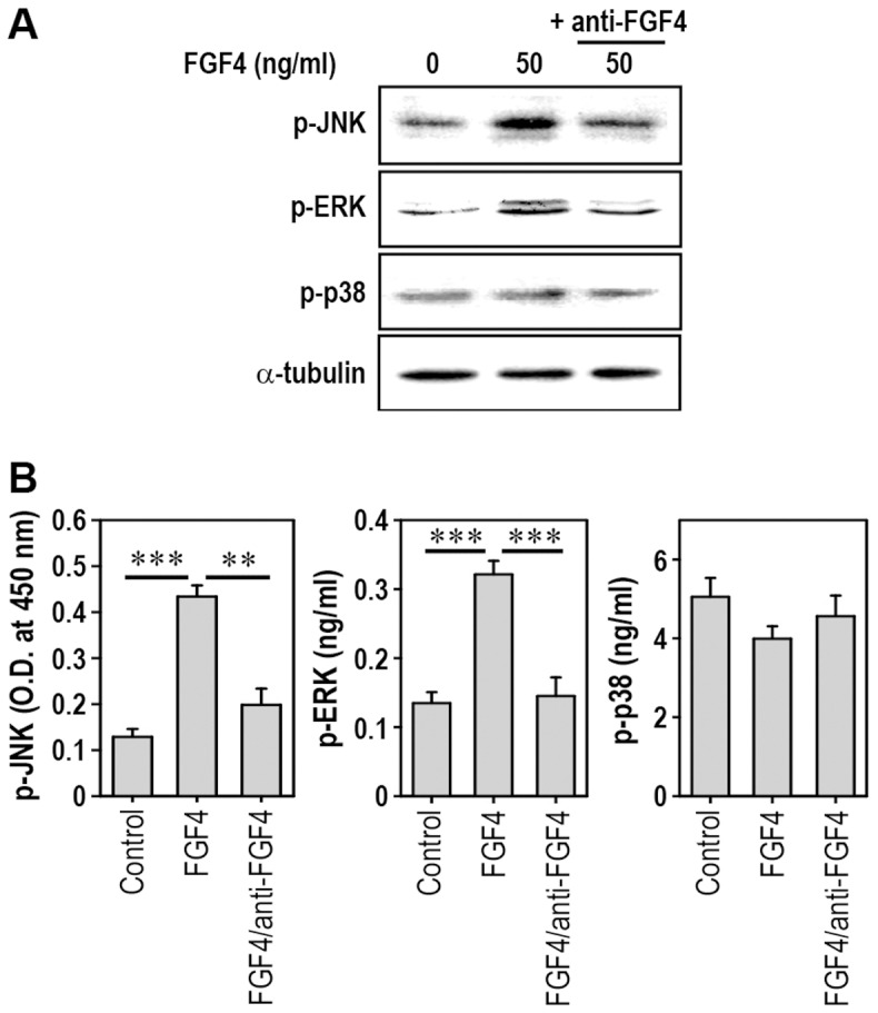
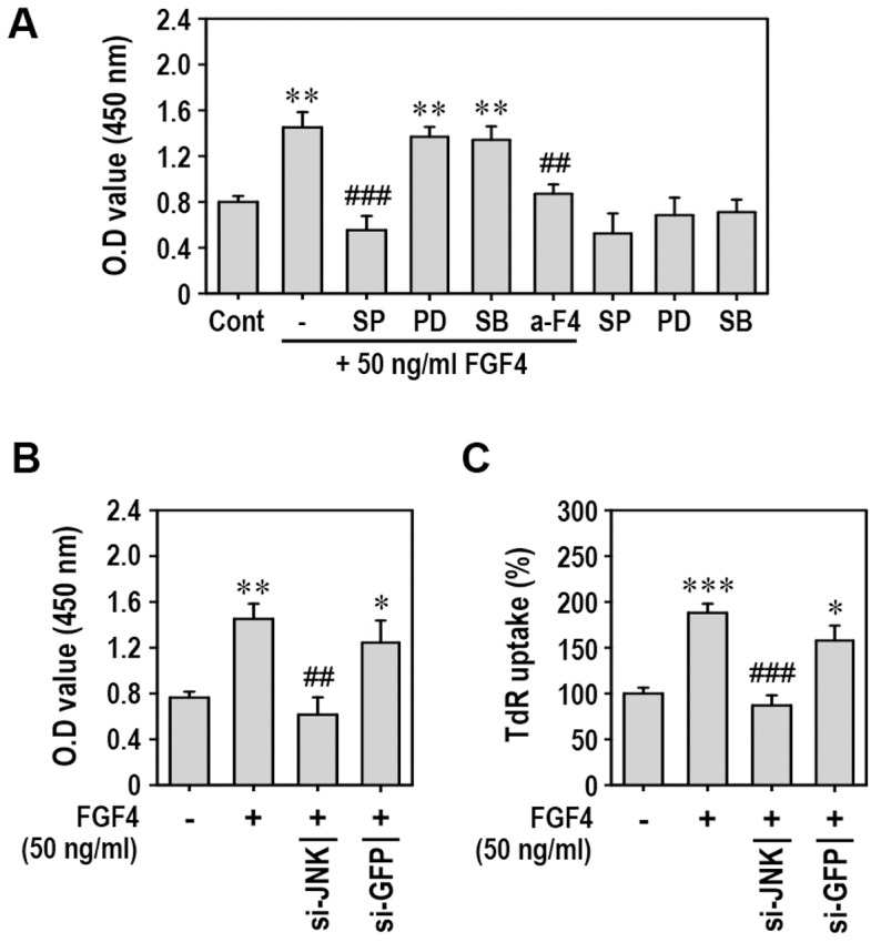
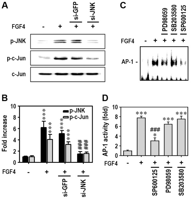
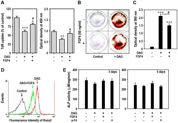
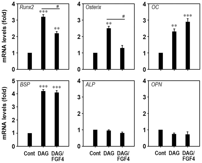
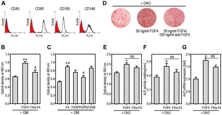
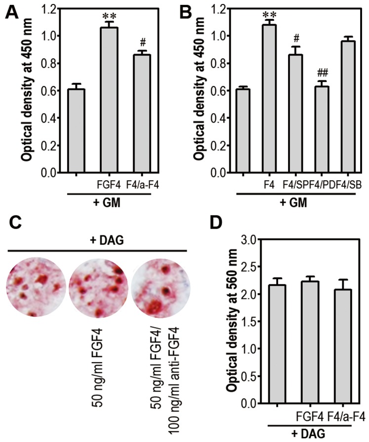
Fibroblast growth factor-4 (FGF4) is expressed in embryonic stages and in adult tissues, where it plays critical roles in modulating multiple cellular functions. However, the exact roles of FGF4 on proliferation and differentiation of embryonic stem cells (ESCs) are not completely understood. Exogenous addition of FGF4 stimulated proliferation of mouse ESCs (mESCs), as proven by the increases in DNA synthesis and cell cycle regulatory protein induction. These increases were almost completely inhibited by pre-treating cells with anti-FGF4 antibody. FGF4 also activated c-Jun N-terminal kinase (JNK) and extracellular-signal regulated kinase (ERK) signaling, but not p38 kinase. Blockage of JNK signaling by SP600125 or by transfection with its specific siRNA significantly inhibited FGF4-stimulated cell proliferation through the suppression of c-Jun induction and activator protein-1 (AP-1) activity. However, ERK or p38 kinase inhibitor did not affect FGF4-stimulated proliferation in mESCs. FGF4 suppressed osteogenic differentiation of mESCs by inhibiting expression of transcription factors involved in bone formation. Further, exogenous FGF4 addition stimulated proliferation of human periodontal ligament stem cells (hPDLSCs) and bone marrow mesenchymal stem cells (BMMSCs) via activation of ERK signaling. FGF4 also augmented mineralization of hPDLSCs, but not of BMMSCs. Collectively, it is suggested that FGF4 triggers proliferation of stem cells by activating MAPK-mediated signaling, while it affects differently osteogenic differentiation according to the origins of stem cells.


Conflict of interest statement

Competing Interests: The authors have declared that no competing interests exist.

Figures

Figure 1
Figure 1. FGF4 increases proliferation of mESCs.
mESCs were incubated in the presence of 0 to 200 ng/ml FGF4 in 96-multiwell plates or with the indicated concentration of FGF4 in 6-well culture plates for 48 h and then further processed for BrdU incorporation assay (A) and hematoxylin staining (B), respectively. Experimental results shown in (A) represent the mean ± SD from three separate experiments. **p<0.01 indicates a significant difference between experiments and untreated control cells.
Figure 2
Figure 2. Effects of anti-FGF4 antibody on FGF4-induced cell proliferation and protein expression associated with cell cycle progression.
mESCs cultured in 6-well culture plates were incubated in the presence of 50 ng/ml FGF4 and/or 100 ng/ml anti-FGF4 antibody and then proliferation rates were analyzed using 3H-TdR incorporation (A, left panel) and Cell Counting Kit-8 (A, right panel) 48 h after treatment. *p<0.05, **p<0.01, and ***p<0.001 indicate significant differences between experiments. (B) Expression patterns of cell cycle regulatory proteins 24 h after treatment were analyzed by Western blotting. (C) Data from Western blot analysis were quantified by densitometry after normalizing bands to α-tubulin levels. **p<0.01 and ***p<0.001 vs. untreated cells. ##p<0.01 and ###p<0.001 vs. cells treated with FGF4 only. (D) mESCs cultured under the same conditions as (C) were also processed for analysis of PCNA expression by flow cytometry. a-F4, anti-FGF4 antibody.
Figure 3
Figure 3. Addition of FGF4 activates JNK and ERK, but not p38 kinase, in mESCs.
mESCs cultured in 6-well culture plates were incubated in the presence of 50 ng/ml FGF4 and/or 100 ng/ml anti-FGF4 antibody for 1 h and then processed to determine levels of phosphorylated MAPK by immunoblotting (A) or by immunometric analyses (B). **p<0.01 and ***p<0.001 indicate significant differences between experiments.
Figure 4
Figure 4. Inhibition of JNK suppresses FGF4-mediated proliferation of mESCs.
(A) mESCs were pretreated with MAPK inhibitor or anti-FGF4 antibody 30 min before addition of 50 ng/ml recombinant FGF4, and cell proliferation rate was determined 48 h after treatment using a cell proliferation assay kit. In addition, siRNAs directed against JNK or GFP were transfected into mESCs. At 24 h post-transfection, cells were exposed to 50 ng/ml FGF4 and cell proliferation rate (B) and 3H-TdR incorporation (C) were measured 48 h after treatment. *p<0.05, **p<0.01, and ***p<0.001 vs. untreated cells. ##p<0.01 and ###p<0.001 vs. cells treated with FGF4 only.
Figure 5
Figure 5. JNK acts as an upstream mediator of c-Jun phosphorylation and AP-1 activation.
(A) mESCs transfected with siJNK were incubated in the presence or absence of 50 ng/ml FGF4 and then processed for Western blot analysis 1 h after treatment. (B) Data were quantified from triplicate experiments by densitometry after normalizing bands to total c-Jun protein. mESCs were also treated with each MAPK-specific inhibitor (10 µM) 30 min before addition of 50 ng/ml FGF4, and cells were adjusted to analyze AP-1-DNA binding activity by EMSA (C) or AP-1 activity using a luciferase assay kit (D) after 2 h of incubation. *p<0.05 and ***p<0.001 vs. untreated controls. ###p<0.001 vs. cells treated with 50 ng/ml FGF4 only.
Figure 6
Figure 6. Exogenous FGF4 decreases osteogenic differentiation of mESCs.
(A) mESCs were incubated in the presence of DAG and/or 50 ng/ml FGF4. After 5 days of incubation, DNA synthesis (left panel) and cell proliferation (right panel) rates were determined. At the same time, cells were processed for Alizarin red staining (B) and absorbance of the dye (C) was also determined at 560 nm. (D) Runx2 protein in mESCs cultured with DAG and/or 50 ng/ml FGF4 for 5 days was analyzed by flow cytometry. (E) ALP activity in the cells was measured after 3 and 5 days of differentiation. *p<0.05, **p<0.01 and ***p<0.001 vs. untreated control cells. #p<0.05 vs. cells treated with DAG only.
Figure 7
Figure 7. Addition of FGF4 diminishes DAG-induced expression of Runx2 and osterix in mESCs.
Cells were incubated in the presence or absence of DAG and 50 ng/ml FGF4 for 24 h, and expression patterns of bone-specific genes were analyzed by real-time RT-PCR. **p<0.01 and ***p<0.001 vs. untreated control cells. #p<0.05 vs. cells treated with DAG only.
Figure 8
Figure 8. FGF4 stimulates proliferation and mineralization of hPDLSCs.
(A) hPDLSCs at passage 3 were labeled with the indicated primary antibodies and then analyzed by flow cytometer. The cells were incubated for 48 h with 50 ng/ml FGF4 and/or 100 ng/ml (B), or with each of MAPK inhibitors (C) in 96-multiwell plates. The cells were processed for cell proliferation assay. **p<0.01 vs. GM alone. #p<0.05 vs. FGF4 treatment alone. (D) hPDLSCs were incubated in the presence of DAG with and without 50 ng/ml FGF4 and 100 ng/ml anti-FGF4 for 14 days and then were processed for Alizarin red staining. (E) Absorbance of the dye was determined at 560 nm. hPDLSCs were also processed for analysis of ALP activity (F) and mRNA expression (G) after 5 days of differentiation. *p<0.05 vs. DAG alone. F4, FGF4; GM, growth medium; NC, non-significant.
Figure 9
Figure 9. FGF4 stimulates proliferation of mBMMSCs via activation of MAPK.
mBMMSCs were incubated with 50 ng/ml FGF4 and/or 100 ng/ml (A), or each of MAPK inhibitors (B) in 96-multiwell plates. After 48 h, these cells were processed for cell proliferation assay. **p<0.01 vs. GM alone. #p<0.05 and ##p<0.01 vs. FGF4 treatment alone. (C) mBMMSCs were incubated in the presence of DAG with and without 50 ng/ml FGF4 and 100 ng/ml anti-FGF4 for 14 days and were then stained with Alizarin red. (D) The absorbance of the dye was measured at 560 nm.

References

    1. Barrientos S, Stojadinovic O, Golinko MS, Brem H, Tomic-Canic M (2008) Growth factors and cytokines in wound healing. Wound Repair and Regeneration 16: 585–601. - PubMed
    1. D’Andrea LD, Del Gatto A, De Rosa L, Romanelli A, Pedone C (2009) Peptides targeting angiogenesis related growth factor receptors. Current Pharmaceutical Design 15: 2414–2429. - PubMed
    1. Quarto N, Wan DC, Longaker MT (2008) Molecular mechanisms of FGF-2 inhibitory activity in the osteogenic context of mouse adipose-derived stem cells (mASCs). Bone 42: 1040–1052. - PubMed
    1. Ahn HJ, Lee WJ, Kwack K, Kwon YD (2009) FGF2 stimulates the proliferation of human mesenchymal stem cells through the transient activation of JNK signaling. FEBS Letters 583: 2922–2926. - PubMed
    1. Ito T, Sawada R, Fujiwara Y, Tsuchiya T (2008) FGF-2 increases osteogenic and chondrogenic differentiation potentials of human mesenchymal stem cells by inactivation of TGF-beta signaling. Cytotechnology 56: 1–7. - PMC - PubMed

Publication types

MeSH terms

Substances