. 2013 Nov;24(11):1882-8.
doi: 10.1016/j.jnutbio.2013.05.004. Epub 2013 Aug 19.

3,3'-diindolylmethane rapidly and selectively inhibits hepatocyte growth factor/c-Met signaling in breast cancer cells

Affiliations

3,3'-diindolylmethane rapidly and selectively inhibits hepatocyte growth factor/c-Met signaling in breast cancer cells

Holly L Nicastro et al. J Nutr Biochem. 2013 Nov.

Abstract

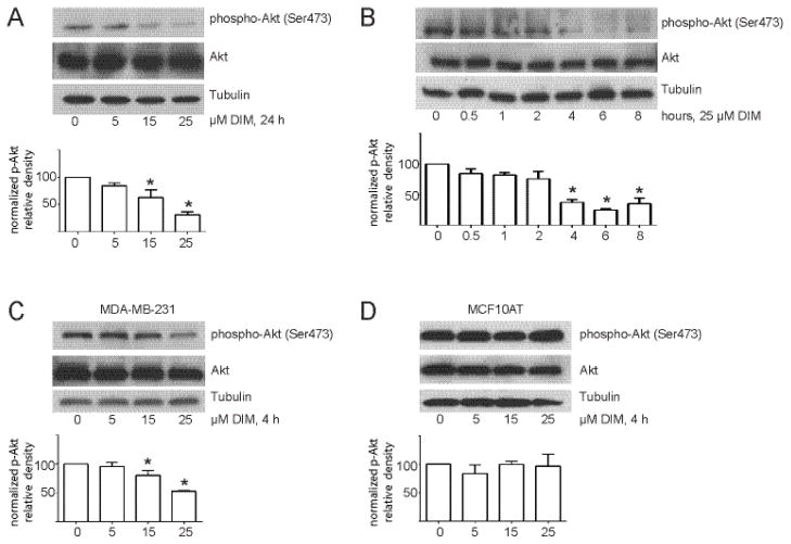
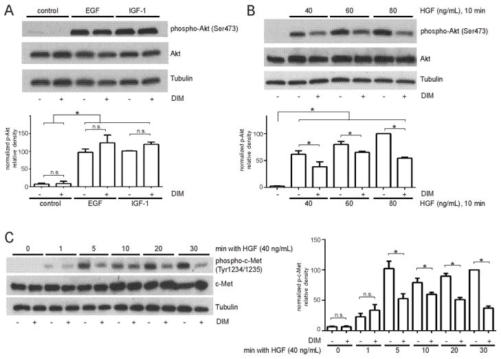
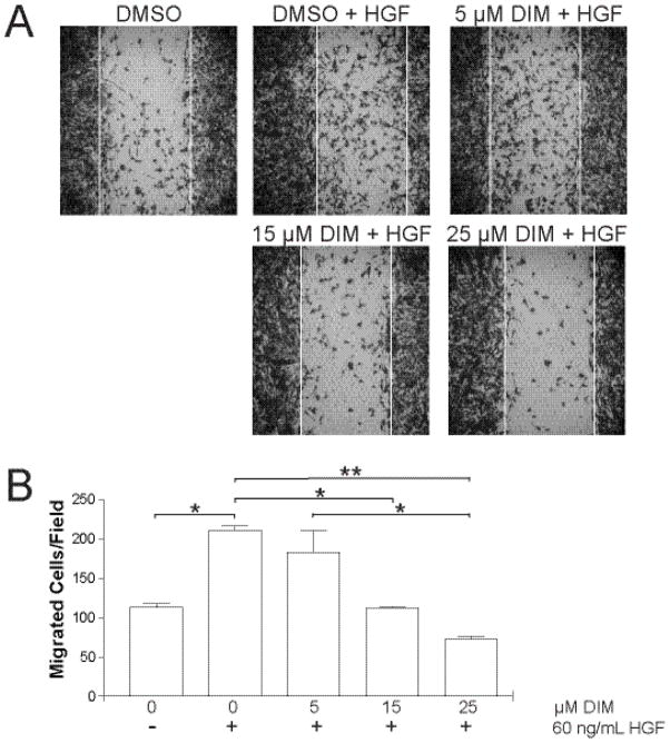
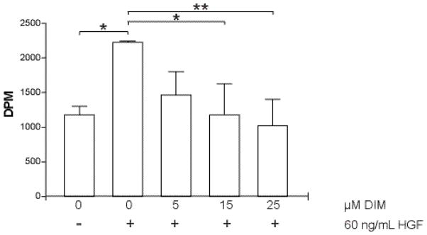
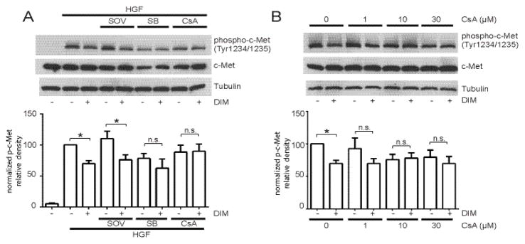
3,3'-Diindolylmethane (DIM), an indole derivative from vegetables of the Brassica genus, has antiproliferative activity in breast cancer cells. Part of this activity is thought to be due to DIM inhibition of Akt signaling, but an upstream mechanism of DIM-induced Akt inhibition has not been described. The goals of this study were to investigate the kinetics of inhibition of Akt by physiologically relevant concentrations of DIM and to identify an upstream factor that mediates this effect. Here we report that DIM (5-25 μM) inhibited Akt activation from 30 min to 24h in tumorigenic MDA-MB-231 cells but did not inhibit Akt activation in non-tumorigenic preneoplastic MCF10AT cells. DIM inhibited hepatocyte growth factor (HGF)-induced Akt activation by up to 46%, cell migration by 66% and cell proliferation by up to 54%, but did not inhibit induction of Akt by epidermal growth factor or insulin-like growth factor-1. DIM decreased phosphorylation of the HGF receptor, c-Met, at tyrosines 1234 and 1235, indicating decreased activation of the receptor. This decrease was reversed by pretreatment with inhibitors of p38 or calcineurin. Our results demonstrate the important role of HGF and c-Met in DIM's anti-proliferative effect on breast cancer cells and suggest that DIM could have preventive or clinical value as an inhibitor of c-Met signaling.

Keywords: 3,3′-diindolylmethane; Akt; Breast cancer; c-Met.


Conflict of interest statement

Conflicts of interest: None

Figures

Fig. 1
Fig. 1
DIM inhibits Akt activation in MDA-MB-231 cells but not in non-tumorigenic MCF10AT cells. (A) Western blot analysis of phosphorylated and total Akt protein from cells incubated in 1% FBS overnight and then treated with increasing concentrations of DIM for 24 h. (B) Western blot analysis of phosphorylated and total Akt protein from cells treated with 25 μM DIM for 0–8 h. (C) and (D) Same as A except MDA-MB-231 cells (C) or MCF10AT cells (D) were treated with DIM for 4 h. Tubulin was used as a loading control. Histograms depict phospho-Akt band density normalized to total Akt band density expressed as percent of control. Bars represent mean ± SEM of 3 independent experiments. *, p<0.05.
Fig. 2
Fig. 2
DIM inhibits Akt activation by HGF and decreases phosphorylation of c-Met. (A) MDA-MB-231 cells were incubated overnight in serum-free medium, treated with DIM for 4 h, and then epidermal growth factor (EGF, 25 ng/mL) or insulin-like growth factor-1 (IGF-1, 25 ng/mL) for 10 min. (B) Cells were incubated overnight in serum-free medium, treated with DIM for 4 h, and HGF (40 ng/mL) for 10 minutes. Total cell lysates were collected and analyzed by Western blotting for phospho-Akt Ser473 and total Akt. (C) Western blot analysis of phospho-c-Met Tyr1234/1235 and total c-Met from cells incubated in serum-free medium overnight and then treated with DMSO or 25 μM DIM for 4 h, followed by HGF treatment for 0–30 min. Tubulin was used as a loading control. Histograms depict phospho-Akt or phospho-c-Met band density normalized to total Akt or total c-Met band density expressed as percent of control. Bars represent mean ± SEM of 3 independent experiments. *, p<0.05; n.s., not statistically significant.
Fig. 3
Fig. 3
DIM decreases Akt activity in cells. (A) Cells were incubated in medium containing 1% FBS overnight and then treated with increasing concentrations of DIM for 4 h. (B) Cells were serum-starved overnight and then treated with increasing concentrations of DIM for 4 h and then 40 ng/mL HGF was added for 10 min. In both experiments, total cell lysates were collected and analyzed by Western blotting for phospho and total GSK-3α/β, and the loading control tubulin. Histograms depict phospho- GSK-3α/β band density normalized to total GSK-3α/β band density expressed as percent of control. Bars represent mean ± SEM of 3 independent experiments. *, p<0.05.
Fig. 4
Fig. 4
DIM inhibits HGF-activated cell motility. (A) Confluent monolayers of MDA-MB-231 cells were scratched with a sterile pipette tip, creating gaps of fixed width. Floating cells and debris were washed away with PBS, and serum-free medium or serum-free medium containing HGF (60 ng/mL) and 0, 5, 15, or 25 μM DIM was added. After 16 h, the wounds were photographed. Photographs are representative of 2 independent fields from duplicate wells. (B) Cells that migrated across the original wound borders were counted. Results are representative of three independent experiments. *, p < 0.05; **, p< 0.001.
Fig. 5
Fig. 5
DIM inhibits HGF-stimulated cellular proliferation. Cells were serum-starved overnight and then treated with DMSO or DIM for 4 hours. HGF (60 ng/mL) was added to cells for 1 hour and the cells were then washed with PBS. Fresh serum-free medium containing [3H]-thymidine was added for an additional 4 hours. Excess [3H]-thymidine was removed and cells were lysed in 0.3 N NaOH. Incorporation of [3H]-thymidine into DNA, as a measure of proliferation, was determined by scintillation counting of the lysates. *, p < 0.05; **, p< 0.001.
Fig. 6
Fig. 6
Involvement of tyrosine phosphatases, p38, and calcineurin in DIM’s effects on c-Met phosphorylation. (A) MDA-MB-231 cells were serum-starved overnight, then pretreated with 100 μM SOV, 10 μM SB or 10 μM CsA for 30 minutes, followed by DMSO or 25 μM DIM for 4 h and 40 ng/mL HGF for 10 min. Because total c-Met levels changed with some pretreatments, band intensities are normalized to tubulin and not total c-Met. (B) Same as (A) except cells were pretreated with increasing concentrations of CsA before addition of DIM and HGF. In both experiments, total cell lysates were collected and analyzed by Western blotting for phospho-c-Met Tyr1234/1235, total c-Met, and tubulin. Histograms depict phospho-c-Met band density normalized to tubulin band density expressed as percent of control. Bars represent mean ± SEM of 3 independent experiments. *, p<0.05; n.s., not statistically significant.

References

    1. Doll R, Peto R. The causes of cancer: quantitative estimates of avoidable risks of cancer in the United States today. Oxford; New York: Oxford University Press; 1981. - PubMed
    1. WCRF/AICR. Food, Nutrition, Physical Activity and the Prevention of Cancer: a Global Perspective. Washington, DC: 1997.
    1. Negri E, Lavecchia C, Franceschi S, Davanzo B, Parazzini F. Vegetable and Fruit Consumption and Cancer Risk. International Journal of Cancer. 1991;48:350–4. - PubMed
    1. Terry P, Wolk A, Persson I, Magnusson C. Brassica vegetables and breast cancer risk. JAMA. 2001;285:2975–7. - PubMed
    1. Kristal AR, Lampe JW. Brassica vegetables and prostate cancer risk: a review of the epidemiological evidence. Nutr Cancer. 2002;42:1–9. - PubMed

Publication types

MeSH terms