. 2015 Jan;25(1):167-79.
doi: 10.1093/cercor/bht209. Epub 2013 Aug 22.

A forward genetic screen in mice identifies mutants with abnormal cortical patterning

Affiliations

A forward genetic screen in mice identifies mutants with abnormal cortical patterning

Seungshin Ha et al. Cereb Cortex. 2015 Jan.

Abstract

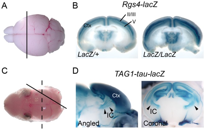
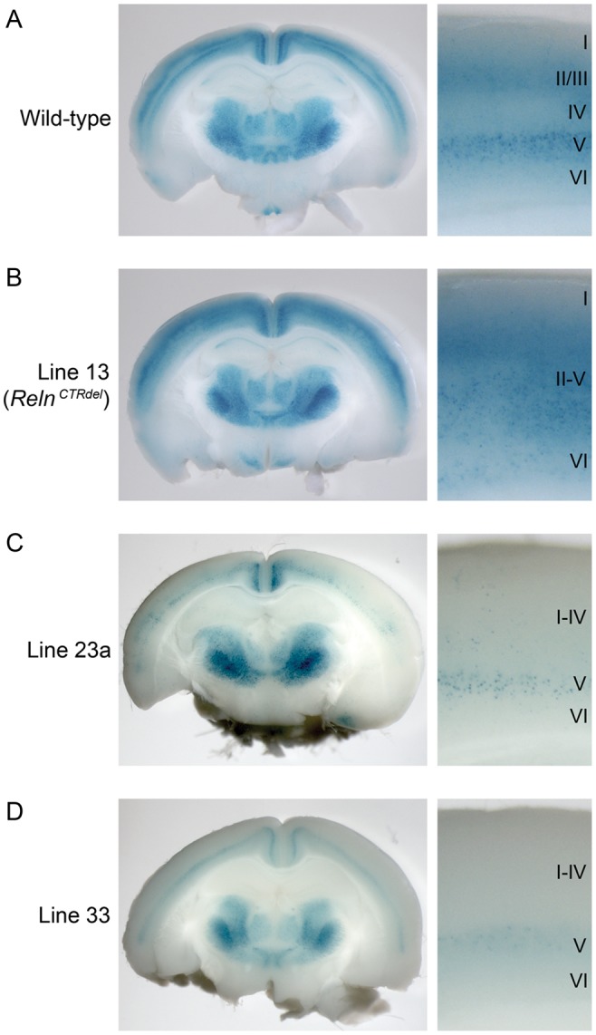
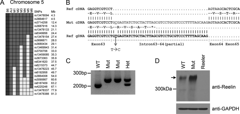
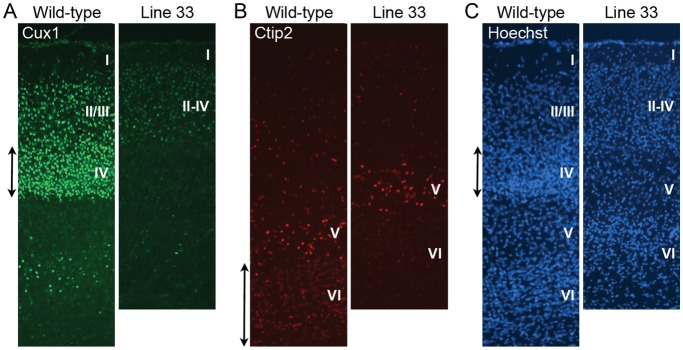
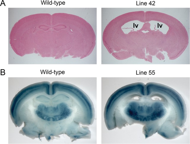
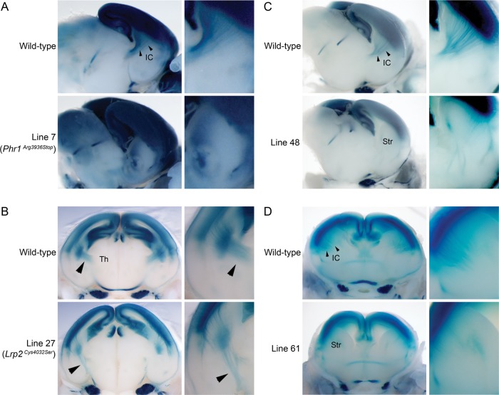
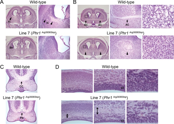
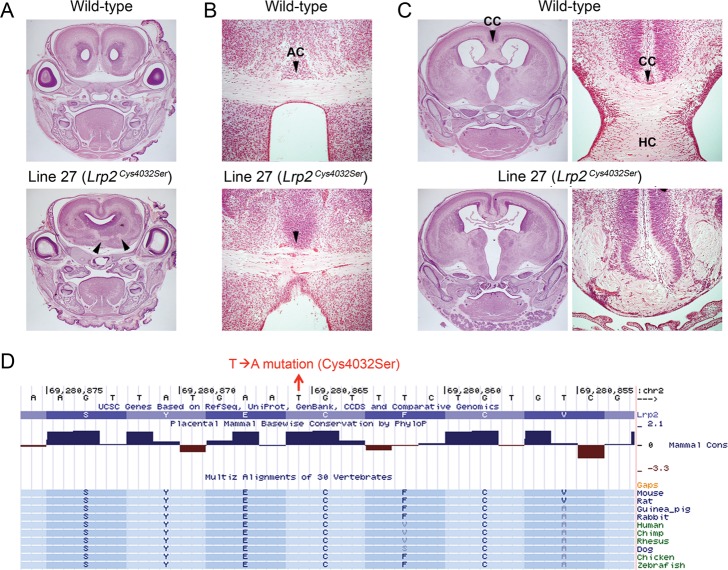
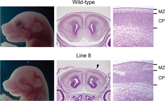
Formation of a 6-layered cortical plate and axon tract patterning are key features of cerebral cortex development. Abnormalities of these processes may be the underlying cause for a range of functional disabilities seen in human neurodevelopmental disorders. To identify mouse mutants with defects in cortical lamination or corticofugal axon guidance, N-ethyl-N-nitrosourea (ENU) mutagenesis was performed using mice expressing LacZ reporter genes in layers II/III and V of the cortex (Rgs4-lacZ) or in corticofugal axons (TAG1-tau-lacZ). Four lines with abnormal cortical lamination have been identified. One of these was a splice site mutation in reelin (Reln) that results in a premature stop codon and the truncation of the C-terminal region (CTR) domain of reelin. Interestingly, this novel allele of Reln did not display cerebellar malformation or ataxia, and this is the first report of a Reln mutant without a cerebellar defect. Four lines with abnormal cortical axon development were also identified, one of which was found by whole-genome resequencing to carry a mutation in Lrp2. These findings demonstrated that the application of ENU mutagenesis to mice carrying transgenic reporters marking cortical anatomy is a sensitive and specific method to identify mutations that disrupt patterning of the developing brain.

Keywords: ENU mutagenesis; cerebral cortex; cortical lamination; corticofugal axon; reelin.


Figures

Figure 1.
Figure 1.
LacZ reporters used for ENU screening. (A) A brain from a P21 mouse was dissected out and coronally sectioned to expose the cross section of the cortex. (B) Reporter expression pattern in the cortex (Ctx) of Rgs4-lacZ mice visualized by whole-mount X-gal staining. Note that superficial layers II/III and deeper layer V shows strong X-gal staining. (C) A brain from P0 pups were dissected out and sectioned in an angled plane (solid line) or coronally (dashed line) to expose the cross section of the cortex and striatum. (D) Reporter expression pattern in the brain of TAG1-tau-lacZ mice. TAG1-tau-lacZ marks the corticofugal axons of the cortex (Ctx) which fasciculate at the internal capsule (IC). Arrowheads indicate the internal capsule.
Figure 2.
Figure 2.
Mutants with cortical lamination defects discovered using an Rgs4-lacZ transgenic reporter. Coronal-sections of whole-mount X-gal stained P21 brains and high-magnification images of the cortical layers are shown. (A) In wild-type cortex, layers II/III and V were stained as 2 distinct bands, and this allows identification of all 6 layers. (B) In line 13 mutant (RelnCTRdel), reporter expression pattern in the cortex was disorganized, and there was no distinction between superficial and deep layers (II–V). (C) Line 23a mutant brain, which has weak reporter expression, with the superficial layers more severely affected. Layers I–IV cannot be distinguished, and layer V is weakly stained. (D) Line 33 mutant brain, which has a similar phenotype as line 23a.
Figure 3.
Figure 3.
Line 13 has a mutation in Reln. (A) Haplotype-based mapping using a whole-genome SNP panel located the causal mutation on chromosome 5 between the centromere and 27.4 Mb. Genotypes of 6 G3 mutants (M1–M6) in the proximal region of chromosome 5 are shown. SNPs are color-coded: Gray, A/J; black, C57BL/6N (B6); white, heterozygous. (B) Sequencing of Reln cDNA identified a mutation at the exon–intron junction of exon 63. An alignment of Reln cDNA sequence from the mutant (middle) against the sequences of reference cDNA (top) and genomic DNA (bottom) is shown. Between exon 63 and 64, an insertion of intronic sequence was found. Predicted protein sequence from the mutant transcript is shown. (C) Reverse transcription-polymerase chain reaction analysis shows that mutants do not express wild-type transcripts. Mutant transcripts (254 bp) are larger than wild-type transcripts (212 bp). (D) Western blot analysis using anti-reelin antibodies shows that reelin is expressed in the mutant cortex. Arrow indicates full-length reelin. GAPDH was used as loading controls.
Figure 4.
Figure 4.
A complementation assay and cerebellar morphology of RelnCTRdel. (Left) A compound heterozygous mutant of line 13 and reeler (RelnCTRdel/Relnrl) develops lamination defect that appears similar to a homozygous mutant from line 13 (RelnCTRdel/RelnCTRdel), with undistinguishable layers II–V. As controls, brains from heterozygote (Relnrl/Reln+) and homozygote reeler mutants (Relnrl/Relnrl) are shown. (Right) RelnCTRdel/RelnCTRdel and RelnCTRdel/Relnrl show normal cerebellar size and foliation in contrast to Relnrl/Relnrl with abnormal cerebellum.
Figure 5.
Figure 5.
Layer-specific marker analysis of line 33 mutant at P7. (A) Immunohistochemistry using a marker for the superficial layers II–IV, Cux1, is shown. The mutant shows less strong staining and layer IV neurons that are most brightly stained in wild type (double-headed arrow) are missing. (B) Deep layer neurons are visualized using layer V/VI maker Ctip2. There are some layer VI neurons lightly stained with this marker in the wild type (double-headed arrow), and few Ctip2-positive neurons are detected in this layer in the mutant. (C) Nuclear stain using Hoechst is shown. Cortical layers (I–VI) are labeled.
Figure 6.
Figure 6.
Ventriculomegaly mutants were identified during the screening using a Rgs4-lacZ reporter. (A) Histology of P21 brains from line 42. “lv” indicates lateral ventricles. (B) Whole-mount X-gal stained brains from line 55. Mutant brain showed ventriculomegaly with normal lamination.
Figure 7.
Figure 7.
Mutants with corticofugal axon defects discovered using a TAG1-tau-lacZ reporter. Cross sections of whole-mount X-gal stained E18.5 brains and high-magnification images of the axon tracts are shown. (A) Wild-type and mutant brains from line 7 (Phr1Arg3936Stop) were sectioned in an angled plane. The internal capsule (IC) in the wild-type brain is indicated (arrowheads). In the mutant striatum, aberrant axons are apparent. (B) Wild-type and mutant brains from line 27 (Lrp2Cys4032Ser) were sectioned coronally. In the wild-type brain, corticofugal axon tracts extend toward the thalamus (Th). In the mutant brain, the direction of axon tracts is different from wild type (arrowheads). (C) Wild-type and mutant brains from line 48 were sectioned in an angled plane. High-magnification image of the striatum (Str) shows fewer and misguided corticofugal axons. (D) Wild-type and mutant brains from line 61 were sectioned coronally at E17. In the mutant striatum (Str), fewer axons with aberrant patterning are observed.
Figure 8.
Figure 8.
Histology of wild-type and mutant E18.5 brains from line 7 (Phr1Arg3936Stop). (A) The internal capsule (IC, arrowheads in the wild-type images) is absent in the striatum (Str) of the mutant. Enlarged images of striatum are shown on the right. (B, left and middle) Anterior commissure (arrows) is absent in the mutant brain. Instead, cross sections of the aberrant axon tracts (arrowheads) are seen. (B, right) The boxed region in the striatum in B (left) is enlarged and shown. In the wild type, the round-shaped cross sections of the axon tracts are seen, while they are absent in the mutant. (C) Midline-crossing defect of corpus callosum (CC, arrow) is seen in the mutant (Pb; Probst bundle, arrow). (D) Abnormally bundled axon tracts (double-headed arrow) are observed in the mutant cortex. This disturbed the ventricular zone (VZ) and made it thinner in the mutant compared with the wild type. Enlarged images of the VZ are shown on the right.
Figure 9.
Figure 9.
Abnormal midline crossing of commissural axons in line 27 (Lrp2Cys4032Ser) brain. Histology of the wild-type and mutant E18.5 brains from line 27. (A) In a mutant brain with fused cortical hemispheres, anterior commissure is often observed in abnormal planes of section. The aberrant anterior commissure displayed a discontinuous pattern (arrowheads). (B) An example of a mutant brain with a midline-crossing defect (arrowheads) in the anterior commissure (AC). (C) A mutant brain with a midline crossing defect in the corpus callosum (CC). High-magnification images of the corpus callosum are shown on the right. In addition, the hippocampal commissure (HC) is absent, and hyperplasia of the choroid plexus is evident. (D) Image capture of the basewise conservation and multiz alignment tracks from UCSC genome browser. A cysteine (Cys) residue, which changes to serine (Ser) by the missense mutation at 69 280 866 bp (T → A), is conserved between species.
Figure 10.
Figure 10.
Cranial morphology and histology of the wild-type and mutant E18.5 brains from line 8. Histological analysis of the anterior cerebral cortex revealed the disorganization of cortical plate (CP) and invasion of the marginal zone (MZ), a phenotype called as a cobblestone-like cortical malformation. The arrowhead indicates where the high-magnification image is taken.

References

    1. Anderson KV. Finding the genes that direct mammalian development : ENU mutagenesis in the mouse. Trends Genet. 2000;16:99–102. - PubMed
    1. Andersen TE, Finsen B, Goffinet AM, Issinger OG, Boldyreff B. A reeler mutant mouse with a new, spontaneous mutation in the reelin gene. Brain Res Mol Brain Res. 2002;105:153–156. - PubMed
    1. Beckstead WA, Bjork BC, Stottmann RW, Sunyaev S, Beier DR. SNP2RFLP: a computational tool to facilitate genetic mapping using benchtop analysis of SNPs. Mamm Genome. 2008;19:687–690. - PMC - PubMed
    1. Beier DR, Herron BJ. Genetic mapping and ENU mutagenesis. Genetica. 2004;122:65–69. - PubMed
    1. Bloom AJ, Miller BR, Sanes JR, DiAntonio A. The requirement for Phr1 in CNS axon tract formation reveals the corticostriatal boundary as a choice point for cortical axons. Genes Dev. 2007;21:2593–2606. - PMC - PubMed

Publication types

MeSH terms