

Neural processing correlates of assaultive violence exposure and PTSD symptoms during implicit threat processing: a network-level analysis among adolescent girls
- PMID: 23969000
- PMCID: PMC3852193
- DOI: 10.1016/j.pscychresns.2013.06.003
Neural processing correlates of assaultive violence exposure and PTSD symptoms during implicit threat processing: a network-level analysis among adolescent girls
Erratum in
- Psychiatry Res. 2014 Sep 30;223(3):271-2
Abstract
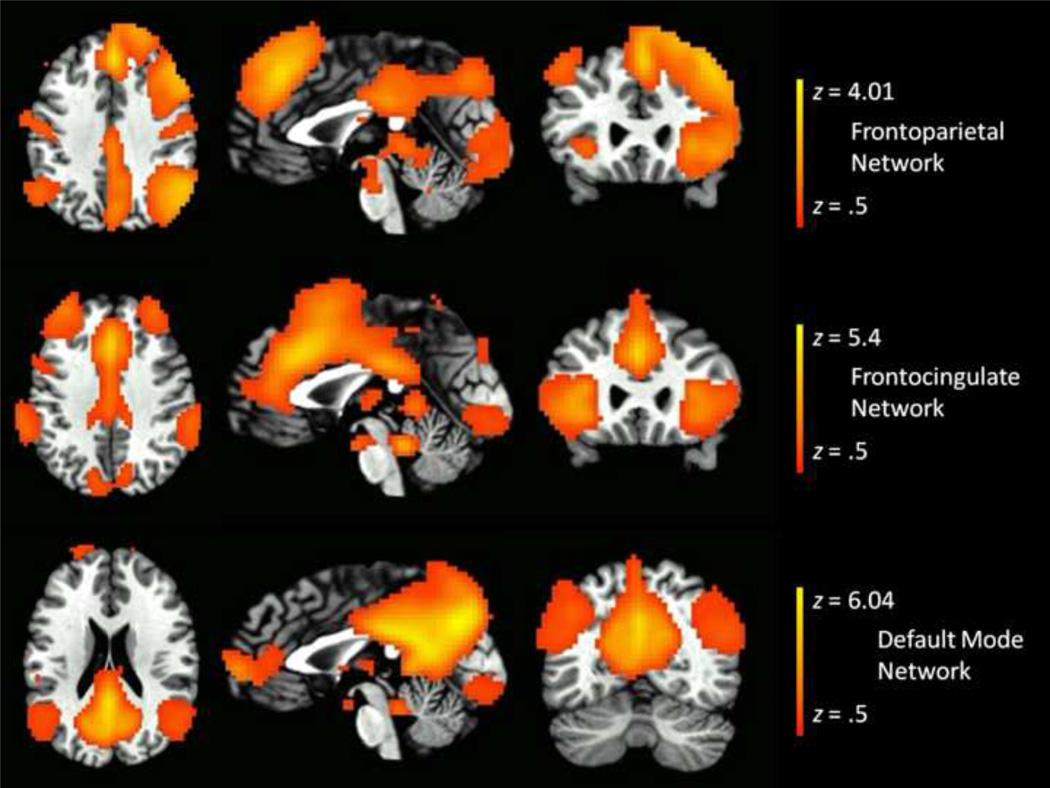
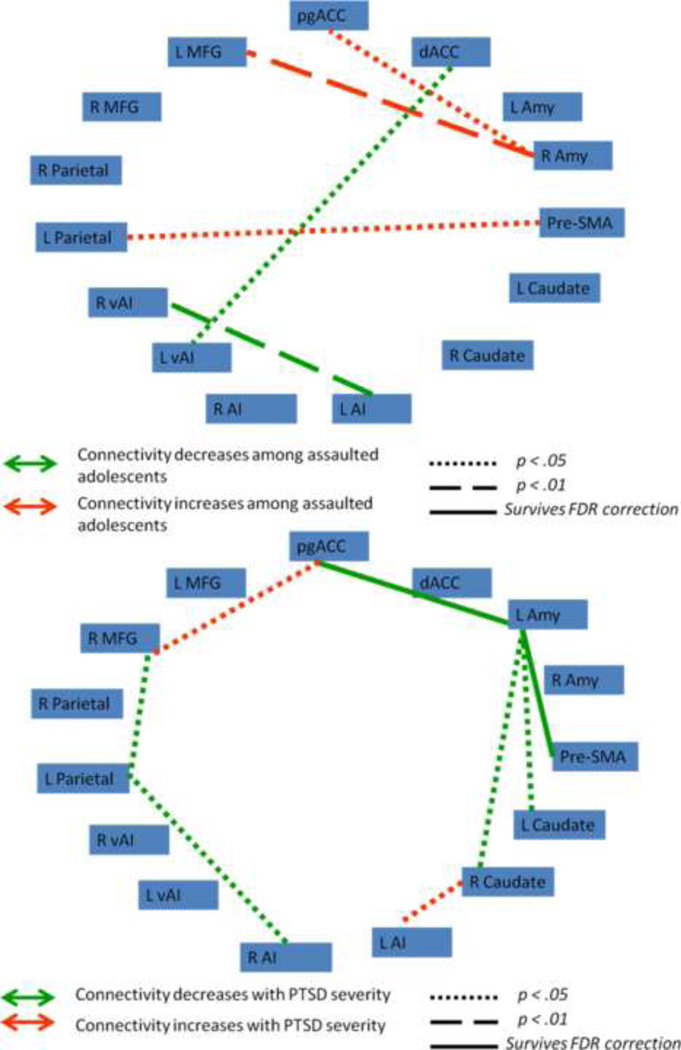
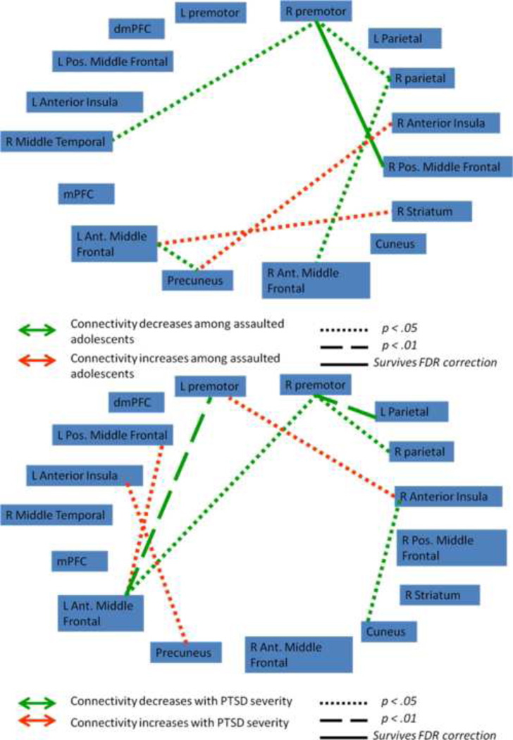
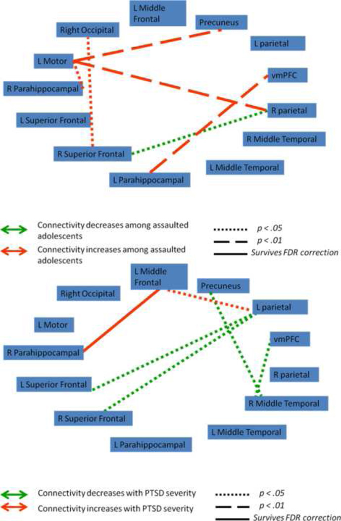
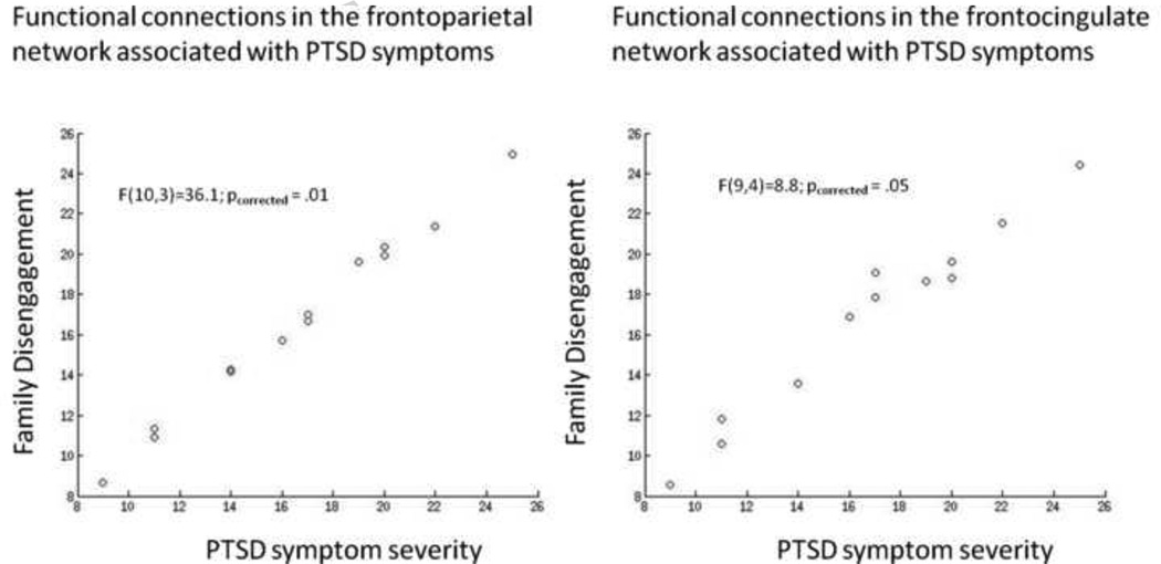
Assaultive violence exposure during childhood is a significant risk factor for posttraumatic stress disorder (PTSD). The purpose of the present study was to characterize the relationships of assault and PTSD severity with the organization of large-scale networks identified during emotion processing. Adolescent girls aged 12-16 with (N=15) and without (N=15) histories of assault underwent functional magnetic resonance imaging (fMRI) while engaged in a task that presented images of fearful or neutral facial expressions. Independent component analysis (ICA) identified a frontocingulate network, a frontoparietal network, and a default mode network. Assault exposure was associated with significantly greater activation of the frontocingulate network for fear versus neutral faces. Within the frontocingulate network, Posttraumatic stress disorder (PTSD) severity was associated with weakened functional connectivity between the left amygdala and the perigenual anterior cingulate. Within the frontoparietal network, assaulted girls demonstrated weakened connectivity of the premotor cortex with the right middle frontal gyrus. Within the default mode network, assault exposure and PTSD severity were associated with strengthening functional connectivity of the parahippocampus with the medial and lateral prefrontal cortex, respectively. Individual differences in functional connections within the frontocingulate network and frontoparietal network among the assaulted group were strongly associated with caregiver-rated family disengagement. These results demonstrate associations between assault and PTSD symptoms with the functional organization of large-scale frontoparietal, frontocingulate, and default mode networks during emotion processing. The relationship with caregiver-rated family disengagement suggests the impact of family support on the neural processing correlates of assault and PTSD symptoms.
Keywords: Adolescence; Early life trauma; Emotion processing; Neuroimaging; PTSD.
Copyright © 2013 Elsevier Ireland Ltd. All rights reserved.
Conflict of interest statement
Financial Disclosures
All authors report no financial conflicts of interest.
Figures
References
-
- Achenbach TM. Integrative Guide to the 1991 CBCL/4-18, YSR, TRF Profiles. Burlington, VT: University of Vermont, Department of Psychology; 1991.
-
- Barlow DH. Anxiety and its disorders: the nature and treatment of anxiety and panic. New York: Guilford Press; 2002.
-
- Bressler SL, Menon V. Large-scale brain networks in cognition: emerging methods and principles. Trends in Cognitive Science. 2010;14:277–290. - PubMed
Publication types
MeSH terms
Grants and funding
LinkOut - more resources
Full Text Sources
Other Literature Sources
Medical
Miscellaneous
