

Review
doi: 10.1038/nrc3589.
Zebrafish cancer: the state of the art and the path forward
Affiliations
- PMID: 23969693
- PMCID: PMC6040891
- DOI: 10.1038/nrc3589
Item in Clipboard
Review
Zebrafish cancer: the state of the art and the path forward
Nat Rev Cancer.
2013 Sep.
Abstract
The zebrafish is a recent addition to animal models of human cancer, and studies using this model are rapidly contributing major insights. Zebrafish develop cancer spontaneously, after mutagen exposure and through transgenesis. The tumours resemble human cancers at the histological, gene expression and genomic levels. The ability to carry out in vivo imaging, chemical and genetic screens, and high-throughput transgenesis offers a unique opportunity to functionally characterize the cancer genome. Moreover, increasingly sophisticated modelling of combinations of genetic and epigenetic alterations will allow the zebrafish to complement what can be achieved in other models, such as mouse and human cell culture systems.
Figures
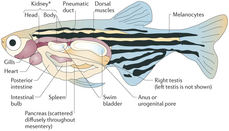
An adult zebrafish is shown with the anatomical structures labelled. Zebrafish share most of their organs with mammalian counterparts, including the brain, heart, liver, spleen, pancreas, gallbladder, intestines, kidney, testis and ovaries. *The kidney is also the site of haematopoiesis in zebrafish.
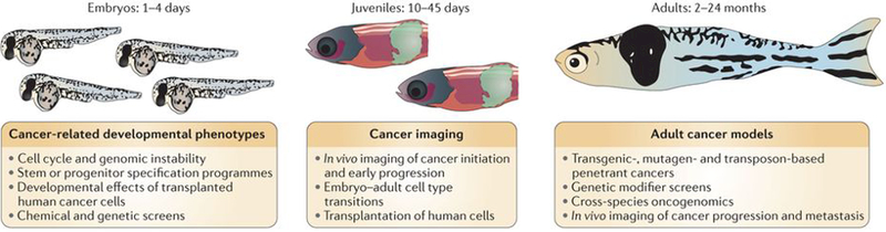
Differently aged animals each offers distinct advantages for cancer-relevant phenotypes. Embryos can be used to identify phenotypes that are highly relevant to cancer biology, such as defects in the cell cycle or genomic instability. Other embryo phenotypes may include stem or progenitor cells that act as cell of origin of the tumour or changes in embryo morphology on transplantation of human cells. Any of these phenotypes can then be used as the basis of chemical or genetic screens to find modifiers, which can be tested for their relevance to human cancer. Juvenile fish have the capacity for modelling early tumorigenesis and remain optically fairly translucent, lending themselves to detailed in vivo imaging. These cancers can be either from transgenic models or can arise via transplantation of tumour cells, and confocal imaging can be used to assess the tumour–stroma interaction at single-cell resolution. Adult fish develop fully penetrant and advanced cancers, both through transgenic techniques and through the transplantation of either zebrafish or human tumour cells. These animals are ideally suited to cross-species oncogenomics, either by directly testing candidate human genomic changes in the fish (by rapid transgenesis) or by comparing the profiles (DNA or RNA) of the mature tumour in the fish to that of the human to look for evolutionarily conserved events. Both the wild-type fish and the transparent casper model add improved capacities compared to mouse models for in vivo imaging and analysis of tumour stem cells and tumour progression and metastasis.
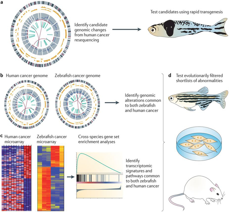
Candidate genomic changes in human cancers (part a) are being identified through consortiums such as The Cancer Genome Atlas and the International Cancer Genome Consortium (ICGC), which have revealed thousands of potential mutations, copy number changes and structural variants, most of which have not been functionally analysed in vivo. By testing a proportion of the high-confidence, recurrent events in transgenic zebrafish models, a direct readout of their effect on actual tumour biology can be rapidly achieved. This type of analysis can be carried out in the transient transgenic setting, allowing for thousands of animals or variants to be tested in a single experiment. Because there are now numerous models of cancer in the zebrafish (parts b,c), the genomic profile of these fish tumours can be directly compared with that of the human tumours to look for events that are common between the two species. This can be done either at the level of DNA (part b) by looking for common copy-number changes, mutations and structural variants, or at the RNA level (part c) by looking for transcriptomic commonalities. This will essentially act as a ‘filter’ to provide much shorter lists of highly penetrant changes that are conserved across millions of years of evolution. These abnormalities can then be efficiently tested in cell culture, zebrafish and mouse models (part d). Part c is reproduced, with permission, from REF. © (2011) Macmillan Publishers Ltd. All rights reserved.
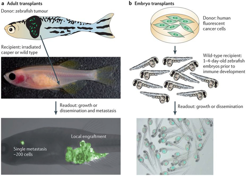
a | Transplantation of primary zebrafish tumour cells (top of the figure) into an irradiated, immunocompromised adult casper or wild-type recipient (middle) allows for detailed assessment of tumour growth and metastasis at single-cell resolution (bottom). b | Transplantation of well-characterized, fluorescently labelled human cell lines or primary human tumours (top) into the zebrafish embryo (middle) reliably leads to engraftment in recipients owing to a lack of immune system development at this time point. Hundreds to thousands of recipients (bottom) can be transplanted in this manner, which can be easily imaged and used for chemical or genetic screening approaches. Images in part a are reproduced, with permission, from REF. © (2008) Cell Press.
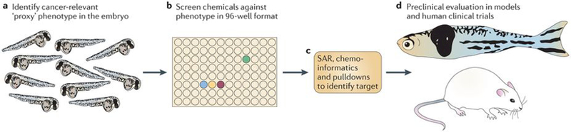
These types of screens are most efficiently carried out on embryos, given their amenability to large-scale, high-throughput manipulation and analysis. a | Identification of an embryonic phenotype that is highly relevant to cancer is a key step in this process. An embryo stained for the neural crest marker crestin is shown; neural crest cells give rise to pigmented melanocytes but also to melanoma in the zebrafish. b | After a relevant embryo phenotype is found, the embryos can be distributed in their chorions to plates, most typically the 96-well format. Each well will receive a distinct small molecule, either manually or with the aid of a liquid-handling robot. This method has been applied to screens ranging from 1,000 to 26,000 molecules. c | Identifying the mechanism of hits is shown. This will primarily depend on the nature of the library used. Molecules with unknown function may require methods such as structure–activity relationships (SARs), chemoinformatics (using algorithms and databases such as PubChem, ChemBank or DiscoveryGate) or pulldowns using tagged versions of drugs and mass spectrometry. For libraries biased towards chemicals with US Food and Drug Administration-approved or known mechanisms, this step can often be rapid, whereas for molecules of unknown function it can take up to 1 year or more. d | While mechanistic evaluation is ongoing, chemicals can be tested for their effects on cancer in multiple downstream assays, including zebrafish cancer models or mouse transgenic and xenograft models. Depending on potency and safety, some of these hits will be amenable to testing in clinical trials in humans.
References
-
- Driever W et al. A genetic screen for mutations affecting embryogenesis in zebrafish. Development 123, 37–46 (1996). - PubMed
-
- Haffter P et al. The identification of genes with unique and essential functions in the development of the zebrafish, Danio rerio. Development 123, 1–36 (1996). References 4 and 5 are landmark papers describing the use of the zebrafish in a phenotypic forward genetic screen. - PubMed
Publication types
MeSH terms
Grants and funding
LinkOut - more resources
Full Text Sources
Other Literature Sources
