. 2013 Aug 22;4(8):e778.
doi: 10.1038/cddis.2013.294.

Withaferin A: a proteasomal inhibitor promotes healing after injury and exerts anabolic effect on osteoporotic bone

Affiliations

Withaferin A: a proteasomal inhibitor promotes healing after injury and exerts anabolic effect on osteoporotic bone

V Khedgikar et al. Cell Death Dis. .

Abstract

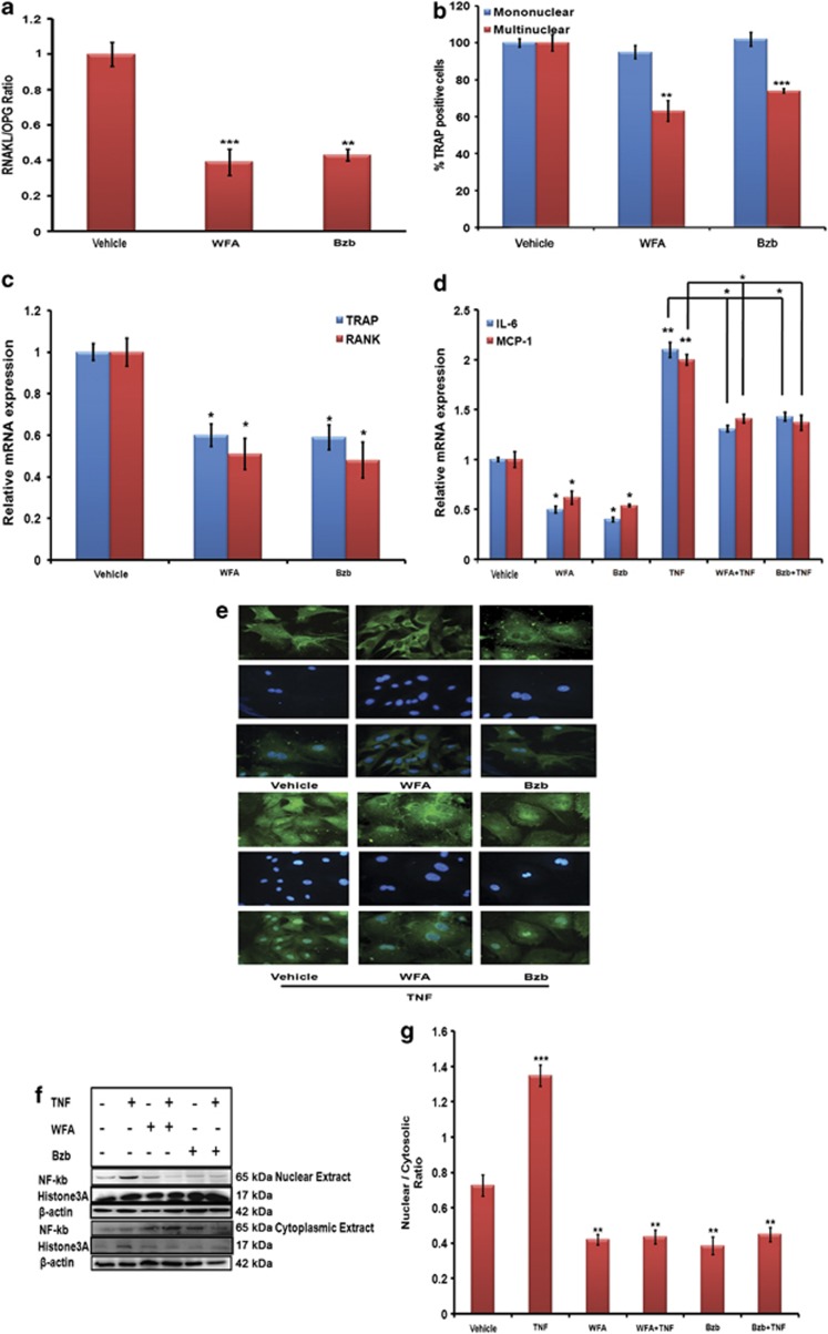
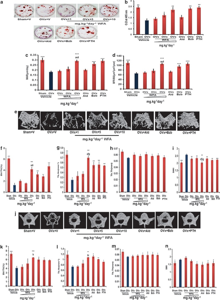
Withania somnifera or Ashwagandha is a medicinal herb of Ayurveda. Though the extract and purified molecules, withanolides, from this plant have been shown to have different pharmacological activities, their effect on bone formation has not been studied. Here, we show that one of the withanolide, withaferin A (WFA) acts as a proteasomal inhibitor (PI) and binds to specific catalytic β subunit of the 20S proteasome. It exerts positive effect on osteoblast by increasing osteoblast proliferation and differentiation. WFA increased expression of osteoblast-specific transcription factor and mineralizing genes, promoted osteoblast survival and suppressed inflammatory cytokines. In osteoclast, WFA treatment decreased osteoclast number directly by decreasing expression of tartarate-resistant acid phosphatase and receptor activator of nuclear factor kappa-B (RANK) and indirectly by decreasing osteoprotegrin/RANK ligand ratio. Our data show that in vitro treatment of WFA to calvarial osteoblast cells decreased expression of E3 ubiquitin ligase, Smad ubiquitin regulatory factor 2 (Smurf2), preventing degradation of Runt-related transcription factor 2 (RunX2) and relevant Smad proteins, which are phosphorylated by bone morphogenetic protein 2. Increased Smurf2 expression due to exogenous treatment of tumor necrosis factor α (TNFα) to primary osteoblast cells was decreased by WFA treatment. This was corroborated by using small interfering RNA against Smurf2. Further, WFA also blocked nuclear factor kappa-B (NF-kB) signaling as assessed by tumor necrosis factor stimulated nuclear translocation of p65-subunit of NF-kB. Overall data show that in vitro proteasome inhibition by WFA simultaneously promoted osteoblastogenesis by stabilizing RunX2 and suppressed osteoclast differentiation, by inhibiting osteoclastogenesis. Oral administration of WFA to osteopenic ovariectomized mice increased osteoprogenitor cells in the bone marrow and increased expression of osteogenic genes. WFA supplementation improved trabecular micro-architecture of the long bones, increased biomechanical strength parameters of the vertebra and femur, decreased bone turnover markers (osteocalcin and TNFα) and expression of skeletal osteoclastogenic genes. It also increased new bone formation and expression of osteogenic genes in the femur bone as compared with vehicle groups (Sham) and ovariectomy (OVx), Bortezomib (known PI), injectible parathyroid hormone and alendronate (FDA approved drugs). WFA promoted the process of cortical bone regeneration at drill-holes site in the femur mid-diaphysis region and cortical gap was bridged with woven bone within 11 days of both estrogen sufficient and deficient (ovariectomized, Ovx) mice. Together our data suggest that WFA stimulates bone formation by abrogating proteasomal machinery and provides knowledge base for its clinical evaluation as a bone anabolic agent.


Figures

Figure 1
Figure 1
WFA has osteogenic effect in vitro. (a) Structure of WFA. (b) Effect of WFA on primary osteoblast proliferation. Primary osteoblasts were cultured in increasing concentrations of WFA and Bzb for 24 h and harvested for cell proliferation direct cell count for cell proliferation assay (I) and BrdU incorporation cell proliferation assay (II). The results were expressed as relative cell growth in percentage, which was compared with control group. We set the control group as 100. Values represents mean±S.E. of three independent experiments (n=3). *P<0.05, **P<0.01 when compared with control. (c) WFA treatment of MCOs for 48 h in osteoblast differentiation medium significantly increased ALP production compared with control. Values represent mean±S.E. of three independent experiments (n=3). **P<0.01, ***P<0.001 compared with control vehicle group. (d) Photomicrographs show that treatment of MCOs with WFA in osteoblast differentiation medium significantly increased mineralized nodules compared with control, as assessed by alizarin Red-S staining. (e) Quantitation of mineralization (Alizarin Red-S stain) data are shown as OD at 405 nm. Values represent mean±S.E. of three independent experiments (n=3). ***P<0.001 compared with control vehicle group. (f) WFA (10 nM) treatment of MCOs increased expression of osteogenic genes. Figure shows increased expression of RunX2, OCN and ColI with WFA treatment. Expression was normalized to GAPDH internal control. Values represent mean±S.E. of three independent experiments (n=3). *P<0.05, ***P<0.001 compared with vehicle control group. (g) WFA exerts anti-apoptotic effects in osteoblasts. Using Becton Dickinson FACS and FL-H channel (annexin-V) and FL2-H channel (PI) data show that WFA treatment inhibited apoptosis of osteoblast cells. Shown are representative dot plots. (h) Quantification of flow cytometry data are shown as percent of total cells. Values represents mean ±S.E. of three independent experiments (n=3). *P<0.05 compared with control for early apoptosis and ##P<0.01 compared with control for late apoptosis
Figure 2
Figure 2
Effect of WFA on osteoblast signaling. (a) Proteasomal effect of WFA in calvarial osteoblast cells. 20S proteasome activities were measured 1, 3, 6 h after treatment with WFA. Values represent mean±S.E. of three (n=3) independent experiments. ***P<0.001, *P<0.001 compared with control. (b) Lower panel shows that 6 h of treatment with WFA induces ubiquitination of proteins through western blot. Protein ladders were detected by anti-ubiquitin antibody. (c) qPCR data of the osteogenic genes after WFA treatment. WFA targets BMP2 signaling by inducing mRNA levels of BMP2 responsive genes in osteoblast cells after 24 h of treatment. Data represent mean±S.E. of three independent experiments (n=3). *P<0.05, **P<0.01 ***P<0.001 when compared with control. (d) Western blot analysis of various osteogenic genes BMP2, Smad1 and its phosphorylation state in the presence of WFA. Expression of Smurf1, Smurf2 a negative regulator of osteogenesis is also shown. All blots were normalized with β-actin. (e) Densitometric analyses of western blot (d) showing fold change. Fold increase was calculated relative to control vehicle-treated cells. The values are the mean±S.E. of three experiments. ***P<0.001 versus vehicle-treated group. (f) Interaction of Smurf2 with Smad1 and RunX2 in the presence of WFA. Immuno-precipitation (IP) assays were performed using anti-Smurf antibody followed by western blot using anti-Smad1 and anti-RunX2 antibodies. (g) WFA prevents RunX2 ubiquitination and proteosomal degradation. Primary osteoblast cells were treated with 5 ng/ml TNFα for 24 h in the presence or absence of WFA, and endogenous RunX2 was immunoprecipated by anti- RunX2 antibody. Ubiquitinated RunX2 (Ub-RunX2) protein ladders were detected by anti-ubiquitin antibody (upper panel). After stripping the antibody, total un-ubiquitinated RunX2 protein levels were determined by anti- RunX2 antibody (lower panel). MW, molecular mass. (h) Western blot of RunX2 and Smurf2 in the presence of various treatments. All blots were normalized with housekeeping gene β-actin. (i) Densitometric analyses of western blot (h) showing fold change. Fold increase was calculated relative to control vehicle-treated cells. The values are the mean±S.E. of three experiments. *P<0.05, **P<0.01 ***P<0.001 versus vehicle -treated group. (j) Relative expression of Smurf2 SiRNA. Osteoblast cells were treated with Smurf2 siRNAs and mRNA levels were measured by real-time RT-PCR. The values are the mean±S.E. of three experiments. **P<0.01versus the vehicle-treated group. (k) Smurf2 knockdown reduced degradation of RunX2. Expression of RunX2 and Smurf2 was determined by western blot analysis after the osteoblast cells were treated with Smurf2 siRNA in the presence and absence of WFA and Bzb. (l) Densitometric analyses of western blot (k) showing fold change. Fold increase was calculated relative to control vehicle-treated cells. The values are the mean±S.E. of three experiments. *P<0.05, **P<0.01 ***P<0.001 versus vehicle (vehicle-treated) group. (m) Smurf2 siRNA blocks TNF-induced RunX2 degradation. Osteoblast cells were treated with Smurf2 siRNA and then treated with (5 ng/ml) TNF for 24 h. Smurf2 and RunX2 expression was determined by western blot analysis. (n) TNF-induced ubiquitination of RunX2 is reversed by Smurf2 knockdown. Endogenous Smurf2 was knocked down in primary osteoblast cells then treated with 5 ng/ml TNFα for 24 h and endogenous RunX2 was immunoprecipated by anti-RunX2 antibody. Ubiquitinated RunX2 (Ub-RunX2) protein ladders were detected by anti-ubiquitin antibody (upper panel). After stripping the antibody, total un-ubiquitinated RunX2 protein levels were determined by anti-RunX2 antibody (lower panel). MW, molecular mass. (o) Smurf2 knockdown prevents RunX2 ubiquitination and proteosomal degradation in presence of WFA. Primary osteoblast cells were treated with WFA for 24 h, and endogenous RunX2 immunoprecipated by anti- RunX2 antibody. Ubiquitinated RunX2 (Ub-RunX2) protein ladders were detected by anti-ubiquitin antibody. After stripping the antibody, total un-ubiquitinated RunX2 protein levels were determined by anti-RunX2 antibody (lower panel). (p) Smurf2 knockdown increased ALP activity in the presence of WFA and TNFα. WFA treatment of MCOs for 48 h in osteoblast differentiation medium significantly increased ALP production compared with control that was abrogated in the presence of TNFα. Treatment with Smurf2 siRNA in the presence of TNFα reversed the effect. Values represent mean±S.E. of three independent experiments (n=3). *P<0.05, ***P<0.001 compared with control vehicle group. (q) Smurf2 knockdown increased expression of osteogenic genes. mRNA expression of BMP2 and RunX2 was assessed by real-time PCR. The values are the mean±S.E. of three experiments. *P<0.05, ***P<0.001 versus control (vehicle) group
Figure 3
Figure 3
Effect of WFA on osteoclastogenesis indirectly through osteoblasts. (a) WFA increases mRNA levels of OPG but decreases mRNA levels of RANKL in primary osteoblasts. Ratio of RANKL: OPG were determined and quantified with qPCR and normalized with GAPDH. Values represents mean±S.E. ***P<0.001 and **P<0.01 of three independent experiments(n=3) when compared with vehicle-treated cells. (b) WFA treatment to OVx mice reduces osteoclastogenesis and increases osteoprogenitor cells in bone marrow. BMCs from mice of various experimental groups were seeded into 48-well plates and osteoclast differentiation was induced as described. Cells were stained for TRAP activity for osteoclast formation. Figure shows quantitative representation of TRAP +ive mononuclear and multinuclear cells. (c) WFA increases mRNA levels of OPG but decreases mRNA levels of TRAP and RANK in osteoclast culture. BMCs were isolated from 4- to 6-week-old mice. After overnight culture cells were cultured for 6 days in the presence of MCSF and RANKL. mRNA levels of TRAP and RANK related to osteoclastogenesis was determined by qPCR from the total RNA made from BMC's. Data represent mean±S.E.M.; n=3. **P<0.01 and ***P<0.001 compared with vehicle-treated cells. (d) WFA treatment decreases expression of inflammatory cytokines in osteoblasts. qPCR data show mRNA levels IL-6, MCP-1. Values represents mean±S.E. **P<0.01 and ***P<0.001 of three independent experiments (n=3) when compared with control. (e) WFA inhibits TNFα-induced NF-kB nuclear translocation in osteoblast cells. Representative photomicrograph of sub cellular localization of p65 was determined by immuno fluorescence (magnification × 40) under control and treatment conditions from three independent experiments (n=3). (f) WFA treatment abolished the nuclear translocation of the NF-kB in osteoblasts. Osteoblasts were treated with TNFα and WFA. Cytoplasmic or nuclear extracts were prepared and NF-kB protein levels were detected by immunoblotting. TNFα enhanced the nuclear translocation of NF-kB, WFA abolished it. Histone H3 was used as a loading control for nuclear extract, β-actin was used as loading controls for cytoplasmic fraction. (g) This figure shows the densitometric analysis of NF-kB from three independent blots. Values represents mean±S.E. of three independent experiments (n=3), ***P<0.001, **P<0.01 when compared with control
Figure 4
Figure 4
Plasma concentration time profile of WFA after oral administration at the dose of 10 mg/kg. The data are represented as mean±S.D. of three individual experiments
Figure 5
Figure 5
WFA promotes bone regeneration in the drill-hole site in sham-operated (ovary intact) and OVx mice. (a) Representative confocal images (magnification= × 100) of calcein labeling shown in the drill-hole site of various groups 0, 11 and 21 days after injury without and with WFA treatment(5 and 10 mg/kg/day). (b) Data show the quantification of the mean intensity of calcein labeling. Values represent mean±S.E. **P<0.01, ***P<0.001 compared with Sham vehicle. #P<0.05, ##P<0.01, ###P<0.001 compared with OVx vehicle group. Inter-dose comparison shows that cP<0.05 when 10 mg/kg/day dose was compared with 5 mg/kg/day dose. *P<0.05 when OVx+V compared with Sham+V. (c) Representative μCT images from the center of the bony hole (defect region) on days 0, 11 and 21 in the sham and OVx groups. (d and e) Graph represents μCT analysis in the defect and intra-medulla regions of sham and sham+ WFA-treatment group. Data show BV/TV (%) at 5 and 10 mg/kg/day dose in both the regions.Values represent mean±S.E. **P<0.01, ***P<0.001 compared with Sham vehicle. ##P<0.01compared with OVx vehicle. Inter-dose comparison shows aP<0.05 when 10 mg/kg/day dose is compared with 5 mg/kg/day dose in sham group on day 11. (f and g) Graph represents μCT analysis in the defect and intra-medulla regions of OVx and OVx+ WFA treatment groups. Values represent Mean±S.E. *P<0.05, ***P<.001 compared with Sham vehicle. #P<0.05 and ##P<0.01 compared with OVx vehicle. Inter-dose comparison shows aP<0.05 when 10 mg/kg/day dose is compared with 5 mg/kg/day dose in sham group on day 11. (hj) Effect of WFA on osteogenic genes after drill-hole injury. mRNA profile of RunX2, BMP2 and OCN extracted from callus of drill site of bone. Values represent mean±S.E. of three independent experiments n=3. **P<0.01, ***P<0.001 compared with Sham vehicle. #P<0.05, ##P<0.01, ###P<0.001 compared with OVx vehicle. Inter-dose comparison shows aP<0.05 when 10 mg/kg/day dose is compared with 5 mg/kg/day dose in sham group on day 11. bP<0.05 when 10 mg/kg/day dose is compared with 5 mg/kg/day in sham group on day 21. cP<0.05 when 10 mg/kg/daydose iseffective than 5 mg/kg/day in OVx group on day 11. dP<0.05 when 10 mg/kg/day dose is effective than 5 mg/kg/day in OVx group at day 21for RunX2 gene
Figure 6
Figure 6
WFA has anabolic effect in osteopenic bones. (a and b). Oral supplementation of WFA to OVx mice increases mineralized nodule formation in BMCs as assessed by alizarin red-S staining. Values represent Mean±S.E. of three independent experiments n=3. *P<0.05, **P<0.01 as compared with OVx vehicle group. (c and d). WFA supplementation increases mineral apposition rate and bone formation rate in OVx mice. Dynamic histomorphometric parameters (MAR and BFR) at the femur mid-diaphysis was calculated from the double fluorochrome labeling experiments in various groups.Values represent Mean±S.E. of three independent experiments n=3. ***P<0.001 compared with OVx vehicle group. aP<0.001 when 10 mg/kg/day dose compared with 1 mg/kg/day. dP<0.001 when 10 mg/kg/day dose compared with 5 mg/kg/day. (e) WFA supplementation restores the trabecular micro-architecture of the femur epiphysis. Representative μCT images of the femur epiphysis of various experimental groups. (fi) μCT analysis of various trabecular parameters of the femur epiphysis, including BV/TV, Tb.No, Tb.Th and SMI are presented. All values are expressed as mean±S.E.M. *P<0.05, **P<0.01, ***P<0.001 aP<0.001 when 10 mg/kg/day dose compared with 1 mg/kg/day. dP<0.001 when 10 mg/kg/day dose compared with 5 mg/kg/day dose, fP<0.05 when 10 mg/kg/day dose compared with 5 mg/kg/day dose, gP<0.05 when 10 mg/kg/day dose compared with Ald 3 mg/kg/day dose, iP<0.05 when 10 mg/kg/day dose compared with Bzb 0.3 mg/kg/day dose WFA supplementation has a significant restorative effect on the trabecular micro-architecture of L5 vertebrae. Figure shows representative μCT images of various treated group. (kn) Trabecular parameters of L5 vertebrae of various treatment groups. All values are expressed as mean±S.E.M. *P<0.05, **P<0.01, ***P<0.001 compared with OVx group, bP<0.01 when 10 mg/kg/day dose compared with 1 mg/kg/day dose, iP<0.05 when 10 mg/kg/day dose compared with Bzb 0.3 mg/kg/day dose
Figure 7
Figure 7
WFA inhibits bone turnover and has direct effect on osteoblast-specific genes in femurs of OVx mice. (a) Serum OCN levels as measured at the end of experiment from various treatment groups. Data show that WFA inhibits bone turnover in OVx mice. All values are expressed as mean±S.E.M. *P<0.05, **P<0.01, ***P<0.001 compared with OVx group. aP<0.001 when 10 mg/kg/day dose was compared with 1 mg/kg/day dose, eP<0.01 when 10 mg/kg/day dose compared with 5 mg/kg/day dose. (b) Circulating TNFα levels from serum of various groups were measured by ELISA at the end of the experiment period. All values are expressed as mean±S.E. of three independent experiments (n=3). **P<0.01 compared with OVx group. (c) Direct effect of WFA at the end of experiment on long bones (femur). Data show qPCR of osteogenic genes RunX2, ColI and OCN in RNA isolated from bone. All values are expressed as mean±S.E. of three independent experiments (n=3). *P<0.05, **P<0.01, ***P<0.001 compared with OVx group. aP<0.001 when 10 mg/kg/day dose was compared with 1 mg/kg/day dose, bP<0.01 when 10 mg/kg/day dose compared with 1 mg/kg/day dose, dP<0.001 when 10 mg/kg/day dose compared with 5 mg/kg/day dose, eP<0.01 when 10 mg/kg/day dose compared with 5 mg/kg/day dose, gP<0.05 when 10 mg/kg/day dose compared with Ald 3 mg/kg/day dose, hP<0.01 when 10 mg/kg/day dose compared with PTH mg/kg/day dose, iP<0.05 when 10 mg/kg/day dose compared with Bzb 0.3 mg/kg/day dose for RunX2 gene expression. (d) Effect of WFA on mRNA expression of Smurf2 in femur at the end of experimental period. All values are expressed as mean±S.E.M. of three independent experiments (n=3). *P<0.05, **P<0.01, ***P<0.001 when compared with OVx group, bP<0.01 when 10 mg/kg/day dose was compared with 1 mg/kg/day dose, iP<0.05 when 10 mg/kg/day dose compared with Bzb 0.3 mg/kg/day dose
Figure 8
Figure 8
Schematic diagram outlining the potential molecular targets and in vivo effect of WFA leading to bone anabolic effect in osteogenic cells and osteoclast precursors induced by proteasome inhibition. BMP signaling induced by WFA prevents degradation of Smad receptors. The transcription factor Runx2, which is induced by BMP2, is further stabilized by preventing completion of proteasomal degradation by E3 ubiquitin ligase, Smurf2. WFA decreases tumor necrosis factor α (TNFα), induced NF-Kb and Smurf2 expression resulting in increased Smad and Runx2 levels. In vivo proteasome inhibition simultaneously increases osteoblastogenesis by stabilizing RunX2 and reduces osteoclast numbers directly by inhibiting RANKL: OPG ratio. Thus, osteoclast differentiation is suppressed by inability of RANKL to bind with RANK. This results in reduced number of TRAP-positive cells. Simultaneous induction of osteoblastogenesis and suppression of osteoclastogenesis results in increased bone mass. Our in vivo data demonstrate that WFA exerted an osteogenic effect in osteopenic OVx mice and accelerated the bone-healing process after bone marrow injury of long bones (See text for details

Similar articles

Cited by

References

    1. Katagiri T, Takahashi N. Regulatory mechanisms of osteoblast and osteoclast differentiation. Oral Dis. 2002;8:147–159. - PubMed
    1. Kawamura N, Kugimiya F, Oshima Y, Ohba S, Ikeda T, Saito T, et al. Akt1 in osteoblasts and osteoclasts controls bone remodeling. PLoS ONE. 2007;2:e1058. - PMC - PubMed
    1. Hie M, Tsukamoto I. Increased expression of the receptor for activation of NF-kappaB and decreased runt-related transcription factor 2 expression in bone of rats with streptozotocin-induced diabetes. Int J Mol Med. 2010;26:611–618. - PubMed
    1. Canalis E, Economides AN, Gazzerro E. Bone morphogenetic proteins, their antagonists, and the skeleton. Endocr Rev. 2003;24:218–235. - PubMed
    1. Garrett IR, Chen D, Gutierrez G, Zhao M, Escobedo A, Rossini G, et al. Selective inhibitors of the osteoblast proteasome stimulate bone formation in vivo and in vitro. J Clin Invest. 2003;111:1771–1782. - PMC - PubMed

Publication types

MeSH terms