

ABIN1 dysfunction as a genetic basis for lupus nephritis
- PMID: 23970121
- PMCID: PMC3810087
- DOI: 10.1681/ASN.2013020148
ABIN1 dysfunction as a genetic basis for lupus nephritis
Abstract
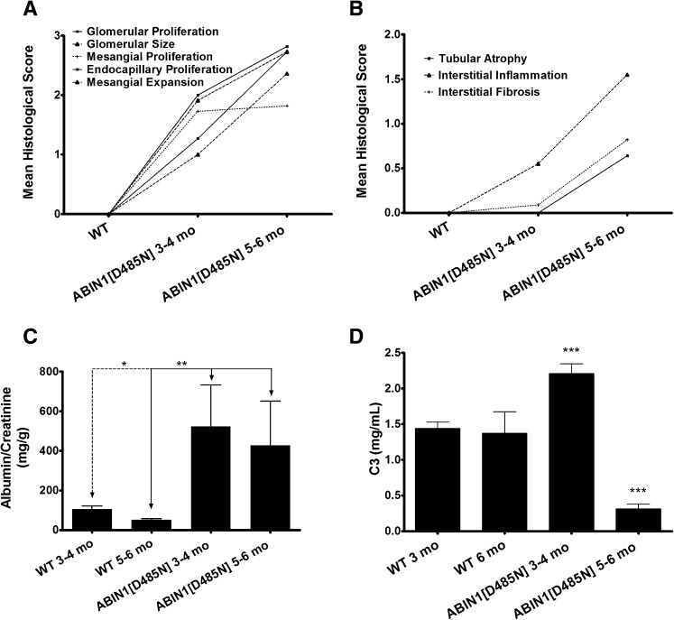
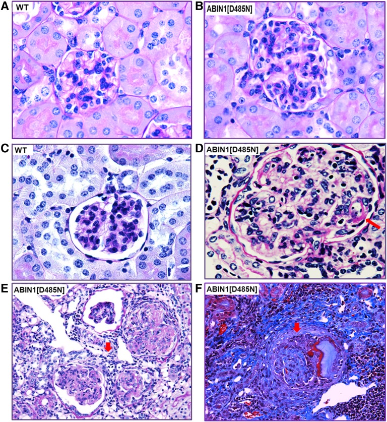
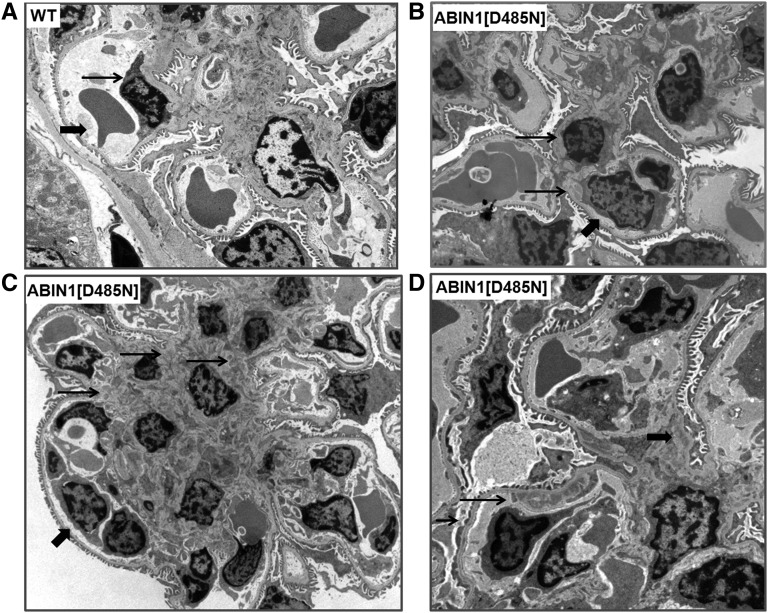
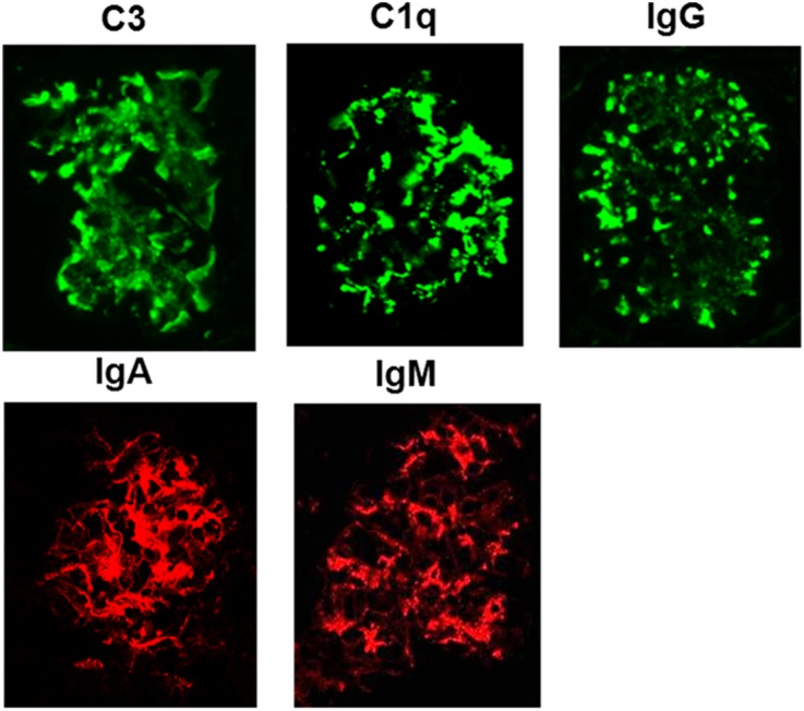
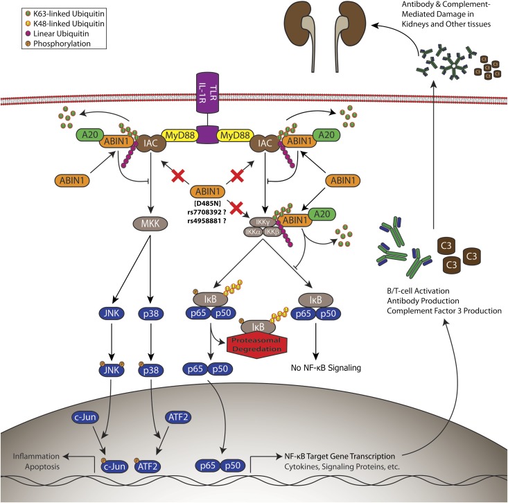
The genetic factors underlying the pathogenesis of lupus nephritis associated with systemic lupus erythematosus are largely unknown, although animal studies indicate that nuclear factor (NF)-κB is involved. We reported previously that a knockin mouse expressing an inactive form of ABIN1 (ABIN1[D485N]) develops lupus-like autoimmune disease and demonstrates enhanced activation of NF-κB and mitogen-activated protein kinases in immune cells after toll-like receptor stimulation. In the current study, we show that ABIN1[D485N] mice develop progressive GN similar to class III and IV lupus nephritis in humans. To investigate the clinical relevance of ABIN1 dysfunction, we genotyped five single-nucleotide polymorphisms in the gene encoding ABIN1, TNIP1, in samples from European-American, African American, Asian, Gullah, and Hispanic participants in the Large Lupus Association Study 2. Comparing cases of systemic lupus erythematosus with nephritis and cases of systemic lupus erythematosus without nephritis revealed strong associations with lupus nephritis at rs7708392 in European Americans and rs4958881 in African Americans. Comparing cases of systemic lupus erythematosus with nephritis and healthy controls revealed a stronger association at rs7708392 in European Americans but not at rs4958881 in African Americans. Our data suggest that variants in the TNIP1 gene are associated with the risk for lupus nephritis and could be mechanistically involved in disease development via aberrant regulation of NF-κB and mitogen-activated protein kinase activity.
Figures
Comment in
-
Lupus nephritis: Ancestry, genetic risk and health disparities.Nat Rev Nephrol. 2013 Dec;9(12):699-700. doi: 10.1038/nrneph.2013.210. Epub 2013 Oct 15. Nat Rev Nephrol. 2013. PMID: 24126589
References
-
- Tsokos GC: Systemic lupus erythematosus. N Engl J Med 365: 2110–2121, 2011 - PubMed
-
- Weening JJ, D’Agati VD, Schwartz MM, Seshan SV, Alpers CE, Appel GB, Balow JE, Bruijn JA, Cook T, Ferrario F, Fogo AB, Ginzler EM, Hebert L, Hill G, Hill P, Jennette JC, Kong NC, Lesavre P, Lockshin M, Looi LM, Makino H, Moura LA, Nagata M: The classification of glomerulonephritis in systemic lupus erythematosus revisited. J Am Soc Nephrol 15: 241–250, 2004 - PubMed
-
- Borchers AT, Leibushor N, Naguwa SM, Cheema GS, Shoenfeld Y, Gershwin ME: Lupus nephritis: A critical review. Autoimmun Rev 12: 174–194, 2012 - PubMed
-
- Buhaescu I, Covic A, Deray G: Treatment of proliferative lupus nephritis—a critical approach. Semin Arthritis Rheum 36: 224–237, 2007 - PubMed
Publication types
MeSH terms
Substances
Grants and funding
- AR049084/AR/NIAMS NIH HHS/United States
- AR043814/AR/NIAMS NIH HHS/United States
- UL1 TR000150/TR/NCATS NIH HHS/United States
- UL1 TR000165/TR/NCATS NIH HHS/United States
- GM103456/GM/NIGMS NIH HHS/United States
- MC_EX_UU_G0800765/MRC_/Medical Research Council/United Kingdom
- P30 CA046934/CA/NCI NIH HHS/United States
- R01 AR033062/AR/NIAMS NIH HHS/United States
- UL1 RR029882/RR/NCRR NIH HHS/United States
- AR063124/AR/NIAMS NIH HHS/United States
- AI024717/AI/NIAID NIH HHS/United States
- P60 AR030692/AR/NIAMS NIH HHS/United States
- P20GM103456/GM/NIGMS NIH HHS/United States
- P60 AR053308/AR/NIAMS NIH HHS/United States
- R01 AI063274/AI/NIAID NIH HHS/United States
- R56 AI063274/AI/NIAID NIH HHS/United States
- P20 GM103456/GM/NIGMS NIH HHS/United States
- AI083194/AI/NIAID NIH HHS/United States
- AI103980/AI/NIAID NIH HHS/United States
- UL1 TR000154/TR/NCATS NIH HHS/United States
- MC_EX_G0800765/MRC_/Medical Research Council/United Kingdom
- 100294/WT_/Wellcome Trust/United Kingdom
- MC_U127084348/MRC_/Medical Research Council/United Kingdom
- UL1 RR025741/RR/NCRR NIH HHS/United States
- AR158959/AR/NIAMS NIH HHS/United States
- R21 AI103980/AI/NIAID NIH HHS/United States
- RR025741/RR/NCRR NIH HHS/United States
- AI063274/AI/NIAID NIH HHS/United States
- R01 AR043814/AR/NIAMS NIH HHS/United States
- R37 AI024717/AI/NIAID NIH HHS/United States
- R01 AI024717/AI/NIAID NIH HHS/United States
- PR094002/PR/OCPHP CDC HHS/United States
- P60AR062755/AR/NIAMS NIH HHS/United States
- TR000165/TR/NCATS NIH HHS/United States
- AR 49084/AR/NIAMS NIH HHS/United States
- I01 BX001834/BX/BLRD VA/United States
- UL1 TR001082/TR/NCATS NIH HHS/United States
- R01 AR063124/AR/NIAMS NIH HHS/United States
- K24 AI078004/AI/NIAID NIH HHS/United States
- AR33062/AR/NIAMS NIH HHS/United States
- MC_UU_12016/11/MRC_/Medical Research Council/United Kingdom
- DK176743/DK/NIDDK NIH HHS/United States
- K24 AR002138/AR/NIAMS NIH HHS/United States
- R21 AI070304/AI/NIAID NIH HHS/United States
- P01 AI083194/AI/NIAID NIH HHS/United States
- P01 AR049084/AR/NIAMS NIH HHS/United States
- AR002138/AR/NIAMS NIH HHS/United States
- UL1RR029882/RR/NCRR NIH HHS/United States
- P60 AR062755/AR/NIAMS NIH HHS/United States
LinkOut - more resources
Full Text Sources
Other Literature Sources
