. 2013 Nov;24(11):1806-19.
doi: 10.1681/ASN.2013030281. Epub 2013 Aug 22.

Tubular von Hippel-Lindau knockout protects against rhabdomyolysis-induced AKI

Affiliations

Tubular von Hippel-Lindau knockout protects against rhabdomyolysis-induced AKI

Michael Fähling et al. J Am Soc Nephrol. 2013 Nov.

Abstract

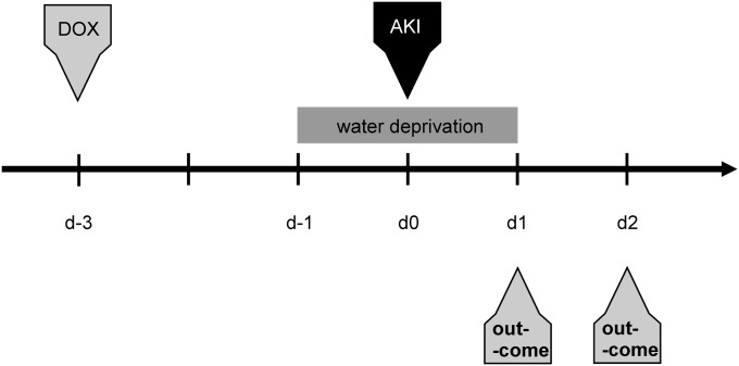
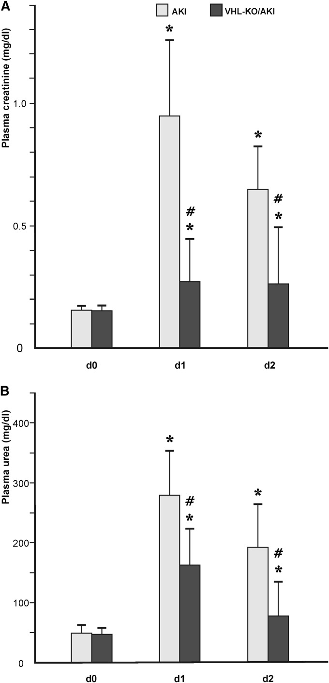
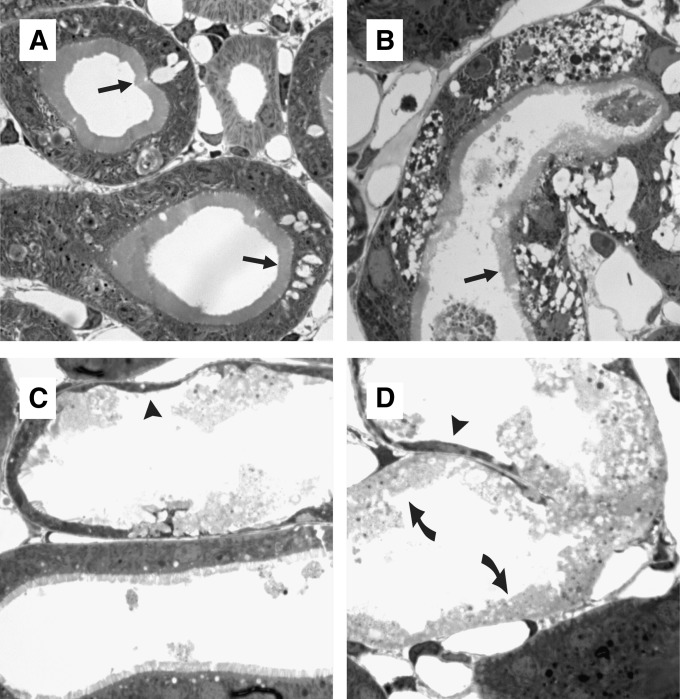
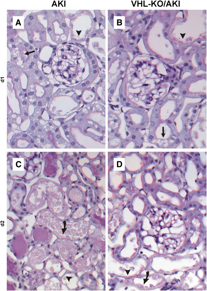
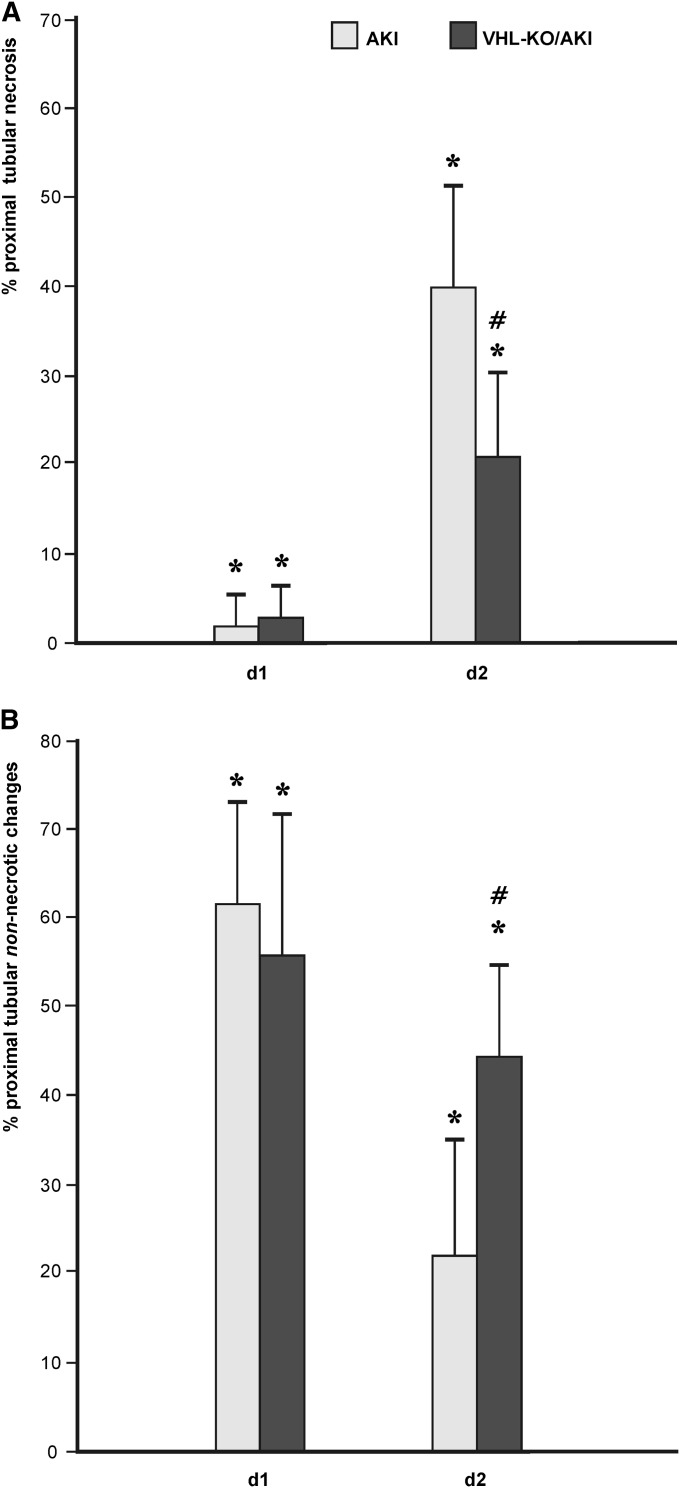
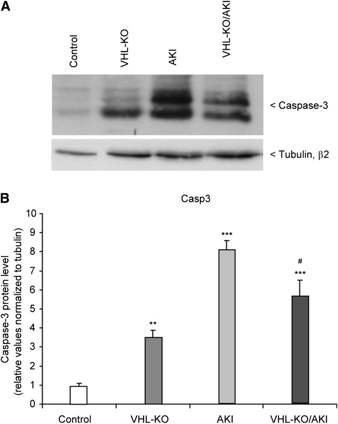
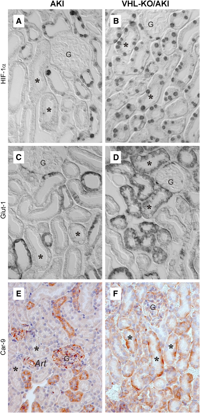
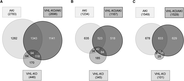
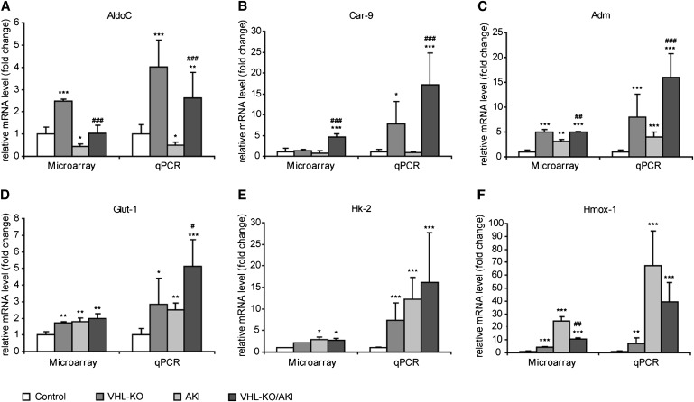
Renal hypoxia occurs in AKI of various etiologies, but adaptation to hypoxia, mediated by hypoxia-inducible factor (HIF), is incomplete in these conditions. Preconditional HIF activation protects against renal ischemia-reperfusion injury, yet the mechanisms involved are largely unknown, and HIF-mediated renoprotection has not been examined in other causes of AKI. Here, we show that selective activation of HIF in renal tubules, through Pax8-rtTA-based inducible knockout of von Hippel-Lindau protein (VHL-KO), protects from rhabdomyolysis-induced AKI. In this model, HIF activation correlated inversely with tubular injury. Specifically, VHL deletion attenuated the increased levels of serum creatinine/urea, caspase-3 protein, and tubular necrosis induced by rhabdomyolysis in wild-type mice. Moreover, HIF activation in nephron segments at risk for injury occurred only in VHL-KO animals. At day 1 after rhabdomyolysis, when tubular injury may be reversible, the HIF-mediated renoprotection in VHL-KO mice was associated with activated glycolysis, cellular glucose uptake and utilization, autophagy, vasodilation, and proton removal, as demonstrated by quantitative PCR, pathway enrichment analysis, and immunohistochemistry. In conclusion, a HIF-mediated shift toward improved energy supply may protect against acute tubular injury in various forms of AKI.


Figures

Figure 1.
Figure 1.
Protocol for AKI induction through rhabdomyolysis. AKI is induced under inhalation anesthesia by a single intramuscular injection of 50% glycerol into the left hind limb. Selective tubular HIF activation is induced by a single subcutaneous injection of doxycycline (DOX) 3 days before AKI induction. This time lag is necessary for establishment of maximum HIF activation via Pax8-rtTA–based knockout of VHL.
Figure 2.
Figure 2.
Tubular VHL knockout improves renal function during AKI. Both serum creatinine (A) and urea (B) are augmented at day 1 after induction of AKI. At day 2, both parameters decline, but are still elevated with respect to baseline. Animals with transgenic HIF activation in all renal tubules (VHL-KO/AKI, compare Figure 7B) exhibit less pronounced elevations of serum creatinine/urea than their littermates with scant and exclusively distal tubular HIF (AKI, compare Figure 7A). *P<0.05 versus control; #P<0.05 versus AKI.
Figure 3.
Figure 3.
Tubular injury forms in rhabdomyolysis-induced AKI. Semithin plastic sections (0.5 μm). Arrow indicates vacuoles, arrowhead indicates cytoplasmic thinning, and curved arrow indicates necrosis. Tubular injury mainly affects proximal convoluted tubules. (A and B) Clear-cut, but non-necrotic, damage may occur under largely preserved brush border (arrows). (C) Largely normal and severely damaged tubules may occur side by side. (D) Different types of injury may coexist within the same tubular profile. Original magnification, ×1200.
Figure 4.
Figure 4.
Tubular VHL knockout improves renal morphology during AKI. Paraffin sections (2 μm) stained with periodic acid–Schiff. Arrow indicates vacuoles, arrowhead indicates cytoplasmic thinning, and curved arrow indicates necrosis. Necrosis is rare at day 1 and is similar in AKI (A) and VHL-KO/AKI (B). By contrast, tubular necrosis is prominent at day 2, and is more extensive in AKI (C) than in VHL-KO/AKI (D). Original magnification, ×400.
Figure 5.
Figure 5.
Semiquantification of injury in proximal tubules during AKI. At day 1 after AKI, induction tubular injury is mainly non-necrotic, and of similar extent in VHL-KO/AKI animals, which have marked HIF activation in all nephron segments (compare Figure 7B), as well as in AKI mice, which exhibit only scant HIF activation, and exclusively in largely unremarkable distal tubules (compare Figure 7A). By contrast, at day 2 after the onset of AKI, approximately 40% of proximal tubules are necrotic in AKI, and VHL-KO reduces this amount to 20%. *P<0.05 versus control; #P<0.05 versus AKI.
Figure 6.
Figure 6.
Tubular VHL knockout reduces caspase-3 during AKI. (A) Representative original Western blot of caspase-3 protein in mouse kidneys. Untreated controls are compared with VHL-KO, rhabdomyolysis-induced AKI, and VHL-KO/AKI. (B) Statistical analyses. n=5 per experimental group. #P<0.05 versus AKI; **P <0.01 versus control; ***P<0.001 versus control.
Figure 7.
Figure 7.
VHL knockout induces HIF-1α and the HIF target genes Glut-1 and Car-9 in tubules at risk for injury. Immunohistochemistry at day 1 after induction of AKI. At this time point, histologic injury is mainly of the non-necrotic type and is comparable in AKI and VHL-KO/AKI (compare Figure 5). AKI features HIF-1α (A), Glut-1 (C), and Car-9 (E) in distal but not in proximal tubules. By contrast, VHL-KO/AKI reveals all three immunosignals in proximal tubules (asterisks in B, D, and F). Art, arteriole; asterisk, proximal tubule; G, glomerulus. Original magnification, ×400 in A–D; ×250 in E and F.
Figure 8.
Figure 8.
Tubular VHL knockout leads to a largely novel transcriptome during AKI. Venn diagram illustrating the number of significantly regulated candidates (P<0.01) in VHL-KO, AKI, or VHL-KO/AKI relative to controls, respectively (A). Separate illustration of upregulated (B) and downregulated candidates (C).
Figure 9.
Figure 9.
Tubular VHL knockout upregulates HIF target genes with cell-protective potential. Microarray analysis and qPCR are shown side by side. AKI and VHL-KO/AKI are shown at day 1 after induction of injury. *P<0.05 versus control; **P<0.01 versus control; ***P<0.001 versus control; #P<0.05 versus AKI; ##P<0.01 versus AKI; ###P<0.001 versus AKI.

References

    1. Waikar SS, Liu KD, Chertow GM: Diagnosis, epidemiology and outcomes of acute kidney injury. Clin J Am Soc Nephrol 3: 844–861, 2008 - PubMed
    1. Hoste EAJ, Schurgers M: Epidemiology of acute kidney injury: How big is the problem? Crit Care Med 36[Suppl]: S146–S151, 2008 - PubMed
    1. Kellum JA: Acute kidney injury. Crit Care Med 36[Suppl]: S141–S145, 2008 - PubMed
    1. Coca SG, Yusuf B, Shlipak MG, Garg AX, Parikh CR: Long-term risk of mortality and other adverse outcomes after acute kidney injury: A systematic review and meta-analysis. Am J Kidney Dis 53: 961–973, 2009 - PMC - PubMed
    1. Huerta-Alardín AL, Varon J, Marik PE: Bench-to-bedside review: Rhabdomyolysis — an overview for clinicians. Crit Care 9: 158–169, 2005 - PMC - PubMed

Publication types

MeSH terms

Substances