. 2013 Oct;19(10):1405-18.
doi: 10.1261/rna.040428.113. Epub 2013 Aug 22.

Mitochondrial protein BmPAPI modulates the length of mature piRNAs

Mitochondrial protein BmPAPI modulates the length of mature piRNAs

Shozo Honda et al. RNA. 2013 Oct.

Abstract

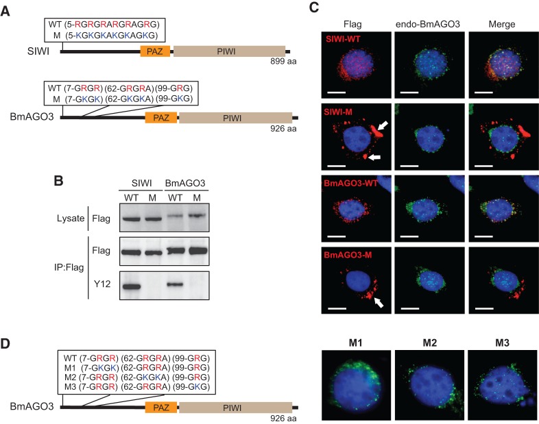
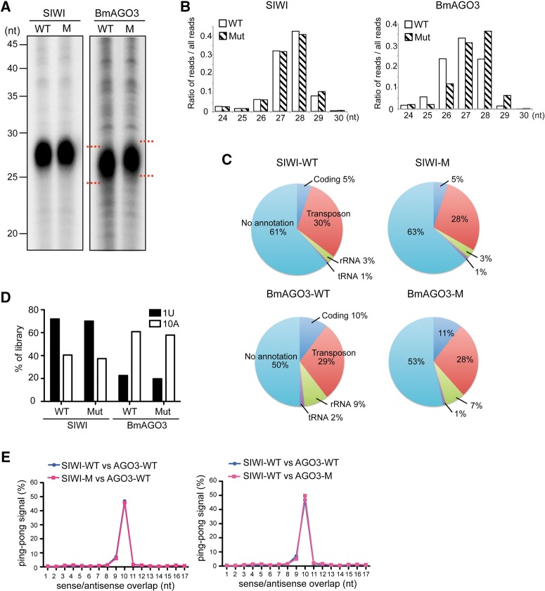
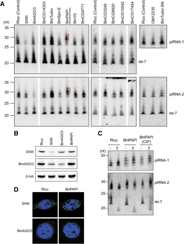
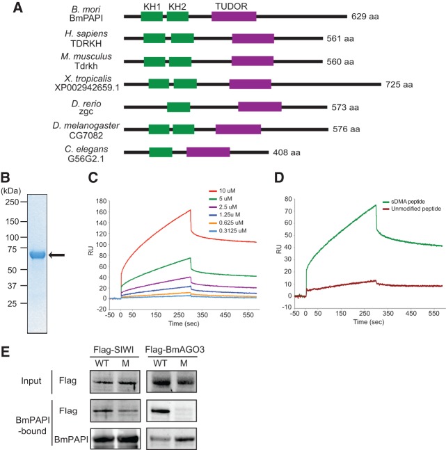
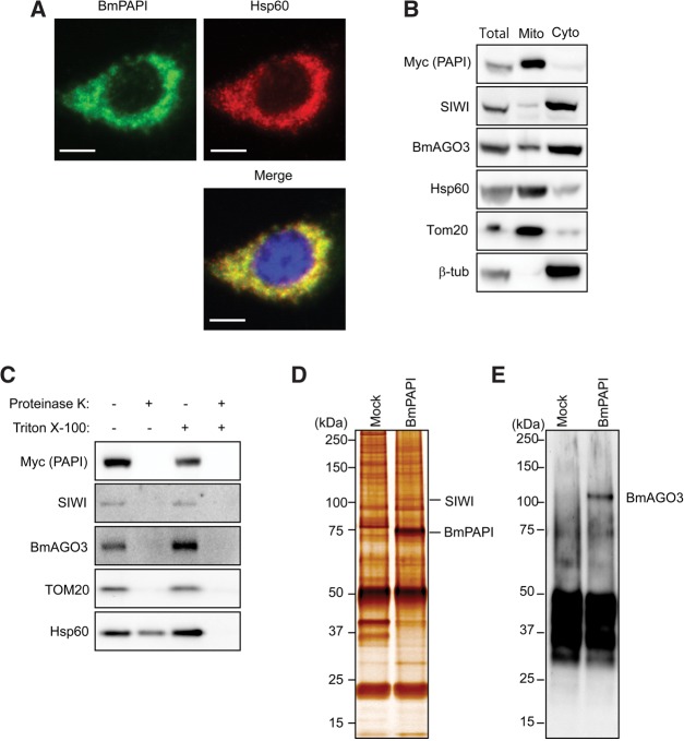
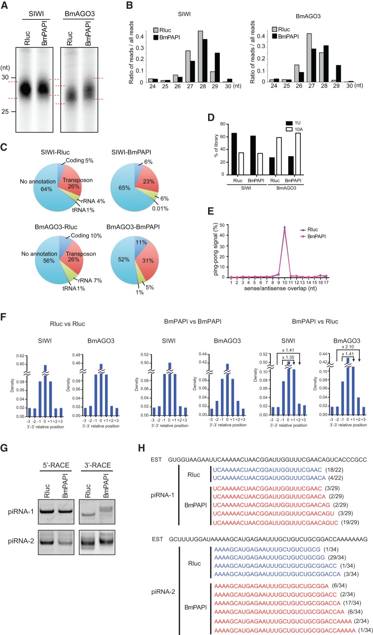
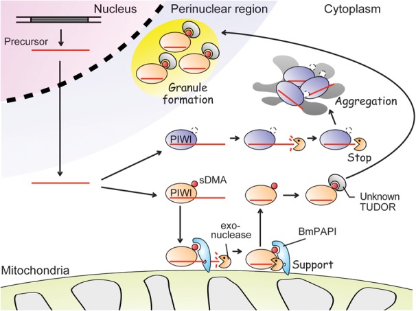
PIWI proteins and their associated PIWI-interacting RNAs (piRNAs) protect genome integrity by silencing transposons in animal germlines. The molecular mechanisms and components responsible for piRNA biogenesis remain elusive. PIWI proteins contain conserved symmetrical dimethylarginines (sDMAs) that are specifically targeted by TUDOR domain-containing proteins. Here we report that the sDMAs of PIWI proteins play crucial roles in PIWI localization and piRNA biogenesis in Bombyx mori-derived BmN4 cells, which harbor fully functional piRNA biogenesis machinery. Moreover, RNAi screenings for Bombyx genes encoding TUDOR domain-containing proteins identified BmPAPI, a Bombyx homolog of Drosophila PAPI, as a factor modulating the length of mature piRNAs. BmPAPI specifically recognized sDMAs and interacted with PIWI proteins at the surface of the mitochondrial outer membrane. BmPAPI depletion resulted in 3'-terminal extensions of mature piRNAs without affecting the piRNA quantity. These results reveal the BmPAPI-involved piRNA precursor processing mechanism on mitochondrial outer membrane scaffolds.

Keywords: BmPAPI; PIWI; TUDOR; arginine methylation; mitochondria; piRNA.


Figures

FIGURE 1.
FIGURE 1.
Arginine methylation is involved in PIWI localization. (A) Wild-type (WT) and mutant (M) PIWI protein sequences showing the arginines in sDMA motifs are substituted by lysines in the mutants. (B) Total protein lysates or anti-FLAG immunoprecipitates from BmN4 stable cell lines expressing wild-type or mutant PIWI were probed on Western blots with anti-FLAG or Y12 antibody (anti-sDMA). (C) Immunofluorescence staining of wild-type or mutant PIWI (red, left), endogenous BmAGO3 (green, middle), and DNA (blue) in BmN4 stable cell lines. A merged image of the three channels is shown on the right. Cytoplasmic aggregates of mutant PIWI are shown by arrows. Scale bar, 10 μm. (D) Sequences of wild-type (WT) BmAGO3 and its mutants (M1, M2, and M3), and immunostaining of the mutants (green) and DNA (blue) in BmN4 cells. M1–M3 mutants are localized in perinuclear granules.
FIGURE 2.
FIGURE 2.
Absence of BmAGO3 arginine methylations increases length of bound piRNAs. (A) piRNAs bound to wild-type or mutant PIWI were 5′-end-radiolabeled and analyzed by denaturing urea-PAGE. (B) Comparison of piRNA read-length distributions between wild-type and mutant SIWI (left graph) and BmAGO3 (right graph). (C) Pie charts summarizing the annotations of wild-type and mutant SIWI- and BmAGO3- bound piRNAs. (D) The preference for a first position U (1U) or a tenth position A (10A) in the piRNAs from wild-type and mutant SIWI and BmAGO3. (E) The distances between 5′-ends of SIWI- and BmAGO3-bound piRNAs across opposite genomic strands were plotted (Ping-pong analysis). Approximately 50% of reads conformed to a separation preference of 10 nt (Ping-pong signal).
FIGURE 3.
FIGURE 3.
RNAi knockdowns of genes encoding TUDOR domain-containing proteins identified BmPAPI as a factor affecting piRNA length. (A) The indicated genes encoding TUDOR domain-containing proteins, PIWI (positive control), and Renilla luciferase (Rluc, negative control) were silenced by RNAi in BmN4 cells. Total RNAs from the cells were subjected to Northern blots against piRNA-1 (SIWI-bound), piRNA-2 (BmAGO3-bound), and let-7 miRNA (control). Quantifications of mRNA to confirm knockdown efficiencies are shown in Supplemental Figure S2. piRNAs that were extended as a result of BmPAPI knockdown are indicated with red lines. (B) Total cell lysates from BmN4 cells treated with dsRNAs targeting Rluc (control), SIWI, BmAGO3, and BmPAPI were subjected to Western blots with the indicated antibodies. (C) Total RNAs from Rluc- (control) and BmPAPI-silenced BmN4 cells were treated with NaIO4, subjected to β-elimination, and detected by Northern blots. Total RNA treated with phosphatase before the NaIO4 reaction was also analyzed. Let-7 was shortened by β-elimination due to its hydroxyl 3′ terminus, whereas piRNAs were not affected due to 2′-O-methylation at their 3′ termini. (D) Immunofluorescence staining of endogenous SIWI and BmAGO3 (green) and DNA (blue) in Rluc- or BmPAPI-silenced BmN4 cells.
FIGURE 4.
FIGURE 4.
BmPAPI specifically recognizes sDMAs and interacts with PIWI proteins. (A) Domain organizations of BmPAPI and its homologs, showing that the KH and TUDOR domains are evolutionarily conserved. (B) Coomassie-stained SDS-PAGE gel of the purified His-tagged ΔN-BmPAPI recombinant protein (indicated by the arrow). (C) Binding curves of ΔN-BmPAPI to an sDMA-containing peptide (MNLPPNPVIA[sDMA]G[sDMA]G[sDMA]G[sDMA]KPN) measured with an SPR binding assay. Curves represent measurements with an increasing concentration of the protein used from bottom to top as indicated. The vertical and horizontal axes show the response unit (RU) and the time scale (seconds). (D) Binding curves of ΔN-BmPAPI (5 μM) to the sDMA-containing peptide and an unmodified peptide (MNLPPNPVIARGRGRGRKPN). (E) Pull-down experiments to analyze the association of BmPAPI with PIWI proteins. His-BmPAPI immobilized on Ni-Dynabeads was incubated with the lysates from BmN4 stable cells expressing wild-type (WT) or mutant (M) PIWI proteins, and after extensive washing the eluates were probed with anti-FLAG antibody (for PIWI detection) and anti-His antibody (for BmPAPI detection).
FIGURE 5.
FIGURE 5.
BmPAPI is localized to mitochondrial outer membrane. (A) Immunofluorescence staining of stably expressed Myc-tagged BmPAPI (green, left) and Hsp60 (red, right) in BmN4 stable cell lines. Panels with DNA staining in blue were merged (lower panel) to show the overlap of the two fluorescence patterns. Scale bar, 10 μm. (B) The total protein lysate (Total), mitochondrial fraction (Mito), and cytoplasmic fraction (Cyto) were subjected to Western blots with the indicated antibodies. Hsp60 and Tom20 were used as markers for mitochondria, and β-tubulin was used as a cytoplasmic marker. (C) The mitochondrial fraction was treated with proteinase K in the presence or absence of Triton X-100. Hsp60 is localized in the intermembrane space and thus is digested only in the presence of detergent. Tom20 protrudes from the outer membrane and therefore is digested even without detergent treatment. Anti-Myc immunoprecipitates from the mitochondrial fraction of BmN4 cells (Mock) or Myc-tagged BmPAPI-expressing stable cells were developed by SDS-PAGE and subjected to silver-staining (D) or Western blot with anti-BmAGO3 (E).
FIGURE 6.
FIGURE 6.
Characterization of piRNAs from BmPAPI-depleted cells. (A) BmN4 stable cell lines expressing FLAG-SIWI or FLAG-BmAGO3 were treated with dsRNAs targeting Rluc (control) or BmPAPI. SIWI- and BmAGO3-bound piRNAs were isolated from FLAG-tagged immunoprecipitates of the respective cell lysates. The piRNAs were 5′-end-labeled and analyzed by denaturing urea-PAGE. (B) Comparison of read-length distributions between the piRNAs from Rluc- (control) and BmPAPI-depleted cells. (C) Pie charts summarizing the annotations of SIWI- and BmAGO3-bound piRNAs from Rluc- or BmPAPI-depleted cells. (D) The preference for a first-position U (1U) or a tenth-position A (10A) in the piRNAs. (E) The Ping-pong analysis resulted in ∼50% of reads conforming to a separation preference of 10 nt (Ping-pong signal) in both Rluc- (control) and BmPAPI-depleted cells. (F) Relative terminal position analyses of piRNAs from BmPAPI-depleted cells compared with those from Rluc-depleted (control) cells. The relative position was defined as the 3′ genomic position of the piRNAs from BmPAPI-depleted cells minus the position of those from control cells. Fold changes between positive and negative positions were depicted in the panels. The analyses between the same samples were shown on the left (Rluc vs. Rluc) and middle (BmPAPI vs. BmPAPI) panels as negative controls that showed no density change between positive and negative positions. (G) 5′- and 3′-RACE of piRNA-1 and piRNA-2 in Rluc- (control) and BmPAPI-depleted cells. The PCR products of 3′-RACE in BmPAPI-depleted cells were longer than those in the control cells. (H) The populations of piRNA-1 and piRNA-2 clarified by 3′-RACE in Rluc- (control) and BmPAPI-depleted cells. piRNA-1 and piRNA-2 were aligned to rbmnc26i22 and bmnc17g01, the EST sequences from SilkBase, respectively. The numbers of each sequenced clone per total clones are indicated on the right.
FIGURE 7.
FIGURE 7.
A proposed model for BmPAPI-involved piRNA biogenesis. The transcripts from piRNA clusters are shortened into piRNA precursors. The cleaved precursors are then loaded onto PIWI proteins. The unidentified exonuclease trims the precursor to form mature 3′-ends. BmPAPI interacts directly with PIWI proteins and supports the trimming activity. The sDMAs of PIWI proteins complexed with mature piRNAs are recognized by an unknown TUDOR domain-containing protein and are transferred to perinuclear granules. sDMA-lacking mutant PIWI proteins are not transferred, resulting in their aggregation in the cytoplasm.

References

    1. Anne J, Ollo R, Ephrussi A, Mechler BM 2007. Arginine methyltransferase Capsuleen is essential for methylation of spliceosomal Sm proteins and germ cell formation in Drosophila. Development 134: 137–146 - PubMed
    1. Bedford MT, Clarke SG 2009. Protein arginine methylation in mammals: Who, what, and why. Mol Cell 33: 1–13 - PMC - PubMed
    1. Blackwell E, Ceman S 2012. Arginine methylation of RNA-binding proteins regulates cell function and differentiation. Mol Reprod Dev 79: 163–175 - PMC - PubMed
    1. Brahms H, Raymackers J, Union A, de Keyser F, Meheus L, Luhrmann R 2000. The C-terminal RG dipeptide repeats of the spliceosomal Sm proteins D1 and D3 contain symmetrical dimethylarginines, which form a major B-cell epitope for anti-Sm autoantibodies. J Biol Chem 275: 17122–17129 - PubMed
    1. Brennecke J, Aravin AA, Stark A, Dus M, Kellis M, Sachidanandam R, Hannon GJ 2007. Discrete small RNA-generating loci as master regulators of transposon activity in Drosophila. Cell 128: 1089–1103 - PubMed

Publication types

MeSH terms

LinkOut - more resources