. 2013 Aug 23;341(6148):899-903.
doi: 10.1126/science.1241735.

Conformational motions regulate phosphoryl transfer in related protein tyrosine phosphatases

Affiliations

Conformational motions regulate phosphoryl transfer in related protein tyrosine phosphatases

Sean K Whittier et al. Science. .

Abstract

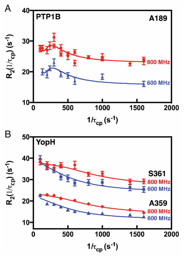
Many studies have implicated a role for conformational motions during the catalytic cycle, acting to optimize the binding pocket or facilitate product release, but a more intimate role in the chemical reaction has not been described. We address this by monitoring active-site loop motion in two protein tyrosine phosphatases (PTPs) using nuclear magnetic resonance spectroscopy. The PTPs, YopH and PTP1B, have very different catalytic rates; however, we find in both that the active-site loop closes to its catalytically competent position at rates that mirror the phosphotyrosine cleavage kinetics. This loop contains the catalytic acid, suggesting that loop closure occurs concomitantly with the protonation of the leaving group tyrosine and explains the different kinetics of two otherwise chemically and mechanistically indistinguishable enzymes.


Figures

Fig. 1
Fig. 1. Comparison of YopH and PTP1B
(A) YopH (cyan) and PTP1B (gray), the phosphate analog, vanadate (gray and red spheres), localizes the active site. (B) Open (light gray) and closed (blue) WPD loops of PTP1B. Vanadate (spheres) binds in the P-loop (orange), with C215 and R221 in stick rendering. Movement of D181 is indicated by dashed lines. (C) Superposition of closed loops for PTP1B and YopH. The three catalytic residues are shown in stick rendering. (D) PTP catalytic reaction of cleavage and hydrolysis. (A) PDB accession numbers are 2I42 and 3I80.
Fig. 2
Fig. 2. NMR detected motions
Apo- PTP1B and YopH WPD loops (A). (B & C) show 15N-CPMG dispersion curves for A189, F182, and W179. In (D) TROSY-detected off-resonance 1H-R dispersion curves for A359 (blue) and S361 (red).
Fig. 3
Fig. 3. Chemical shift differences for open and closed WPD loops
For PTP1B (A,B) and YopH (C,D) the NMR measured (red symbols) and calculated (black open symbols) chemical shifts are shown for residues in the WPD loop.
Fig. 4
Fig. 4. NMR detected motions
15N-CPMG dispersion analyses for peptide bound- PTP1B (A) and YopH (B). (A) shows a global fit at 800 and 600 MHz for A189. (B) shows a global fit at 800 (red) and 600 (blue) MHz for A359 and S361 in YopH.

References

    1. Kamerlin SC, Warshel A. Proteins. 2010;78:1339–1375. - PMC - PubMed
    1. Adamczyk AJ, Cao J, Kamerlin SC, Warshel A. Proc Natl Acad Sci U S A. 2011;108:14115–14120. - PMC - PubMed
    1. Boehr DD, McElheny D, Dyson HJ, Wright PE. Science. 2006;313:1638–1642. - PubMed
    1. Rozovsky S, McDermott AE. Proc Nat Acad Sci (USA) 2007;104:2080–2085. - PMC - PubMed
    1. Eisenmesser EZ, et al. Nature. 2005;438:117–121. - PubMed

Publication types

MeSH terms

Substances