. 2013 Aug 20:7:462.
doi: 10.3389/fnhum.2013.00462. eCollection 2013.

Characterizing aging in the human brainstem using quantitative multimodal MRI analysis

Affiliations

Characterizing aging in the human brainstem using quantitative multimodal MRI analysis

Christian Lambert et al. Front Hum Neurosci. .

Abstract

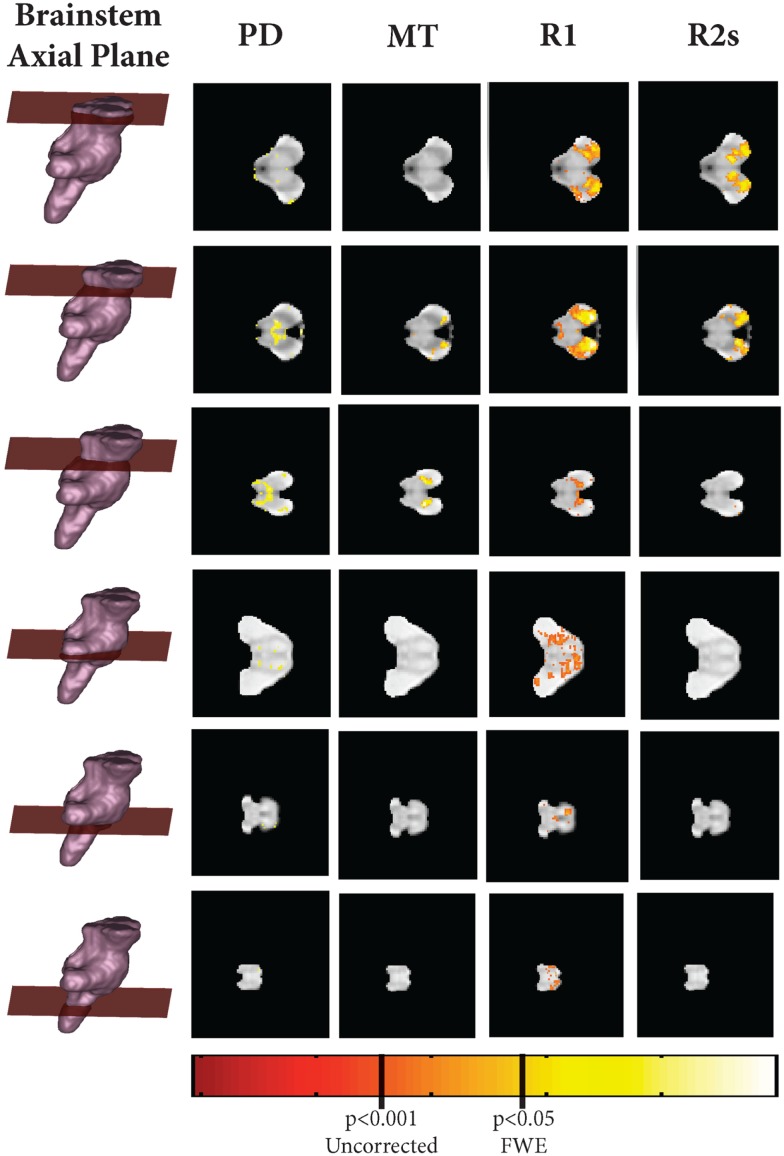
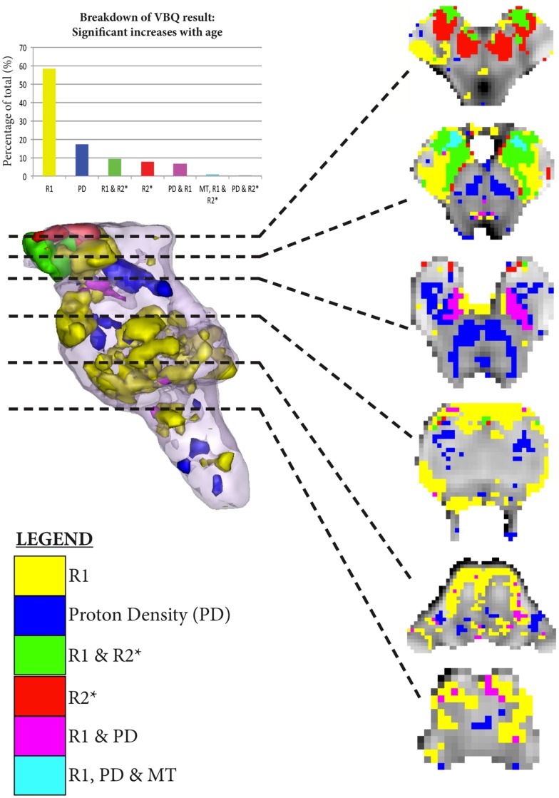
Aging is ubiquitous to the human condition. The MRI correlates of healthy aging have been extensively investigated using a range of modalities, including volumetric MRI, quantitative MRI (qMRI), and diffusion tensor imaging. Despite this, the reported brainstem related changes remain sparse. This is, in part, due to the technical and methodological limitations in quantitatively assessing and statistically analyzing this region. By utilizing a new method of brainstem segmentation, a large cohort of 100 healthy adults were assessed in this study for the effects of aging within the human brainstem in vivo. Using qMRI, tensor-based morphometry (TBM), and voxel-based quantification (VBQ), the volumetric and quantitative changes across healthy adults between 19 and 75 years were characterized. In addition to the increased R2* in substantia nigra corresponding to increasing iron deposition with age, several novel findings were reported in the current study. These include selective volumetric loss of the brachium conjunctivum, with a corresponding decrease in magnetization transfer and increase in proton density (PD), accounting for the previously described "midbrain shrinkage." Additionally, we found increases in R1 and PD in several pontine and medullary structures. We consider these changes in the context of well-characterized, functional age-related changes, and propose potential biophysical mechanisms. This study provides detailed quantitative analysis of the internal architecture of the brainstem and provides a baseline for further studies of neurodegenerative diseases that are characterized by early, pre-clinical involvement of the brainstem, such as Parkinson's and Alzheimer's diseases.

Keywords: aging; brainstem; quantitative MRI; tensor-based morphometry; voxel-based quantification.


Figures

Figure 1
Figure 1
Comparison of brainstem tissue classes against three corresponding ex vivo brainstem sections from “MR microscopy at 9.4T” (taken from Duvernoy’s Atlas of the Human Brain Stem and Cerebellum with permission). Figure reproduced from Lambert et al. (2013) with permission from Elsevier.
Figure 2
Figure 2
Summary of segmentation pipeline. This includes examples of the Multiparameter Maps (MPMs) as shown at the top of the image.
Figure 3
Figure 3
High resolution ex vivo combined MT T2* MRI with anatomical annotations for reference. Figure adapted from Lambert et al. (2013) with permission from Elsevier. Abbreviations: A8, dopaminergic center (approximate location), CP, cerebral peduncle (anterior to posterior: consisting of frontopontine, corticonuclear, corticospinal, and parietotemporal pontine tracts); CST, corticospinal tract; CTT, central tegmental tract; ICP, inferior cerebellar peduncle; ML, medial lemniscus; MLF, medial longitudinal fasciculus; PAG, periaqueductal gray; SCT, spinocerebellar tract; SNpc, substantia nigra pars compacta; SNpr, substantia nigra pars reticulata; TST, tectospinal tract; VTA, ventral tegmental area. *Artifact due to fixation.
Figure 4
Figure 4
Areas of significant regional decreases in tissue volume and magnetization transfer, binarized at p < 0.001 uncorrected.
Figure 5
Figure 5
Scatter plots of mean individual values extracted from binarized T-maps at p < 0.001 uncorrected. Linear fit shown in red. Increases: (A) Proton Density; (B) R1; (C) Magnetization Transfer; (D) R2*. Decreases: (E). Magnetization Transfer; (F). TBM. All Pearson’s correlation coefficients significant at p < 1 × 10−8.
Figure 6
Figure 6
Axial, coronal, and sagittal slice-wise images showing positive correlates with age (R2s, MT, A, R1) projected onto group-average MT map.
Figure 7
Figure 7
Significant regional increases binarized at p < 0.001 uncorrected. The legend classifies the regions of significant increase across the MPM, allowing overlapping significant increases to be simultaneously visualized. The histogram takes the total number of all significant voxels shown in the rendering, and then classifies them according to the MPMs in which those voxels are found in as per the legend.

References

    1. Aanerud J., Borghammer P., Chakravarty M. M., Vang K., Rodell A. B., Jónsdottir K. Y., et al. (2012). Brain energy metabolism and blood flow differences in healthy aging. J. Cereb. Blood Flow Metab. 32, 1177–118710.1038/jcbfm.2012.18 - DOI - PMC - PubMed
    1. Alalade E., Denny K., Potter G., Steffens D., Wang L. (2011). Altered cerebellar-cerebral functional connectivity in geriatric depression. PLoS ONE 6:e20035.10.1371/journal.pone.0020035 - DOI - PMC - PubMed
    1. Alvarez J. C., Díaz C., Suárez C., Fernández J. A., González del Rey C., Navarro A., et al. (2000). Aging and the human vestibular nuclei: morphometric analysis. Mech. Ageing Dev. 114, 149–17210.1016/S0047-6374(00)00098-1 - DOI - PubMed
    1. Andersen B. B., Gundersen H. J., Pakkenberg B. (2003). Aging of the human cerebellum: a stereological study. J. Comp. Neurol. 466, 356–36510.1002/cne.10884 - DOI - PubMed
    1. Armstrong C. L., Traipe E., Hunter J. V., Haselgrove J. C., Ledakis G. E., Tallent E. M., et al. (2004). Age-related, regional, hemispheric, and medial-lateral differences in myelin integrity in vivo in the normal adult brain. AJNR Am. J. Neuroradiol. 25, 977–984 - PMC - PubMed

LinkOut - more resources