. 2013 Aug 23:10:106.
doi: 10.1186/1742-2094-10-106.

Anti-inflammatory and immunomodulatory mechanisms of mesenchymal stem cell transplantation in experimental traumatic brain injury

Affiliations

Anti-inflammatory and immunomodulatory mechanisms of mesenchymal stem cell transplantation in experimental traumatic brain injury

Run Zhang et al. J Neuroinflammation. .

Abstract

Background: Previous studies have shown beneficial effects of mesenchymal stem cell (MSC) transplantation in central nervous system (CNS) injuries, including traumatic brain injury (TBI). Potential repair mechanisms involve transdifferentiation to replace damaged neural cells and production of growth factors by MSCs. However, few studies have simultaneously focused on the effects of MSCs on immune cells and inflammation-associated cytokines in CNS injury, especially in an experimental TBI model. In this study, we investigated the anti-inflammatory and immunomodulatory properties of MSCs in TBI-induced neuroinflammation by systemic transplantation of MSCs into a rat TBI model.

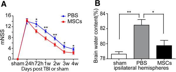
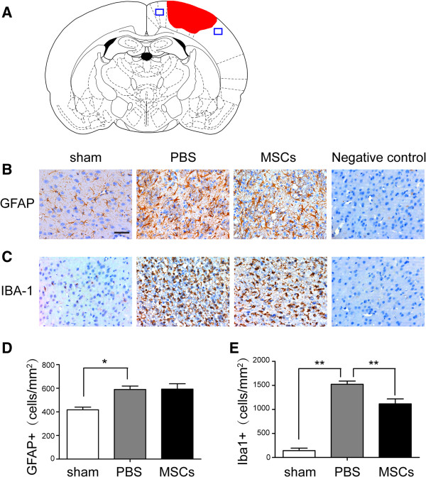
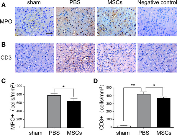
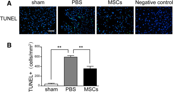
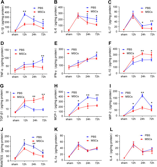
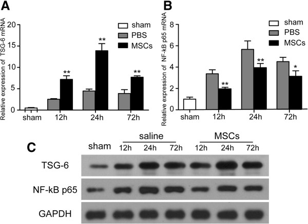
Methods/results: MSCs were transplanted intravenously into rats 2 h after TBI. Modified neurologic severity score (mNSS) tests were performed to measure behavioral outcomes. The effect of MSC treatment on neuroinflammation was analyzed by immunohistochemical analysis of astrocytes, microglia/macrophages, neutrophils and T lymphocytes and by measuring cytokine levels [interleukin (IL)-1α, IL-1β, IL-4, IL-6, IL-10, IL-17, tumor necrosis factor-α, interferon-γ, RANTES, macrophage chemotactic protein-1, macrophage inflammatory protein 2 and transforming growth factor-β1] in brain homogenates. The immunosuppression-related factors TNF-α stimulated gene/protein 6 (TSG-6) and nuclear factor-κB (NF-κB) were examined by reverse transcription-polymerase chain reaction and Western blotting. Intravenous MSC transplantation after TBI was associated with a lower density of microglia/macrophages and peripheral infiltrating leukocytes at the injury site, reduced levels of proinflammatory cytokines and increased anti-inflammatory cytokines, possibly mediated by enhanced expression of TSG-6, which may suppress activation of the NF-κB signaling pathway.

Conclusions: The results of this study suggest that MSCs have the ability to modulate inflammation-associated immune cells and cytokines in TBI-induced cerebral inflammatory responses. This study thus offers a new insight into the mechanisms responsible for the immunomodulatory effect of MSC transplantation, with implications for functional neurological recovery after TBI.


Figures

Figure 1
Figure 1
Surface marker expression in MSCs. MSCs were confirmed by flow cytometry analysis after three passages as positive for CD44 (99.01%), CD90 (99.28%) and CD105 (97.71%), with low positivity for CD14 (0.79%), CD34 (0.78%), CD45 (0.67%) and HLA-DR (1.11%).
Figure 2
Figure 2
Modified neurologic severity score (mNSS) and brain water content. (A) Neurological function was analyzed by mNSS on days 1, 3, 7, 14, 21 and 28 after TBI. Treatment with MSCs significantly lowered mNSS from days 3–28 compared with the PBS group. There was no significant difference in scores between the MSC- and PBS-treated groups only at 24 h post-TBI (n = 6 per group). (B) Brain water content of ipsilateral hemispheres was measured at 72 h after injury. The PBS group had a significantly higher brain water content than the sham-injured control group. MSC treatment significantly reduced brain water content compared with the PBS group (n = 6 per group). Data are presented as the mean ± SD. *p < 0.05, **p < 0.01.
Figure 3
Figure 3
Effect of MSC treatment on GFAP+ astrocytes and Iba-1+ microglia/macrophages. (A) Diagram of a coronal rat brain section showing the relationship of the lesion cavity (red) to the regions photographed (blue squares). The density of astrocytes was not significantly different after MSC treatment (B, D) (n = 6 per group). The number of microglia/macrophages (C, E) (sham: 142.7 ± 45.4 cells/mm2; PBS: 1,524.7 ± 60.1 cells/mm2; MSCs: 1,124.3 ± 104.5 cells/mm2) was significantly decreased after MSC administration at 72 h post-TBI compared with the PBS-treatment group (n = 6 per group). Data are presented as mean ± SD. Bar = 50 μm. *p < 0.05.
Figure 4
Figure 4
Influence of MSC administration on MPO+ neutrophils and CD3+ lymphocytes. MSCs reduced the numbers of infiltrating MPO+ neutrophils (A, C) (PBS: 775.0 ± 55.34 cells/mm2; MSCs: 638.67 ± 72.15 cells/mm2), CD3+ lymphocytes (B, D) (PBS: 421.67 ± 28.15 cells/mm2; MSCs: 367.67 ± 17.5 cells/mm2). Data are presented as the mean ± SD. Bar = 50 μm; n = 6 per group, *p < 0.05.
Figure 5
Figure 5
Effect of MSC transplantation on apoptosis. Apoptotic cells (sham: 18.7 ± 8.1 cells/mm2; PBS: 295 ± 15 cells/mm2; MSCs: 179.3 ± 25.8 cells/mm2) in the injured cortex at 72 h after TBI were reduced in the MSC treatment group compared with the PBS group (A, B) (n = 6 per group). Number of apoptotic cells is presented as the mean ± SD. Bar = 50 μm. **p < 0.01.
Figure 6
Figure 6
Influence of MSC treatment on cytokine concentrations. Levels of the proinflammatory cytokines IL-1β (at 12, 24 and 72 h), IL-6 (at 24 and 72 h), IL-17 (at 24 and 72 h), TNF-α (at 24 and 72 h) and IFN-γ (at 72 h) were significantly decreased in the MSC-treatment group compared with the PBS group (AE). Levels of the anti-inflammatory cytokines IL-10 and TGF-β1 (at 24 and 72 h after TBI) (F, G) were increased in the MSC-treatment group compared with the PBS group. The chemokines MCP-1, MIP-2 and RANTES were reduced at 12, 24 and 72 h after TBI in the MSC group compared with the PBS group (HJ). There were no significant differences in levels of the cytokines IL-1α and IL-4 (K, L) between the two groups. n = 6 in each time point of per group. Data are presented as the mean ± SD. *p < 0.05, **p < 0.01.
Figure 7
Figure 7
MSC treatment upregulates TSG-6 and downregulates NF-κB expression. Upregulation of TSG-6 (A) was observed from 12 to 72 h in the injured cortex after TBI in the MSC-treatment group. mRNA levels of NF-κB (B) decreased from 12 to 48 h after MSC transplantation. Similar results were observed at the protein level (C) for TSG-6 and NF-κB p65. n = 6 in each time point of per group. Data are presented as the mean ± SD. *p < 0.05, **p < 0.01 versus PBS group.

References

    1. Xiong Y, Mahmood A, Chopp M. Neurorestorative treatments for traumatic brain injury. Discov Med. 2010;10:434–442. - PMC - PubMed
    1. Helmy A, Carpenter KL, Menon DK, Pickard JD, Hutchinson PJ. The cytokine response to human traumatic brain injury: temporal profiles and evidence for cerebral parenchymal production. J Cereb Blood Flow Metab. 2011;31:658–670. doi: 10.1038/jcbfm.2010.142. - DOI - PMC - PubMed
    1. Correale J, Villa A. The neuroprotective role of inflammation in nervous system injuries. J Neurol. 2004;251:1304–1316. doi: 10.1007/s00415-004-0649-z. - DOI - PubMed
    1. Ziebell JM, Morganti-Kossmann MC. Involvement of pro- and anti-inflammatory cytokines and chemokines in the pathophysiology of traumatic brain injury. Neurotherapeutics. 2010;7:22–30. doi: 10.1016/j.nurt.2009.10.016. - DOI - PMC - PubMed
    1. Rhodes J. Peripheral immune cells in the pathology of traumatic brain injury? Curr Opin Crit Care. 2011;17:122–130. doi: 10.1097/MCC.0b013e3283447948. - DOI - PubMed

Publication types