. 2013 Nov;13(9):1474-84.
doi: 10.2174/15665240113139990076.

A novel role of the STAT3 pathway in brain inflammation-induced human neural progenitor cell differentiation

Affiliations

A novel role of the STAT3 pathway in brain inflammation-induced human neural progenitor cell differentiation

E Chen et al. Curr Mol Med. 2013 Nov.

Abstract

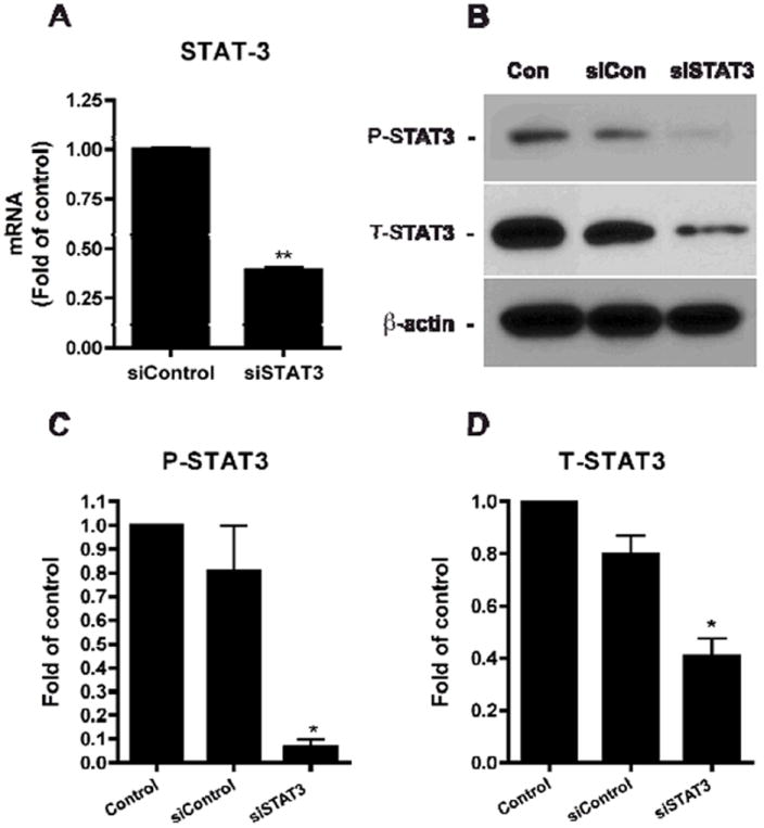
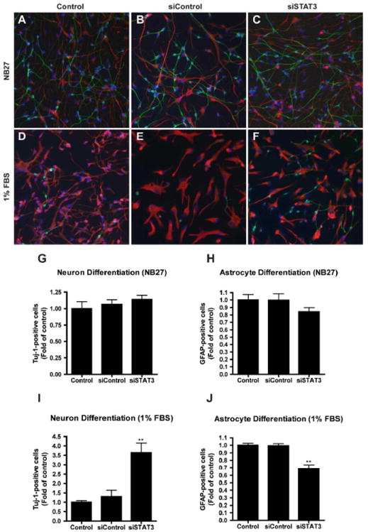
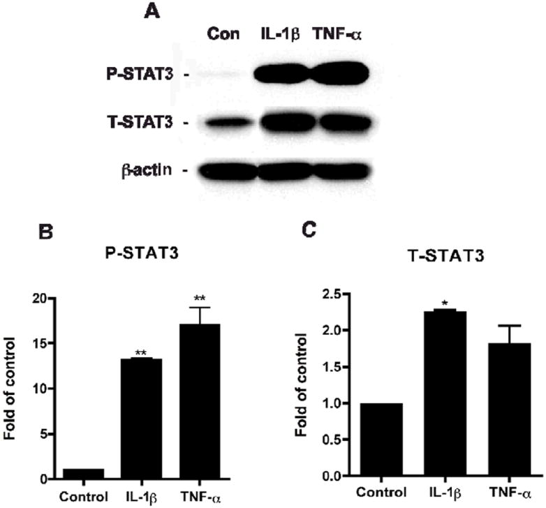
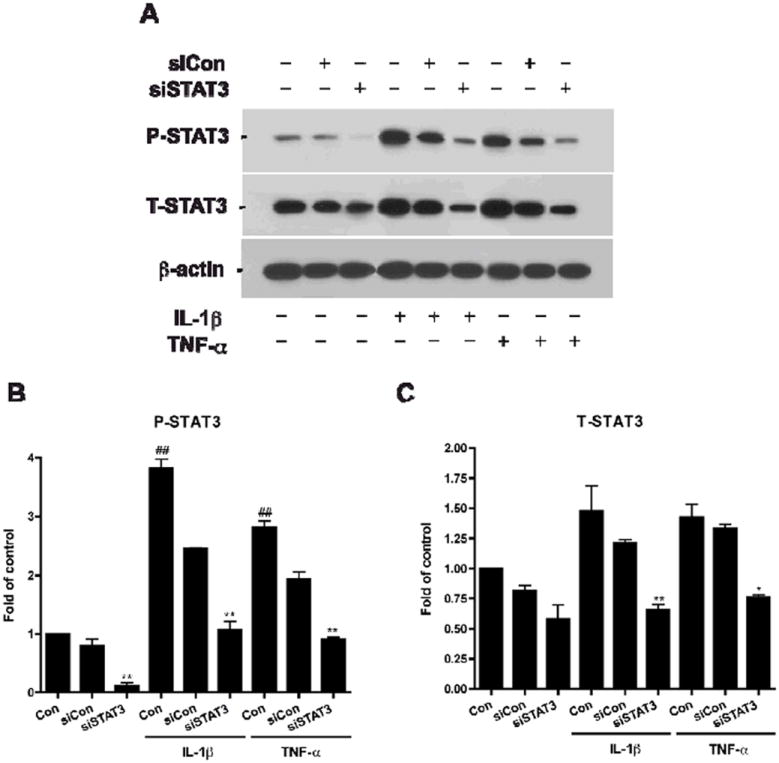
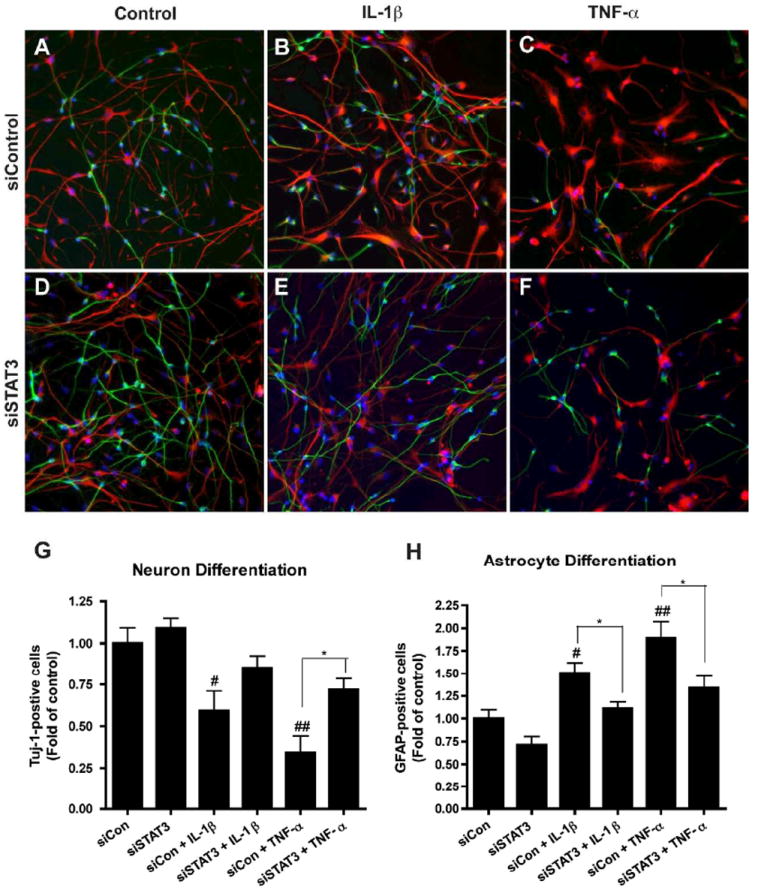
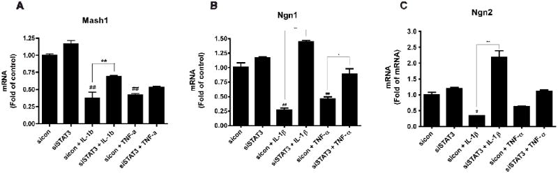
Brain inflammation is a primary pathological driving force of many neurodegenerative disorders. In the destructive process, pro-inflammatory cytokines (IL-1β and TNF-α), are robustly released, affecting normal neural progenitor cell (NPC) differentiation, and resulting in a vast number of astrocytes and a diminished neural population. A counteractive mechanism is still unknown. In this study, we have identified a link between brain inflammation and the signal transducer and activator of transcription 3 (STAT3) pathway: IL-1β and TNF-α induce STAT3 activation in NPCs. Then to investigate STAT3's effects on NPC fate, we observed that an inhibition of STAT3 expression by siRNA inhibited astrocytic differentiation and increased neuronal differentiation of human NPCs in fetal bovine serum (FBS)-induced astrocyte differentiation condition. Furthermore, STAT3-targeting siRNA abrogated IL-1β and TNF-α-induced astrocyte differentiation and partially restored neuronal differentiation. Elimination of STAT3 expression also countered IL-1β and TNF-α-induced inhibition of proneural bHLH genes, mammalian achaete-schute homologue-1 (Mash1), Neurogenin1 (Ngn1), and Neurogenin2 (Ngn2). These data suggest that a suppression of STAT3 during brain inflammation would inhibit astrogliogenesis and promote neurogenesis. Thus, STAT3 could be a potential target of drug therapy for neurodegenerative disorders.


Figures

Figure 1
Figure 1. Characterization of human cortical NPCs
Neurospheres were dissociated to single cell suspension and plated in poly-D-lysine-coated cover slips for 24 h. Cells were fixed and stained for Nestin (green, C) and Sox2 (red, D). Nuclei were stained using Hoechst 33342 (blue, B). Figure 1A shows a merged image of B, C, and D. The original magnification is × 20. Results are representative of three donors.
Figure 2
Figure 2. SiSTAT3 blocks STAT3 expression
A. Human NPCs were transfected with siCon or siSTAT3. Six days later, total mRNA was extracted and real-time RT-PCR was conducted to examine the mRNA expression levels of STAT3. STAT3 expression was normalized to GAPDH as an internal gene expression control and data is presented as fold of siCon. Data were obtained from three independent donors. ** differs significantly compared to siCon (p < 0.01). B-D. Expressions of STAT3 were detected by Western blotting. The films were scanned and the acquired images were analyzed using the public domain NIH image program for data quantification. Expression of P-STAT3 (C) and T-STAT3 (D) were normalized to β-actin. Data is presented as fold of control (NB27). Results are an average of three independent donors. * differs significantly compared to control (p < 0.05).
Figure 3
Figure 3. Suppression of STAT3 reduces astrocytic and increases neuronal proportions
A-F. Human NPCs were transfected with either siCon (B, E) or siSTAT3 (C, F) and differentiated in NB27 (A-C) or 1% FBS (D-F). Six days later, neuron and astrocyte differentiation were evaluated by immunocytochemical staining with antibodies against Tuj-1 (green) and GFAP (red). Representative fluorescence overlay-micrographs display the morphology of neurons (green) and astrocytes (red). G-J. Percentage of Tuj-1/ GFAP-positive cells was calculated based on the total count of cell nuclei, marked by Hoechst (blue). Percentages were then normalized as fold of control (G-J). Data were collected from four independent measurements. */** differs significantly compared to Control and siControl (* p < 0.05; ** p < 0.01). Scale = 200 μm.
Figure 4
Figure 4. Pro-inflammatory cytokines activate STAT3
Human NPCs were treated with IL-1β or TNF-α. Cells were subsequently lysed, and proteins were collected 6 days after treatment for Western blotting (A). P-STAT3 (B) and T-STAT3 (C) expressions were detected, and the films with the acquired images were later scanned and analyzed for data quantification. Expression was normalized to β-actin as a loading control, and data is presented as fold of Control. Results were representative of three independent experiments. */** differs significantly compared to Control (* p < 0.05; ** p < 0.01).
Figure 5
Figure 5. SiSTAT3 blocks STAT3 expression under cytokine treatment
Human NPCs were transfected with siCon or siSTAT3 and then were treated with IL-1β or TNF-α for 24 h. Cells were subsequently lysed, and proteins were collected for Western blotting (A). P-STAT3 (B) and T-STAT3 (C) expressions were detected, and the films with the acquired images were later scanned and analyzed for data quantification. Expression was normalized to β-actin as a loading control, and data is presented as fold of control (NB27). Results were representative of two independent experiments. */** differs significantly compared to respective control (NB27 + IL-1β or NB27 + TNF-α, * p < 0.05; ** p < 0.01)), ## differs significantly compared to Control (## p < 0.01).
Figure 6
Figure 6. Suppression of STAT3 reduces cytokine-induced increase of astrocytes and decrease of neurons
A-F. Human NPCs were transfected with siCon (A-C) or siSTAT3 (D-F) and were differentiated with or without IL-1β (B, E) or TNF-α (C, F) for 6 days. Neuron and astrocyte differentiation expressions were evaluated by immunocytochemical staining with antibodies against Tuj-1 (green) and GFAP (red). Representative fluorescence overlay-micrographs display the morphology of neurons (green) and astrocytes (red). G-H. Percentage of Tuj-1/GFAP-positive cells was calculated based on the total count of cell nuclei, marked by Hoechst (blue). Percentages were then normalized as fold of siCon. Data were collected from four independent measurements. */** differs significantly compared to respective siCon ((siCon + IL-1β or siCon + TNF-α, * p < 0.05; ** p < 0.01), and #/## differs significantly compared to siCon, # p < 0.05; ## p < 0.01). Scale = 200 μm.
Figure 7
Figure 7. Suppression of STAT3 regulates cytokine-mediated bHLH factor expression
Human NPCs were transfected with siCon or siSTAT3 and were then treated with IL-1β or TNF-α for 24 h. Total mRNA was extracted and real-time RT-PCR were conducted to detect transcription factors (Mash1, Ngn 1, and Ngn2) expressions. A-C. Data were obtained from three independent measurements. Data were presented as fold of siCon. */** differs significantly compared to respective siCon (siCon + IL-1β or siCon + TNF-α, * p < 0.05; ** p < 0.01), and #/## differs significantly compared to siCon (# p < 0.05; ## p < 0.01).

References

    1. Gage FH. Mammalian neural stem cells. Science. 2000;287(5457):1433–8. - PubMed
    1. McKay R. Stem cells in the central nervous system. Science. 1997 Apr 4;276(5309):66–71. - PubMed
    1. Ming GL, Song H. Adult neurogenesis in the mammalian central nervous system. Annu Rev Neurosci. 2005;28:223–50. - PubMed
    1. Whitney NP, Eidem TM, Peng H, Huang Y, Zheng JC. Inflammation mediates varying effects in neurogenesis: Relevance to the pathogenesis of brain injury and neurodegenerative disorders. J Neurochem. 2009 Jan 19; - PMC - PubMed
    1. Minagar A, Shapshak P, Fujimura R, Ownby R, Heyes M, Eisdorfer C. The role of macrophage/microglia and astrocytes in the pathogenesis of three neurologic disorders: HIV-associated dementia, Alzheimer disease, and multiple sclerosis. J Neurol Sci. 2002 Oct 15;202(1-2):13–23. - PubMed

Publication types

MeSH terms