. 2013;15(4):R67.
doi: 10.1186/bcr3461.

LOXL2 induces aberrant acinar morphogenesis via ErbB2 signaling

LOXL2 induces aberrant acinar morphogenesis via ErbB2 signaling

Joan Chang et al. Breast Cancer Res. 2013.

Abstract

Introduction: Lysyl oxidase-like 2 (LOXL2) is a matrix-remodeling enzyme that has been shown to play a key role in invasion and metastasis of breast carcinoma cells. However, very little is known about its role in normal tissue homeostasis. Here, we investigated the effects of LOXL2 expression in normal mammary epithelial cells to gain insight into how LOXL2 mediates cancer progression.

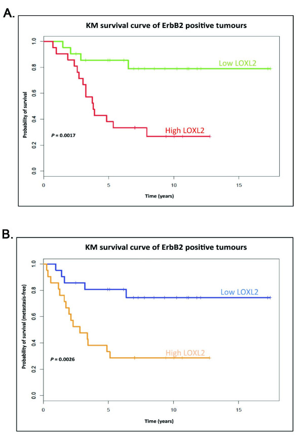
Methods: LOXL2 was expressed in MCF10A normal human mammary epithelial cells. The 3D acinar morphogenesis of these cells was assessed, as well as the ability of the cells to form branching structures on extracellular matrix (ECM)-coated surfaces. Transwell-invasion assays were used to assess the invasive properties of the cells. Clinically relevant inhibitors of ErbB2, lapatinib and Herceptin (traztuzumab), were used to investigate the role of ErbB2 signaling in this model. A retrospective study on a previously published breast cancer patient dataset was carried out by using Disease Specific Genomic Analysis (DSGA) to investigate the correlation of LOXL2 mRNA expression level with metastasis and survival of ErbB2-positive breast cancer patients.

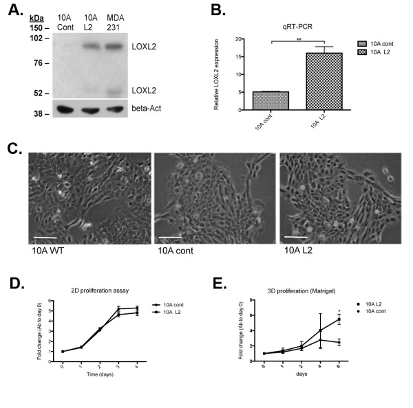
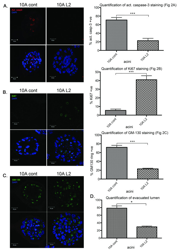
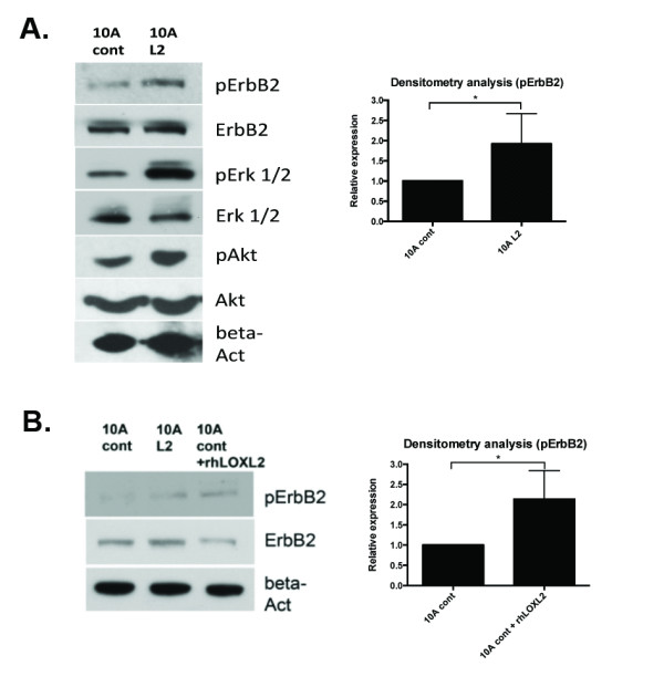
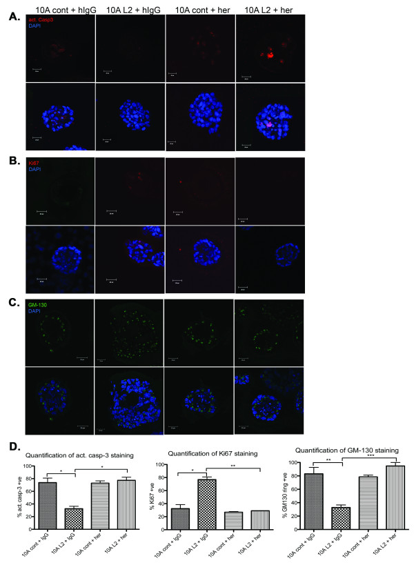
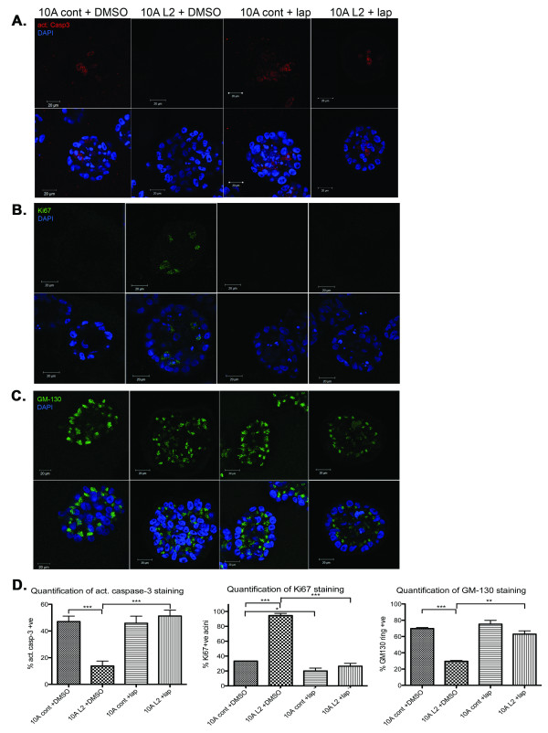
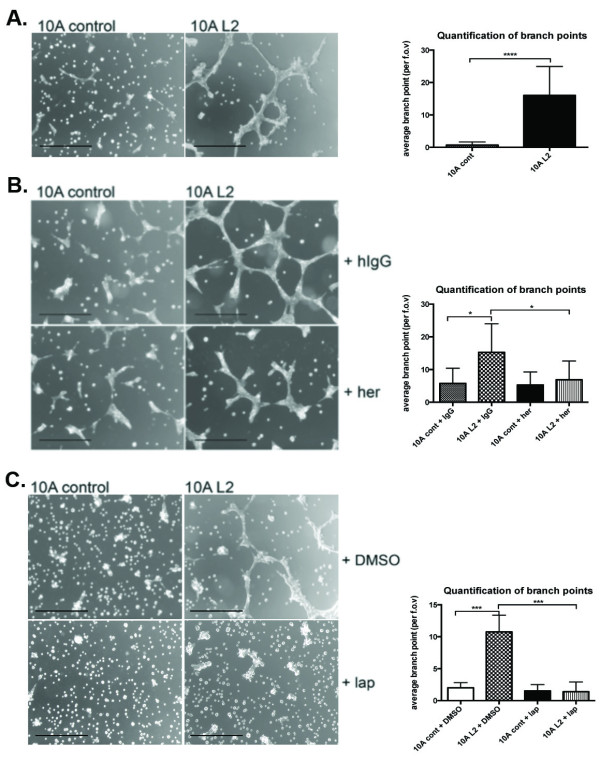
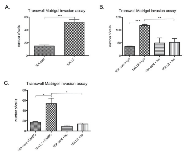
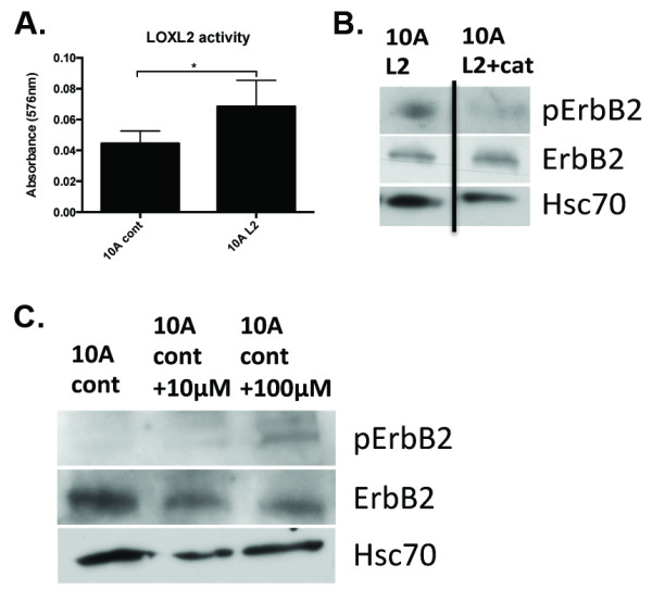
Results: Fluorescence staining of the acini revealed increased proliferation, decreased apoptosis, and disrupted polarity, leading to abnormal lumen formation in response to LOXL2 expression in MCF10A cells. When plated onto ECM, the LOXL2-expressing cells formed branching structures and displayed increased invasion. We noted that LOXL2 induced ErbB2 activation through reactive oxygen species (ROS) production, and ErbB2 inhibition by using Herceptin or lapatinib abrogated the effects of LOXL2 on MCF10A cells. Finally, we found LOXL2 expression to be correlated with decreased overall survival and metastasis-free survival in breast cancer patients with ErbB2-positive tumors.

Conclusions: These findings suggest that LOXL2 expression in normal epithelial cells can induce abnormal changes that resemble oncogenic transformation and cancer progression, and that these effects are driven by LOXL2-mediated activation of ErbB2. LOXL2 may also be a beneficial marker for breast cancer patients that could benefit most from anti-ErbB2 therapy.


Figures

Figure 1
Figure 1
Manipulation of LOXL2 expression levels in human MCF10A normal mammary epithelial cells. (A) Western blot of secreted LOXL2 in CM generated from MDA MB-231 cells (MDA 231), MCF10A cells infected with LOXL2 (10A L2) and vector alone (10A cont) revealed that LOXL2 protein expression was upregulated in the 10A L2 cells to a level similar to that detected in MDA cells. β-Actin was used as a loading control. (B) Quantitative real-time PCR (qRT-PCR) of LOXL2 mRNA levels in manipulated MCF10A cells showed that LOXL2 mRNA levels were upregulated in 10A L2 cells. P = 0.009. (C) Morphologies of the manipulated MCF10A cells compared with WT cells showed that upregulation of LOXL2 did not produce significant alterations on cells plated on 2D tissue-culture plastic. Cells were viewed under a microscope (Leica DM1L), and representative images were taken. (D) 2D MTS proliferation assay of manipulated MCF10A cells suggested that when cultured on plastic, LOXL2 expression did not alter proliferation of the 10A cells. Error bars represent SEM for three independent experiments. (E) 3D MTS proliferation assay of manipulated MCF10A cells with manipulated LOXL2 expression plated within Matrigel suspension suggested that increased LOXL2 expression increases proliferation of the 10A cells in 3D. Error bars represent SEM for three independent experiments. P = 0.034 for day 6.
Figure 2
Figure 2
LOXL2 expression disrupts normal breast epithelial acini formation. 10A cont and 10A L2 cells were plated on top of a thin layer of 50 μl Matrigel in eight-well chamber slides in a suspension of Matrigel/culture media mix to investigate the acinar morphogenesis of these cells. The cultures were allowed to grow for 8, 10, and 13 days, and then fixed and stained with primary antibodies, as described in the different panels. All data were based on at least three independent experimental repeats. Scale bar, 20 μm. (A) Immunofluorescence staining of acini with anti-activated caspase-3 antibody to detect apoptotic cells on day 8 reveals decreased apoptosis in the central cells of the L2 acini. Representative images of activated caspase-3 staining in acini for each cell line are shown. Quantification represented average percentage ± standard error of acini containing activated caspase-3-positive cells (P = 0.0007). (B) Immunofluorescence staining of acini with anti-Ki67 antibody to detect proliferating cells on day 13 reveals that L2 acini had more proliferative cells. Representative images of Ki67-positive acini from each cell line are shown. Quantification represented average percentage ± standard error of acini containing Ki67-positive cells. (P = 0.0007). (C) Immunofluorescence staining of acini with anti-GM130 antibody to assess cell polarity on day 10. Representative images of GM130 staining in acini for each cell line are shown. Quantification represented average percentage ± standard error of acini forming a regular ring structure, as assessed by GM130 staining (P = 0.0005; n = 80 acini for each cell line per repeat). (D) Quantification of average percentage of acini at day 13 with evacuated lumens ± standard error. Acini with evacuated lumens were defined as having no more than 20% of total number of cells, as well as Ki67-positive cells present in the center (P = 0.013).
Figure 3
Figure 3
LOXL2 expression in MCF10A cells increases phosphorylation of ErbB2. Cells were plated on a thin layer of Matrigel and serum-starved for 3 hours before being subjected to serum-blasting and subsequent lysis of cells. (A) Western blotting revealed that phospho-ErbB2 was elevated in 10A L2 cells when compared with 10A cont cells, whereas total ErbB2 levels were equivalent in the two lines, suggesting increased phosphorylation of ErbB2 in the 10A L2 cells. Densitometry analysis was calculated for pErbB2 levels relative to total ErbB2 and revealed a significant increase in 10A L2 cells (P = 0.0241). The levels of phospho-Akt and phospho-Erk1/2 were also elevated in 10A L2 cells. (B) The 10A cont cells were subjected to 16-hour treatment with 50 nM recombinant human LOXL2 (rhLOXL2; R&D Systems) followed by serum-starvation and serum-blasting (10A cont + rhLOXL2). Western-blotting analysis showed that in 10A cont treated with rhLOXL2, phospho-ErbB2 level was increased to a greater extent than 10A L2 cells when compared with sham-treated 10A cont cells. Densitometry analysis was calculated for pErbB2 levels relative to total ErbB2 and revealed significant increase in 10A cont + rhLOXL2 cells (P = 0.0438). This suggested that extracellular recombinant LOXL2 was capable of activating the ErbB2 receptor in 10A cells.
Figure 4
Figure 4
Treatment with the ErbB2-specific inhibitor trastuzumab (Herceptin) restores LOXL2-expressing acini phenotype to a more normal phenotype. Acini were cultured as described in Figure 2; 300 nM Herceptin (Her) dissolved in water was added to cells at day 6 (10A cont+her; 10A L2+her), and an equivalent amount of human IgG was added as a control (10A cont+hIgG; 10A L2+hIgG). Acini were fixed, stained, quantified, and presented as described in Figure 2. All data are based on at least three independent experimental repeats. Scale bar, 20 μm. (A) Immunofluorescence staining of acini with anti-activated caspase-3 antibody to detect apoptotic cells on day 8. Staining revealed increased activation of caspase-3 in 10A L2+her acini when compared with 10A L2+hIgG acini. (B)Immunofluorescence staining of acini with anti-Ki67 antibody to detect proliferating cells on day 13. Staining revealed a decrease in proliferating cells in 10A L2+her acini when compared with 10A L2+hIgG acini. (C) Immunofluorescence staining of acini with anti-GM130 antibody to assess cell polarity on day 10. Staining revealed GM-130 ring-like staining effect in 10A L2+her acini, compared with the scrambled and disorganized staining observed in 10A L2+hIgG acini. (D) Quantitative analysis of activated caspase-3 staining (left panel; P = 0.0364 for 10A cont+hIgG and 10A L2+hIgG; P = 0.0189 for 10A L2 ± her), Ki67 staining (middle panel; P = 0.0268 for 10A cont+hIgG and 10A L2+hIgG; P = 0.0073 for 10A L2 ± her), and GM130-ring structures (right panel; P = 0.0083 for 10A cont+IgG and 10A L2+IgG; P = 0.0007 for 10A L2 ± her) in acini. These results suggest that Herceptin treatment of 10A L2 acini significantly reverted the acinar morphology to a more-normal phenotype.
Figure 5
Figure 5
The dual ErbB1/ErbB2 inhibitor lapatinib reverts LOXL2-mediated acinar morphologic changes to a more-normal phenotype. Acini were cultured as described in Figure 2; 5 μM lapatinib (lap) dissolved in DMSO was added to cells at day 6 (10A cont+lap; 10A L2+lap), and equivalent amounts of DMSO were added as a control (10A cont+DMSO; 10A L2+DMSO). Acini were fixed, stained, and quantified as described. All data are based on at least three independent experimental repeats. Scale bar, 20 μm. (A) Immunofluorescence staining of acini with anti-activated caspase-3 antibody to detect apoptotic cells on day 8. Staining revealed increased activation of caspase-3 in 10A L2+lap acini when compared with 10A L2+DMSO acini. (B) Immunofluorescence staining of acini with anti-Ki67 antibody to detect proliferating cells on day 13. Staining revealed a decrease in proliferating cells in 10A L2+lap acini when compared with 10A L2+DMSO acini. (C) Immunofluorescence staining of acini with anti-GM130 antibody to assess cell polarity on day 10. Staining revealed GM-130 ring-like staining effect in 10A L2+lap acini, compared with the scrambled and disorganized staining observed in 10A L2+DMSO acini. (D) Quantitative analysis of activated caspase-3 staining (left panel; P = 0.00047 for 10A cont+DMSO and 10A L2+DMSO; P = 0.0006 for 10A L2 ± lap), Ki67 staining (middle panel; P = 0.00032 for 10A cont+DMSO and 10A L2+DMSO; P = 0.0257 for 10A cont ± lap; P = 0.00013 for 10A L2 ± lap), and GM130-ring structures (right panel. P = 0.00016 for 10A cont+DMSO and 10A L2+DMSO; P = 0.0013 for 10A L2 ± lap) in acini. These results suggest that lapatinib treatment of 10A L2 acini significantly reverted the acinar morphology to a more-normal phenotype.
Figure 6
Figure 6
LOXL2-expressing MCF10A cells form ErbB2-dependent branching structures on matrix. (A) When plated on Matrigel-coated plates, 10A cont cells remained small and discrete, whereas 10A L2 cells formed extensive branching structures. Quantification of branch-points revealed that 10A L2 cells have significantly more branch-points (right panel; P = 0.00004). (B) Herceptin treatment of the 10A L2 cells significantly decreased the degree of branching of these cells to that seen for controls (right panel; P = 0.0167 and P = 0.0398, respectively). (C) Lapatinib treatment of the 10A L2 cells significantly abrogated the branching ability of these cells to that seen for controls (right panel; P = 0.0007 and P = 0.0003, respectively).
Figure 7
Figure 7
LOXL2 promotes invasion of normal breast epithelial cells; this effect is abrogated with ErbB2 inhibition. (A) The 10A L2 cells exhibited increased invasion through Matrigel compared with 10A cont cells in Transwell invasion assays. (B) Herceptin treatment reduced the invasive ability of the 10A L2 cells to a level comparable to 10A cont cells. Human IgG treatment had no effect on the invasiveness of the manipulated 10A cells. (C) Lapatinib treatment of the 10A L2 cells greatly reduced the invasiveness of the cells to a level comparable to the 10A cont cells. DMSO treatment had no effect on the invasive properties of the manipulated 10A cells.
Figure 8
Figure 8
LOXL2 expression induces ErbB2 activation through production of 2O2. (A)Production of H2O2 on Matrigel was measured as an activity read-out in the 10A cont and 10A L2 cells, by using a commercially available kit and following the manufacturer's instructions (Abcam). 10A L2 cells produced higher levels of H2O2, as expected, because of LOXL2 overexpression. (B) The 200 U/ml catalase (+cat) was added to remove H2O2 produced by LOXL2. Western blotting revealed that pErbB2 was decreased in 10A L2 cells treated with catalase (L2+cat) in comparison with untreated cells (L2). (C) Addition of H2O2 to 10A cont cells strongly induced ErbB2 activation. Taken together, these results indicate that, by removing H2O2, we can significantly abrogate ErbB2 activation in 10A L2 cells, and addition of H2O2 activated ErbB2.
Figure 9
Figure 9
Elevated LOXL2 and ErbB2 are associated with invasive cell behavior and poor patient prognosis. (A) Disease-specific genomic analysis (DSGA) was performed on gene-expression microarray data from the NKI cohort [17]. Of 295 tumors, 51 were found to express high levels of ErbB2, and a Kaplan-Meier survival curve is constructed from this subset of ErbB2-positive tumors. Shown here is the overall survival of patients with Her2/ErbB2-positive tumors (n = 51) separated into low and high LOXL2 expression. P = 0.017. (B) As in Figure 8A, showing metastasis-free survival. P = 0.026.

Similar articles

Cited by

References

    1. Csiszar K. Lysyl oxidases: a novel multifunctional amine oxidase family. Prog Nucleic Acid Res Mol Biol. 2001;15:1–32. - PubMed
    1. Kim YM, Kim EC, Kim Y. The human lysyl oxidase-like 2 protein functions as an amine oxidase toward collagen and elastin. Mol Biol Rep. 2011;15:145–149. doi: 10.1007/s11033-010-0088-0. - DOI - PubMed
    1. Barker HE, Cox TR, Erler JT. The rationale for targeting the LOX family in cancer. Nat Rev Cancer. 2012;15:540–552. doi: 10.1038/nrc3319. - DOI - PubMed
    1. Barker HE, Chang J, Cox TR, Lang G, Bird D, Nicolau M, Evans HR, Gartland A, Erler JT. LOXL2-mediated matrix remodeling in metastasis and mammary gland involution. Cancer Res. 2011;15:1561–1572. doi: 10.1158/0008-5472.CAN-10-2868. - DOI - PMC - PubMed
    1. Akiri G, Sabo E, Dafni H, Vadasz Z, Kartvelishvily Y, Gan N, Kessler O, Cohen T, Resnick M, Neeman M, Neufeld G. Lysyl oxidase-related protein-1 promotes tumor fibrosis and tumor progression in vivo. Cancer Res. 2003;15:1657–1666. - PubMed

Publication types

MeSH terms