. 2013 Aug 23:6:246.
doi: 10.1186/1756-3305-6-246.

Molecular characterization of Trichinella spiralis aminopeptidase and its potential as a novel vaccine candidate antigen against trichinellosis in BALB/c mice

Molecular characterization of Trichinella spiralis aminopeptidase and its potential as a novel vaccine candidate antigen against trichinellosis in BALB/c mice

Ya Zhang et al. Parasit Vectors. .

Abstract

Background: Trichinella spiralis is an intracellular parasite that can cause a serious threat to human health by causing trichinellosis. The aminopeptidase (AP) was found in the proteins produced by T. spiralis infective larvae after in vitro co-culture with intestinal epithelial cells, but its characteristics and function are unknown. The purpose of this study was to identify the T. spiralis aminopeptidase (TsAP) and to investigate its potential as a vaccine candidate antigen against T. spiralis infection.

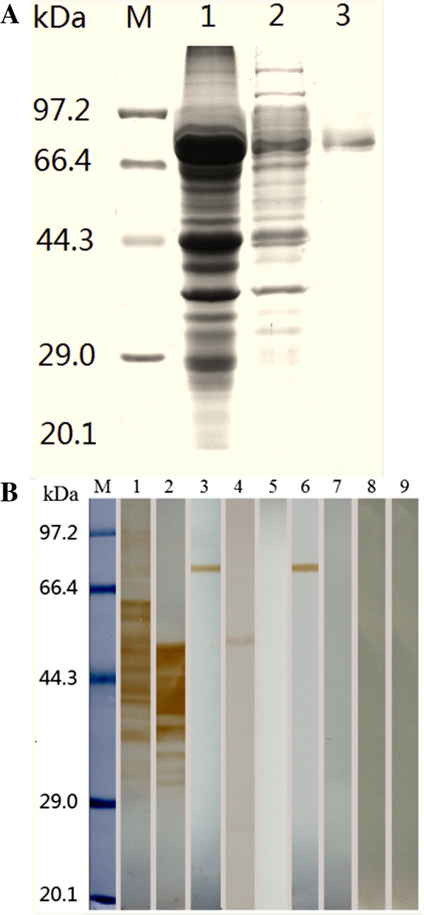
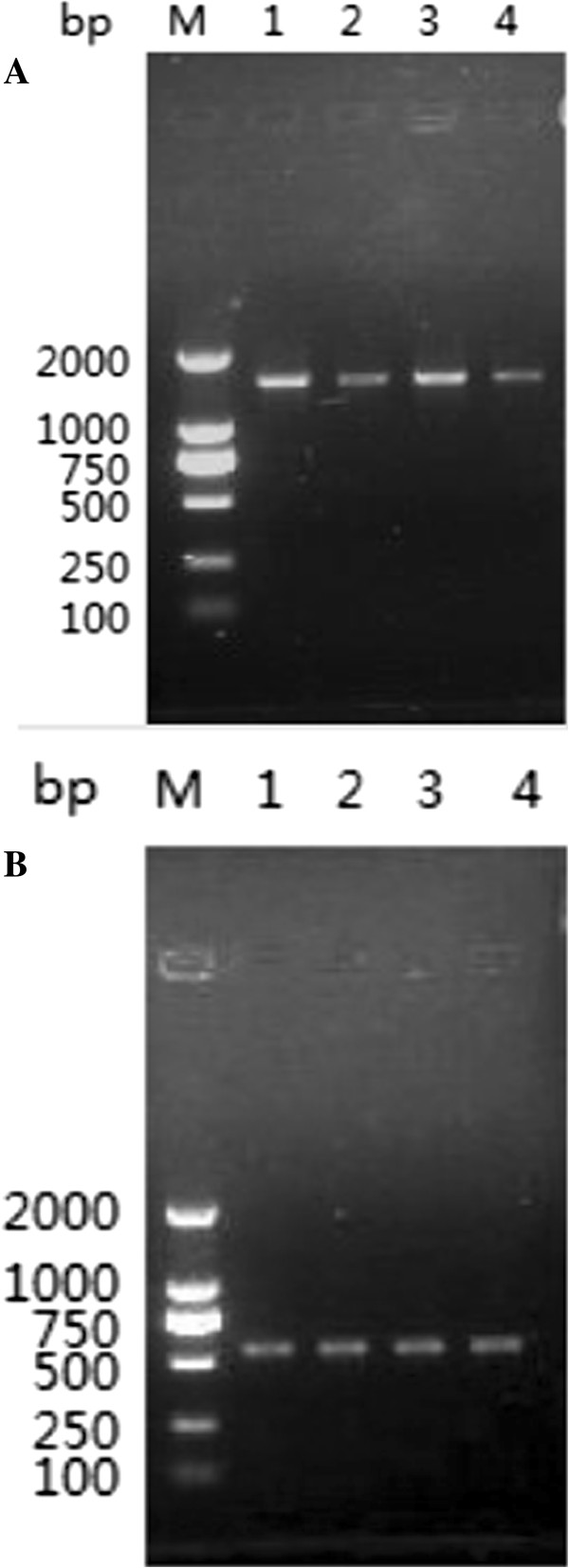
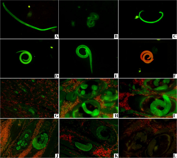
Methods: T. spiralis aminopeptidase (TsAP) gene encoding a 54.7 kDa protein was cloned and expressed in Escherichia coli, and purified recombinant TsAP protein was used to immunize BALB/c mice. The antibodies obtained were used to determine where TsAP was localized in the parasite. Transcription and expression of TsAP in different developmental stages of T. spiralis were observed by RT-PCR and Immunofluorescence test (IFT). The immune protection of recombinant TsAP protein against T. spiralis infection in BALB/c mice was evaluated.

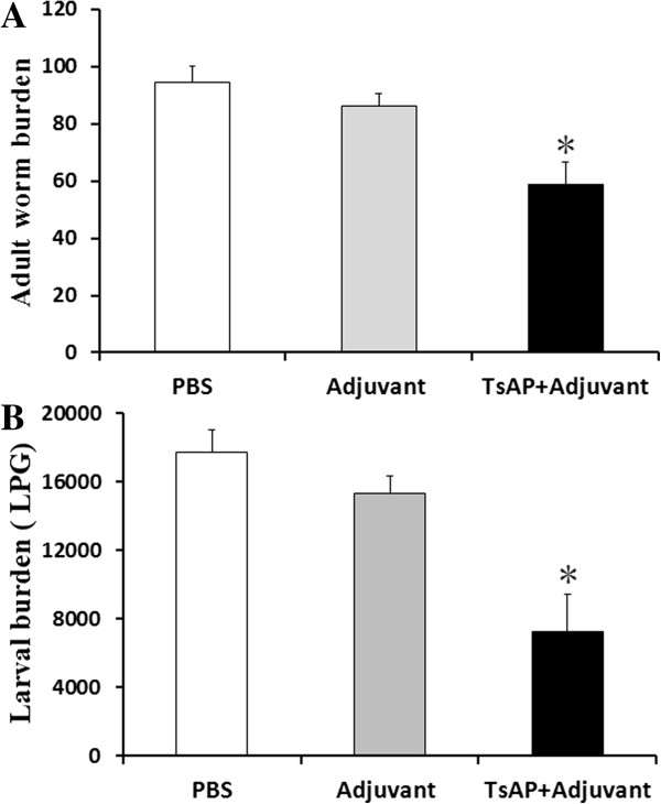
Results: Anti-TsAP antibodies recognized the native protein migrating at 54.7 kDa by Western blotting of the crude antigens from muscle larvae. Transcription and expression of TsAP gene was observed in different developmental stages (adult worms, newborn larvae, pre-encapsulated larvae and muscle larvae). TsAP appears to be a cytoplasmic protein located primarily at the cuticle and internal organs of this parasite. After a challenge infection with T. spiralis infective larvae, mice immunized with the recombinant TsAP protein displayed a 38.1% reduction in adult worm burden and 59.1% reduction in muscle larval burden.

Conclusions: In this study, T. spiralis aminopeptidase (TsAP) was first characterized and will help reveal its potential biological functions. TsAP is a novel potential vaccine candidate antigen that merits further investigation.


Figures

Figure 1
Figure 1
Identification of recombinant TsAP protein. (A) SDS-PAGE analysis of recombinant TsAP protein. M: protein molecular weight marker; 1: the lysis of the induced recombinant bacteria after ultrasonication; 2: TsAP inclusion body dissolved in 8 M urea; 3: recombinant TsAP protein purified by GST Sefinose Resin. (B) Western-blot analysis of recombinant TsAP protein antigenicity. The T. spiralis crude antigens (lane 1), ES antigens (lane 2), and recombinant TsAP protein (lane 3) were recognized by sera of mice infected with T. spiralis at 30 dpi. The native TsAP protein in crude antigens (lane 4) and recombinant TsAP protein (lane 6) were recognized by anti-TsAP serum, but the ES antigens (lane 5) were not recognized by anti-TsAP serum. The T. spiralis crude antigens (lane 7), ES antigens (lane 8), and recombinant TsAP protein (lane 9) were not recognized by sera of normal mice.
Figure 2
Figure 2
The specific IgG antibody levels of mice immunized with recombinant TsAP protein assayed by ELISA using different antigens. The optical density (OD) values shown for each group are the mean ± standard deviation (SD) of antibody levels (n = 13). Asterisks (*) indicate statistically significant differences (P < 0.01) in OD values of one kind of antigens compared to other two kinds of antigens.
Figure 3
Figure 3
RT-PCR analysis of TsAP gene transcript at different stages of T. spiralis. RT-PCR detection of mRNA transcription for the TsAP gene (A) and GAPDH gene (B) at different developmental stages of T. spiralis. M: DNA marker; Lane 1: AW at 3 dpi; Lane 2: ML at 42 dpi; Lane 3: PEL at 19 dpi; Lanes 4: NBL.
Figure 4
Figure 4
Expression of TsAP at different developmental stages and immunolocalization in T. spiralis. A–H: The results of IFT with whole parasite of T. spiralis different stages reacted with anti-TsAP serum. The notable immunostaining is found in the body of AW at 3 dpi (A), NBL (B), PEL at 19 dpi (C) and ML at 42 dpi (D). The ML at 42 dpi reacted with serum of mice infected T. spiralis at 30 dpi (E) as a positive control; ML at 42 dpi did not show recognition by normal serum (F) as a negative control. G–L: The results of IFT with the sections of skeletal muscles of infected mice reacted with anti-TsAP serum. The immunostaining is seen at the cuticle and internal organs of PEL at 19 dpi (G) and ML at 42 dpi (H and I). The ML at 42 dpi reacted with anti-ES serum (J) and serum of mice infected T. spiralis at 30 dpi (K) as a positive control, ML at 42 dpi did not did not show recognition by normal mouse serum (L) as a negative control.
Figure 5
Figure 5
The number of adult worms (A) and larvae per gram (lpg) muscles (B) recovered from vaccinated mice after a challenge infection with 300T. spiralis larvae. Results are presented as the arithmetic mean ± standard deviation (SD) of eight mice in each group. Asterisks (*) indicate statistically significant differences (P < 0.01) in worm recovery of the immunized group compared to both control groups.

Similar articles

Cited by

References

    1. Despommier DD. How does Trichinella spiralis make itself at home? Parasitol Today. 1998;14(8):318–323. doi: 10.1016/S0169-4758(98)01287-3. - DOI - PubMed
    1. Bruschi F, Chiumiento L. Trichinella inflammatory myopathy: host or parasite strategy? Parasit Vectors. 2011;4:42. doi: 10.1186/1756-3305-4-42. - DOI - PMC - PubMed
    1. Bruce RG. Structure of the esophagus of the infective juvenile and adult Trichinella spiralis. J Parasitol. 1970;56(3):540–549. doi: 10.2307/3277622. - DOI - PubMed
    1. Inaba T, Sato H, Kamiya H. Monoclonal IgA antibody-mediated expulsion of Trichinella from the intestine of mice. Parasitology. 2003;126(6):591–598. doi: 10.1017/S003118200300310X. - DOI - PubMed
    1. ManWarren T, Gagliardo L, Geyer J, McVay C, Pearce-Kelling S, Appleton J. Invasion of intestinal epithelia in vitro by the parasitic nematode Trichinella spiralis. Infect Immun. 1997;65(11):4806–4812. - PMC - PubMed

Publication types