. 2013 Dec;34(36):9201-9.
doi: 10.1016/j.biomaterials.2013.08.007. Epub 2013 Aug 22.

Enhancing microvascular formation and vessel maturation through temporal control over multiple pro-angiogenic and pro-maturation factors

Affiliations

Enhancing microvascular formation and vessel maturation through temporal control over multiple pro-angiogenic and pro-maturation factors

Yevgeny Brudno et al. Biomaterials. 2013 Dec.

Abstract

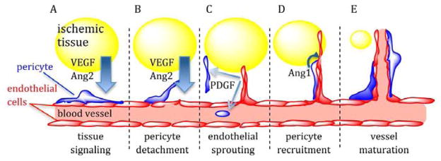
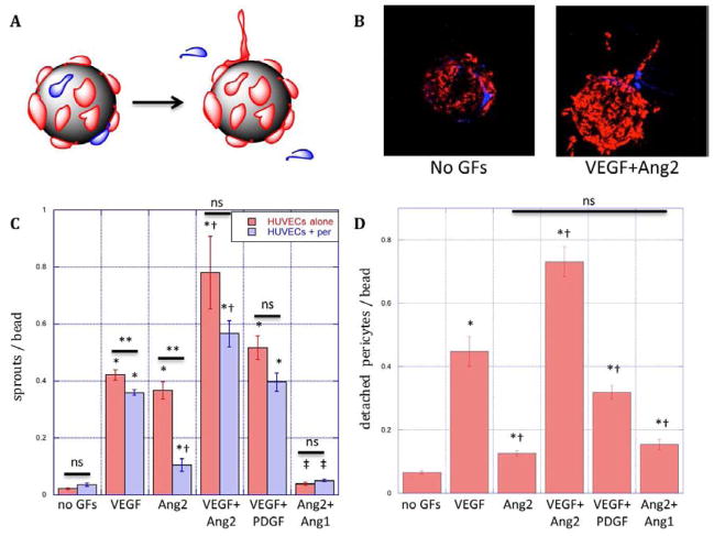
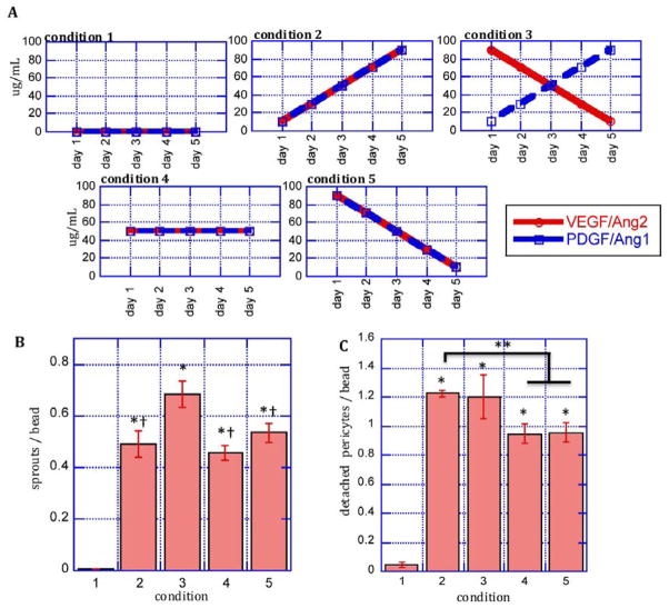
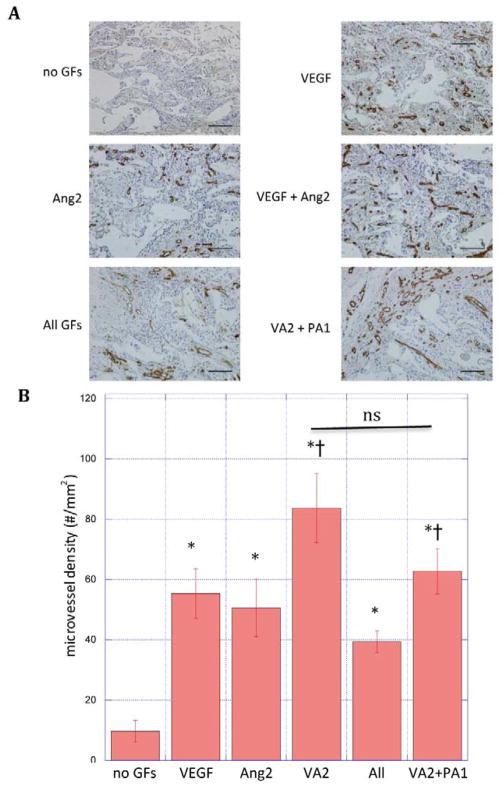
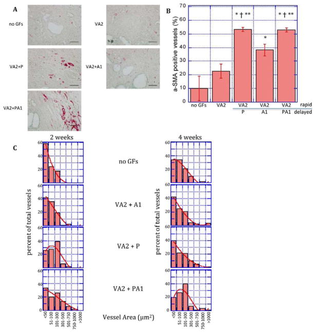
Therapeutic stimulation of angiogenesis to re-establish blood flow in ischemic tissues offers great promise as a treatment for patients suffering from cardiovascular disease or trauma. Since angiogenesis is a complex, multi-step process, different signals may need to be delivered at appropriate times in order to promote a robust and mature vasculature. The effects of temporally regulated presentation of pro-angiogenic and pro-maturation factors were investigated in vitro and in vivo in this study. Pro-angiogenic factors vascular endothelial growth factor (VEGF) and angiopoietin 2 (Ang2) cooperatively promoted endothelial sprouting and pericyte detachment in a three-dimensional in vitro EC-pericyte co-culture model. Pro-maturation factors platelet-derived growth factor B (PDGF) and angiopoietin 1 (Ang1) inhibited the early stages of VEGF- and Ang2-mediated angiogenesis if present simultaneously with VEGF and Ang2, but promoted these behaviors if added subsequently to the pro-angiogenesis factors. VEGF and Ang2 were also found to additively enhance microvessel density in a subcutaneous model of blood vessel formation, while simultaneously administered PDGF/Ang1 inhibited microvessel formation. However, a temporally controlled scaffold that released PDGF and Ang1 at a delay relative to VEGF/Ang2 promoted both vessel maturation and vascular remodeling without inhibiting sprouting angiogenesis. Our results demonstrate the importance of temporal control over signaling in promoting vascular growth, vessel maturation and vascular remodeling. Delivering multiple growth factors in combination and sequence could aid in creating tissue engineered constructs and therapies aimed at promoting healing after acute wounds and in chronic conditions such as diabetic ulcers and peripheral artery disease.

Keywords: Angiogenesis; Controlled drug release; Drug delivery; Endothelial cell; Growth factors; Smooth muscle cell.


Figures

Figure 1
Figure 1
Model of select growth factor signaling during angiogenesis. (A) Ischemic tissues (yellow) release pro-angiogenic factors such as VEGF and Ang2, creating growth factor gradients that signal blood vessels to increase capacity. (B) Pro-angiogenic factor signaling destabilizes EC-pericyte interactions, promoting pericyte detachment from the endothelium and EC sprouting away from existing vessels. (C) As sprouts grow and penetrate hypoxic tissues, they release PDGF, which activates and recruits pericytes from surrounding tissue and progenitor cells from the blood stream to nascent vessels. (D) Pericyte-derived Ang1 antagonizes the Ang2 receptor (Tie2) and serves as a stabilizing factor that strengthens pericyte-EC interactions and promotes vessel maturation and lumen formation (E).
Figure 2
Figure 2
Schematic (A) and microscopy (B) images of R18-labeled endothelial cell (red) sprouting and CMFDA-labeled pericyte (blue) detachment from microcarriers in response to growth factor signals. (C) Endothelial cell sprouting from microcarriers over three days with EC (red bars) or EC/pericytes (blue bars) cultures in fibrin in response to VEGF (50ng/mL), Ang2 (250ng/mL), combined VEGF(50ng/mL) and Ang2(250ng/mL, VA2), combined VEGF and PDGF(both 50ng/mL, VP) or combined Ang2 and Ang1 (both 250ng/mL, Ang1). (D) Pericyte migration away from endothelium on microcarriers with EC/pericyte co-cultures in response to conditions specified in C. *: statistically significant relative to no GF control. †: statistically significant relative to VEGF; ‡: statistically significant relative to Ang2; **: statistically significant between presence and absence of pericytes; ns: not significant by Student’s T-test. Data represent mean and S.E.M.
Figure 3
Figure 3
Early angiogenic responses to temporally regulated growth factor presentation. A: EC/pericyte co-cultured on microcarriers in fibrin gels were subjected to different growth factor conditions over five days. B: Quantification of endothelial cell sprouting and C: quantification pericyte detachment from the endothelium in response to growth factor conditions in A. *: statistically significant relative to condition 1; †: statistically significant relative to condition 3; **: statistically significant relative to condition 2 by Student’s T-test. Data represent mean and S.E.M.
Figure 4
Figure 4
(A) Photomicrographs of hematoxylin and CD31 stained section of scaffolds and (B) quantification of microvessel density in PLG scaffolds subcutaneously implanted in mice for two weeks. Scaffolds (n = 3) containing no growth factors (no GFs), rapidly releasing VEGF, Ang2, VEGF and Ang2 simultaneously (VA2), VEGF, Ang2, PDGF and Ang1 all simultaneously (all GF), or rapidly releasing VEGF and Ang2 followed by delayed release of PDGF and Ang1 (VA2+PA1). Data represent mean and S.E.M. *: statistically significant relative to no GFs; † statistically significant relative to All GFs; ns: not significant by Student’s T-test. scale bar: 100μm
Figure 5
Figure 5
Delayed release of pro-maturation factors increases mural cell association and vascular remodeling. A: Photomicrographs of α-SMA-stained sections of scaffolds implanted subcutaneously releasing no growth factors (no GFs), VEGF/Ang2 (VA2) alone or followed by a delayed release of PDGF (P), Ang1 (A1), or both (PA1) at two weeks. B: Quantification of the percentage of blood vessels associating with smooth muscle cells determined by α-smooth muscle actin staining of tissue sections from subcutaneously-implanted scaffolds C: Cross-sectional area of vessels formed by rapid delivery of VEGF/Ang2 (VA2) followed by delayed release of PDGF (P), Ang1 (A1), or both (PA1) at two and four weeks. Values represent mean and S.E.M. *, †, ** Denote p<0.05 relative to blank, VA2, and VA2+A1, respectively by Student’s T-test. Scale bar = 100μm

References

    1. Attanasio S, Snell J. Therapeutic angiogenesis in the management of critical limb ischemia: current concepts and review. Cardiol Rev. 2009;17:115–20. - PubMed
    1. Freedman S, Isner J. Therapeutic angiogenesis for ischemic cardiovascular disease. J Mol Cell Cardiol. 2001;33:379–93. - PubMed
    1. Losordo D, Dimmeler S. Therapeutic angiogenesis and vasculogenesis for ischemic disease: part II: cell-based therapies. Circulation. 2004;109:2692–7. - PubMed
    1. Silva E, Mooney D. Spatiotemporal control of vascular endothelial growth factor delivery from injectable hydrogels enhances angiogenesis. J Thromb Haemost. 2007;5:590–8. - PubMed
    1. Silva E, Mooney D. Effects of VEGF temporal and spatial presentation on angiogenesis. Biomaterials. 2010;31:1235–41. - PMC - PubMed

Publication types

MeSH terms

Substances