. 2013 Aug 23:10:105.
doi: 10.1186/1742-2094-10-105.

The growth factor progranulin attenuates neuronal injury induced by cerebral ischemia-reperfusion through the suppression of neutrophil recruitment

Affiliations

The growth factor progranulin attenuates neuronal injury induced by cerebral ischemia-reperfusion through the suppression of neutrophil recruitment

Yusuke Egashira et al. J Neuroinflammation. .

Abstract

Background: To improve the clinical outcome of patients who suffered ischemic stroke, cerebral ischemia-reperfusion (I/R) injury is one of the major concerns that should be conquered. Inflammatory reactions are considered a major contributor to brain injury following cerebral ischemia, and I/R exacerbates these reactions. The aim of this study was to investigate the possible ameliorative effects of progranulin (PGRN) against I/R injury in mice.

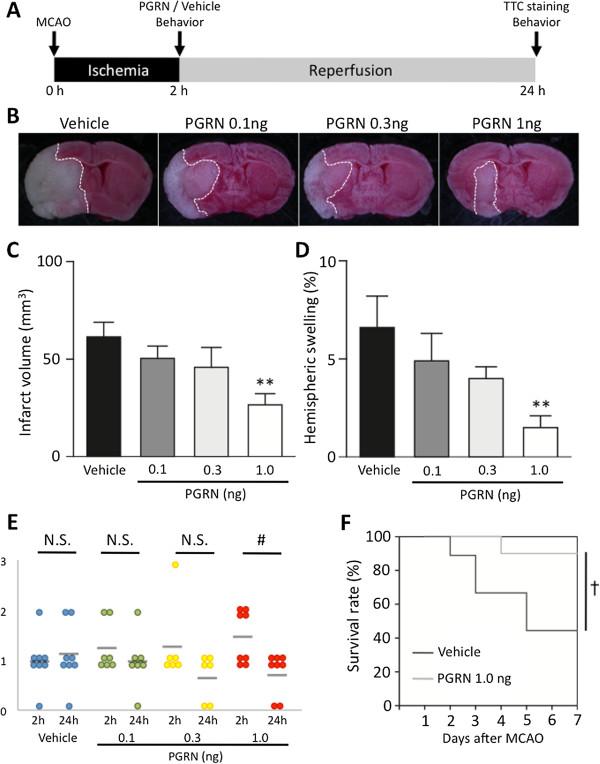
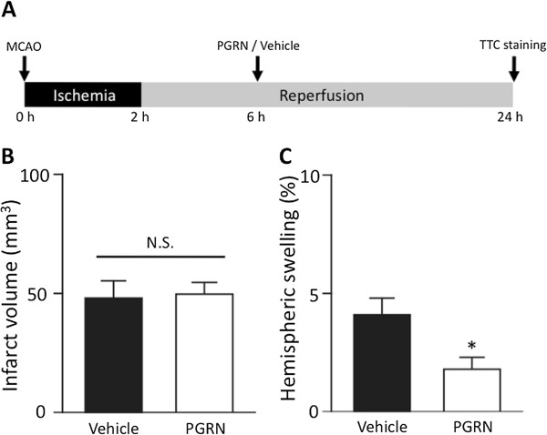
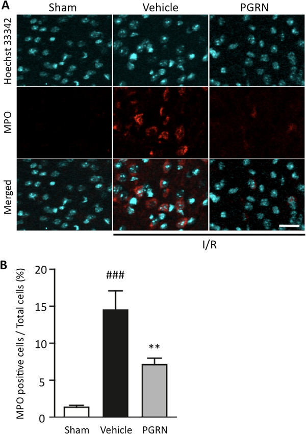
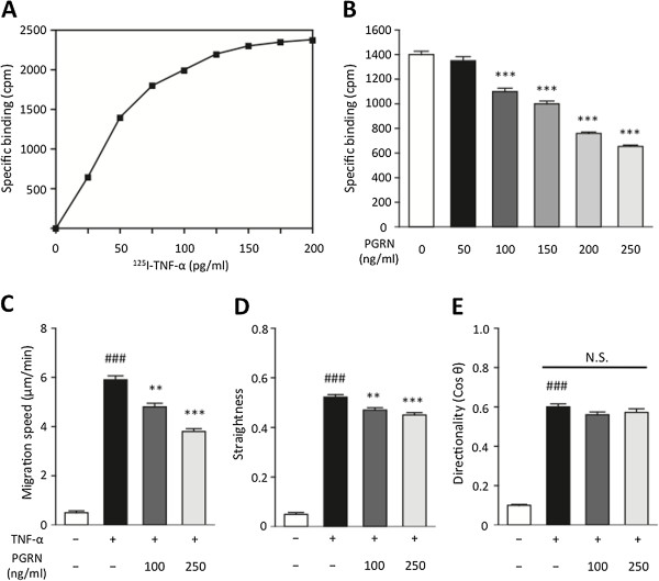
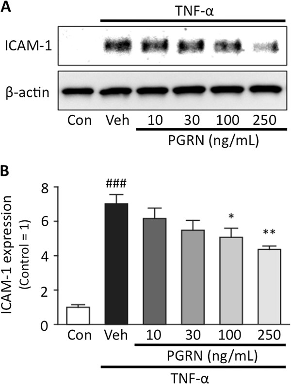
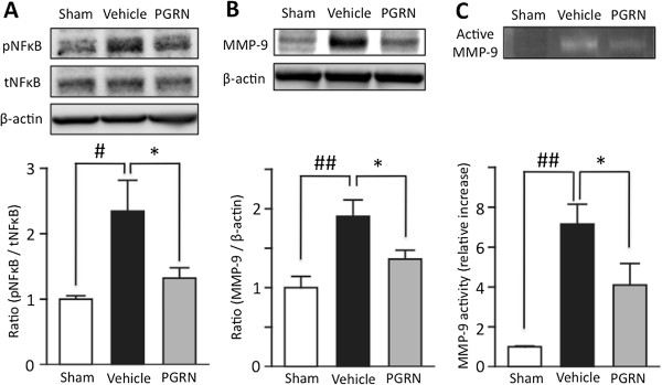
Methods: In vivo I/R was induced in four-week-old male ddY mice by 2 h of MCAO (middle cerebral artery occlusion) followed by 22 h of reperfusion. We evaluate expression of PGRN in I/R brain, efficacy of recombinant-PGRN (r-PGRN) treatment and its therapeutic time-window on I/R injury. Two hours after MCAO, 1.0 ng of r-PRGN or PBS was administered via intracerebroventricular. We assess neutrophil infiltration, expression of tumor necrosis factor (TNF)-α, matrix metalloproteinase-9 (MMP-9) and phosphorylation of nuclear factor-κB (NF-κB) by immunofluorescense staining and Western blotting. We also investigate neutrophil chemotaxis and intercellular adhesion molecule-1 (ICAM-1) expression in vitro inflammation models using isolated neutrophils and endothelial cells.

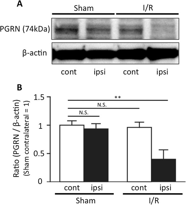
Results: We found that expression of PGRN was decreased in the I/R mouse brain. r-PGRN treatment at 2 h after MCAO resulted in a reduction in the infarct volume and decreased brain swelling; this led to an improvement in neurological scores and to a reduction of mortality rate at 24 h and 7 d after MCAO, respectively. Immunohistochemistry, Western blotting, and gelatin zymography also confirmed that r-PGRN treatment suppressed neutrophil recruitment into the I/R brain, and this led to a reduction of NF-κB and MMP-9 activation. In the in vitro inflammation models, PGRN suppressed both the neutrophil chemotaxis and ICAM-1 expression caused by TNF-α in endothelial cells.

Conclusions: PGRN exerted ameliorative effects against I/R-induced inflammation, and these effects may be due to the inhibition of neutrophil recruitment into the I/R brain.


Figures

Figure 1
Figure 1
Expression of progranulin in the ischemia-reperfusion injured brain. Progranulin (PGRN) expression was significantly decreased following ischemia-reperfusion (I/R) insults. (A) Representative PGRN bands from the Western blotting analysis of brain tissue taken from sham-operated and I/R animals; ipsilateral and contralateral hemispheres to the middle cerebral artery occlusion (MCAO). (B) Optical densitometry quantification of PGRN protein levels, normalized to β-actin. In the I/R brain, the expression of PGRN was significantly decreased 24 h after the induction of transient cerebral ischemia. **P <0.01 vs. sham contralateral brain; one-way ANOVA followed by Dunnett's test; n = 4 for each group.
Figure 2
Figure 2
r-PGRN treatment reduces cerebral infarct volume and brain edema in transient focal cerebral ischemia. (A) Protocol for surgery and r-PGRN administration. Intracerebroventricular (i.c.v.) injections of either vehicle or r-PGRN (0.1 to 1.0 ng) were administered 2 h after middle cerebral artery occlusion (MCAO). All assessments, with the exception of survival rate evaluation, were performed at 24 h after the induction of 2 h of transient MCAO. (B) Representative photograph showing TTC staining of coronal brain sections 24 h after MCAO in each treatment group. (C) Administration of 1 ng of r-PGRN significantly reduced the infarct volume, (D) and reduced brain edema, compared to the vehicle treatment. Although the 0.1 ng r-PGRN- and 0.3 ng r-PGRN-treated groups tended to experience reduced infarct volume and brain edema, the difference was not statistically significant. * P <0.05 vs. vehicle-treated group; one-way ANOVA followed by Dunnett's test; n = 6 to n = 8 for each group. (E) Only the 1.0 ng r-PGRN-treated group had significantly better neurological function at 24 h after MCAO than at 2 h after MCAO. # P <0.05; Wilcoxon signed-rank test. (F) A higher survival rate was observed throughout the follow-up period in the 1.0 ng r-PGRN-treated group. In contrast, a continuous reduction of the survival rate was observed in the vehicle-treated group. The difference between the groups was statistically significant. † P <0.05; Log-rank test; n = 9 or n =10 for each group. r-PGRN, recombinant-progranulin.
Figure 3
Figure 3
The effects of delayed administration of r-PGRN 6 h after transient MCAO. (A) Protocol for surgery and PGRN administration. Injections (i.c.v.) of either vehicle or r-PGRN (1.0 ng) were administered 6 h after the MCAO procedure. All assessments were performed at 24 h after the induction of 2 h of transient MCAO. (B) Administration of 1 ng of r-PGRN 6 h after MCAO did not reduce the infarct volume assessed at 24 h after the induction of 2 h of MCAO; (C) however, it significantly reduced brain edema. N.S. not significant; * P <0.05 vs. vehicle-treated group; Student's t-test; n = 8 or n = 9 for each group. i.c.v., intracerebroventricular; MCAO, middle cerebral artery occlusion; PGRN, progranulin; r-PGRN, recombinant-progranulin.
Figure 4
Figure 4
r-PGRN treatment significantly suppresses neutrophil recruitment into the I/R brain following MCAO. (A) Representative immunohistochemical staining for myeloperoxidase (MPO) in each of the areas of interest in the sham-operation, vehicle-treated and r-PGRN-treated groups. (B) Quantification of MPO-immunoreactive cells. The number of MPO-positive cells was significantly higher in the vehicle-treated mice than in the r-PGRN-treated mice. Scale bar = 20 μm. ## P <0.01 vs. sham-operation mice; ** P <0.01 vs. vehicle-treated mice; Student's t-test. n = 4 or n = 5 for each group. I/R, ischemia-reperfusion; MCAO, middle cerebral artery occlusion; r-PGRN, recombinant-progranulin.
Figure 5
Figure 5
PGRN inhibits 125I-TNF-α binding to neutrophil surfaces and suppresses neutrophil chemotaxis induced by TNF-α. (A) Saturation curve for specific 125I-TNF-α binding to neutrophil surfaces was determined, and in accordance with these results, 50 pg/mL of 125I-TNF-α was used in the subsequent experiments. (B) The 125I-TNF-α binding significantly decreased with increasing concentrations of PGRN. ***P <0.001 vs. 0 ng/mL of PGRN group; one-way ANOVA followed by Dunnett's test. Data were obtained from three independent experiments and presented as mean ± SEM. (C, D, E) Neutrophil chemotaxis was induced by TNF-α, and PGRN was found to significantly suppress this effect in a concentration-dependent manner; at 100 and 250 ng/mL of PGRN attenuates the migration speed and straightness of the route of migration, but did not affect the directionality of migration. ### P <0.001 vs. control group; Student t-test; ** P <0.01, *** P <0.001 vs. TNF-α only group; one-way ANOVA followed by Dunnett's test; n = 5 for each group. PGRN, progranulin; TNF-α, tumor necrosis factor-alpha.
Figure 6
Figure 6
PGRN ameliorates TNF-α-induced inflammation in hBMVECs. (A) Representative bands from the Western blotting analysis of ICAM-1 and β-actin. (B) Optical densitometry quantification of ICAM-1, normalized to β-actin. TNF-α (10 ng/mL) induced an approximately eight-fold increase in ICAM-1 in hBMVECs after a 20-h exposure. ### P <0.001 vs. control group; Student's t-test. PGRN significantly suppressed TNF-α-induced ICAM-1 expression in a concentration-dependent manner. * P <0.05, ** P <0.01 vs. vehicle-treated group; one-way ANOVA followed by Dunnett's test; n = 4 for each group. hBMVECs, human brain microvascular endothelial cells; ICAM-1, intercellular adhesion molecule-1; PGRN, progranulin; TNF-α, tumor necrosis factor-alpha.
Figure 7
Figure 7
PGRN significantly suppresses the expression of MMP-9, and the phosphorylation of NF-κB in I/R brain. (A) Representative bands from Western blotting analysis of phosphorylated and total NF-κB (upper). Optical densitometry quantification for the phosphorylation of NF-κB (p NF-κB), normalized to total NF-κB (tNF-κB) and β-actin (lower). In the I/R brain, phosphorylation of NF-κB was significantly increased. ## P <0.01 vs. sham control group; Student's t-test. PGRN significantly suppressed this increased phosphorylation of NF-κB induced by I/R. * P <0.05 vs. vehicle-treated group; Student t-test. (B) Representative bands from Western blotting analysis of MMP-9 expression (upper). Optical densitometry quantification of MMP-9 expression, normalized to β-actin (lower). MMP-9 expression was significantly increased in the I/R brain. ## P <0.01 vs. sham control group; Student's t-test. PGRN significantly suppressed the expression of MMP-9 induced by I/R. * P <0.05 vs. vehicle-treated group; Student's t-test; n = 5 for each group. (C) Representative bands from gelatin zymography for activated MMP-9 (upper). Optical densitometry quantification of activated MMP-9 (lower). Activated MMP-9 was significantly increased in the I/R brain. ## P <0.01 vs. sham control group; Student's t-test. PGRN significantly suppressed the activation of MMP-9 induced by I/R. * P <0.05 vs. vehicle-treated group; Student's t-test; n = 3 for sham or n = 4 for each treated group. I/R, ischemia-reperfusion; MMP-9, matrix metalloproteinase-9; NF-κB, nuclear factor-κappaB; PGRN, progranulin.

References

    1. Roger VL, Go AS, Lloyd-Jones DM, Benjamin EJ, Berry JD, Borden WB, Bravata DM, Dai S, Ford ES, Fox CS, Fullerton HJ, Gillespie C, Hailpern SM, Heit JA, Howard VJ, Kissela BM, Kittner SJ, Lackland DT, Lichtman JH, Lisabeth LD, Makuc DM, Marcus GM, Marelli A, Matchar DB, Moy CS, Mozaffarian D, Mussolino ME, Nichol G, Paynter NP, Soliman EZ. et al. Heart disease and stroke statistics–2012 update: a report from the American heart association. Circulation. 2012;125:e2–e220. - PMC - PubMed
    1. Iadecola C, Anrather J. The immunology of stroke: from mechanisms to translation. Nat Med. 2011;17:796–808. doi: 10.1038/nm.2399. - DOI - PMC - PubMed
    1. Jean WC, Spellman SR, Nussbaum ES, Low WC. Reperfusion injury after focal cerebral ischemia: the role of inflammation and the therapeutic horizon. Neurosurgery. 1998;43:1382–1396. discussion 1396–1387. - PubMed
    1. Kriz J. Inflammation in ischemic brain injury: timing is important. Crit Rev Neurobiol. 2006;18:145–157. doi: 10.1615/CritRevNeurobiol.v18.i1-2.150. - DOI - PubMed
    1. Lambertsen KL, Biber K, Finsen B. Inflammatory cytokines in experimental and human stroke. J Cereb Blood Flow Metab. 2012;32:1677–1698. doi: 10.1038/jcbfm.2012.88. - DOI - PMC - PubMed

Publication types

MeSH terms