. 2013 Aug 20;105(4):880-6.
doi: 10.1016/j.bpj.2013.07.019.

Human PIEZO1: removing inactivation

Affiliations

Human PIEZO1: removing inactivation

Chilman Bae et al. Biophys J. .

Abstract

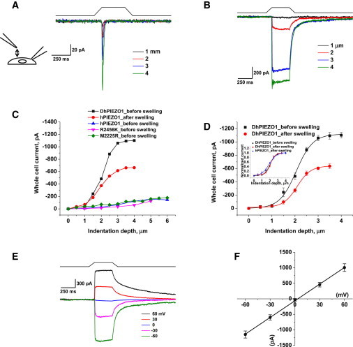
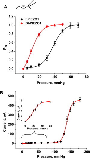
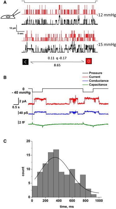
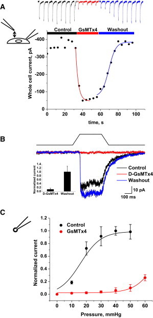
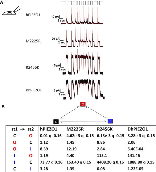
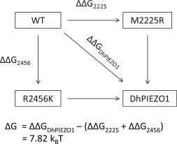
PIEZO1 is an inactivating eukaryotic cation-selective mechanosensitive ion channel. Two sites have been located in the channel that when individually mutated lead to xerocytotic anemia by slowing inactivation. By introducing mutations at two sites, one associated with xerocytosis and the other artificial, we were able to remove inactivation. The double mutant (DhPIEZO1) has a substitution of arginine for methionine (M2225R) and lysine for arginine (R2456K). The loss of inactivation was accompanied by ∼30-mmHg shift of the activation curve to lower pressures and slower rates of deactivation. The slope sensitivity of gating was the same for wild-type and mutants, indicating that the dimensional changes between the closed and open state are unaffected by the mutations. The unitary channel conductance was unchanged by mutations, so these sites are not associated with pore. DhPIEZO1 was reversibly inhibited by the peptide GsMTx4 that acted as a gating modifier. The channel kinetics were solved using complex stimulus waveforms and the data fit to a three-state loop in detailed balance. The reaction had two pressure-dependent rates, closed to open and inactivated to closed. Pressure sensitivity of the opening rate with no sensitivity of the closing rate means that the energy barrier between them is located near the open state. Mutant cycle analysis of inactivation showed that the two sites interacted strongly, even though they are postulated to be on opposite sides of the membrane.


Figures

Figure 1
Figure 1
DhPIEZO1 does not inactivate. (A) Whole-cell currents of WT hPIEZO1 rapidly inactivate. (B) Whole-cell currents of DhPIEZO1 as a function of indentation depth showed no inactivation. Note the slow rate of deactivation after the stimulus is removed. (C) WT channels are sensitive to hypotonic swelling. Prior to swelling, there was less current for the same incremental indentation than after swelling. Also shown is the response of DhPIEZO1 prior to swelling. (D) Comparison of DhPIEZO1 responses before and after hypotonic swelling with 50% osmolarity. The inset shows the same data normalized. The sensitivity to indentation is similar, suggesting that the channels are already near a saturated level of stress in the resting state. (E) Deactivation is extremely slow and voltage independent. Deactivation for this channel may represent the kinetics of domain reformation rather than channel kinetics. (F) The I/V curve of peak currents shows a reversal potential near 0 mV for DhPIEZO1, indicating that the mutations do not affect the pore.
Figure 2
Figure 2
(A) The gating curve as a function of pressure fit to a Boltzmann. p1/2 and the slope sensitivity q are indicated in the table. q is a measure of the dimensional change between closed and open states. q is the same for DhPIEZO1 (n = 12), WT (n = 18), and the single-site mutants, implying that the mutations have no effect on the key activation processes. However, p1/2 was left-shifted relative to WT, representing a change in the intrinsic stress of the channel environment favoring the opening state. (B) To calibrate the absolute stress sensitivity, we cotransfected cells with bacterial MscL (7) and DhPIEZO1. Fit to a sum of two Boltzmanns, the slope sensitivities of both channels were nearly identical (n = 5), meaning that the dimensional changes of both channels are similar and also similar to WT and single-site mutants. The dimensional changes are equivalent to an in-plane area change of 20 nm2.The inset shows an expansion of the region containing DhPIEZO1’s response.
Figure 3
Figure 3
(A) DhPIEZO1 single-channel currents show high pressure sensitivity because of the left shift in the gating curve. The current trace is shown in black and the theoretical fit is in red. With a change of only 3 mmHg, there is a significant increase in the number of open channels. The kinetics are well fit by a two-state model with only the activation rate being pressure dependent. (B) Single-channel currents of DhPIEZO1 have a pronounced latency for activation, and this occurs with no significant change in patch capacitance, suggesting that the latency does not arise from large changes in the patch structure. The capacitance measuring noise level was 0.12 fF RMS, equivalent to ∼0.012 μm2 (assuming a specific capacitance of 1 μF/cm2, or 10 fF/μm2). Note the spontaneous (background) channel openings of DhPIEZO1 during the recording that is a result of its higher absolute sensitivity and tension from the gigaseal. (C) The distribution of latencies fit to a Gaussian gives a mean latency of 344 ± 133 ms. We attribute these latencies to the time required for domain fracture under stress.
Figure 4
Figure 4
DhPIEZO1 channels in whole-cell patches were reversibly inhibited by 10 μM extracellular D-GsMTx4. (A) By fitting exponentials, we extracted a mean association time constant of 3.0 ± 0.6 s and a mean dissociation time constant of 13.4 ± 0.8 s (n = 4). The estimated association and dissociation rates are 2.6 × 104 M−1 s−1 and 0.08 s−1, respectively, and the equilibrium constant calculated from the ratio is KD ∼3 μM. (B) D-GsMTx4 inhibition of DhPIEZO1 in the absence of inactivation. This suggests that the mechanism of action of D-GsMTx4 does not involve inactivation domains. The bar graph (inset) shows the average peak currents ± SD (n = 3) illustrated in Fig. 4B. (C) The dose–response relationship shifts to higher stress with GsMTx4, as expected for a gating modifier (12).
Figure 5
Figure 5
Channel kinetics. (A) Multichannel currents for different types of channels. The stimulus was a series of square pressure pulses applied with varying off intervals (typically 3.0, 2.0, 1.0, 0.5, 0.25, 0.1 s, and the reverse, top trace). Pressure pulses were 0 to −70 mmHg for HPIEZO1, 2555R PIEZO1, and 2456K PIEZO. For DhPIEZO1, they were 0 to –40 mmHg. The data trace is in black and the QuB fit in red. Notice how hPIEZO1 effectively summed currents from the applied stresses at short off-times of the stimulus. However, the mutant M2225R tended to accumulate inactivation in the same part of the stimulus (lower peaks at shortest resting intervals), but this can be accounted for simply by a change in the rate constants and requires no additional states. The kinetic parameters that characterize the behavior of all channels are presented in (B). (B) Tabulation of the quantified kinetics with the three-state loop in detailed balance. The states are named C =closed state, O = open state, and I = inactivated state. Although DhPIEZO1 does not appear to have an inactivated state, we included it for consistency to better compare the models. The pressure dependence for all channel types is contained in the opening rate and the inactivated-closed rate. The pressure sensitivity of the rates is indicated by the parameter q [mmHg−1]. q was consistent across all types of channels at ∼0.15 so that the conformation associated with opening in all channel types is identical. The DhPIEZO1 trace ends with a jump that is probably closure of the last open channel.
Figure 6
Figure 6
Mutant cycle analysis. The energy difference between open and inactivated states for WT, single-site mutants, and DhPIEZO1. The free energy change in DhPIEZO1 (ΔΔGDhPIEZO1) is larger than the sum of free energy changes for the two single mutants by ∼8 kBT, showing that the sites are not independent but exhibit positive cooperativity.

References

    1. Coste B., Mathur J., Patapoutian A. Piezo1 and Piezo2 are essential components of distinct mechanically activated cation channels. Science. 2010;330:55–60. - PMC - PubMed
    1. Coste B., Xiao B., Patapoutian A. Piezo proteins are pore-forming subunits of mechanically activated channels. Nature. 2012;483:176–181. - PMC - PubMed
    1. Kim S.E., Coste B., Patapoutian A. The role of Drosophila Piezo in mechanical nociception. Nature. 2012;483:209–212. - PMC - PubMed
    1. Gottlieb P.A., Sachs F. Piezo1: properties of a cation selective mechanical channel. Channels (Austin) 2012;6:214–219. - PMC - PubMed
    1. Nilius B., Honoré E. Sensing pressure with ion channels. Trends Neurosci. 2012;35:477–486. - PubMed

Publication types

Supplementary concepts