. 2013:115:327-342.
doi: 10.1016/B978-0-12-407757-7.00020-7.

Studying kinetochore-fiber ultrastructure using correlative light-electron microscopy

Affiliations

Studying kinetochore-fiber ultrastructure using correlative light-electron microscopy

Daniel G Booth et al. Methods Cell Biol. 2013.

Abstract

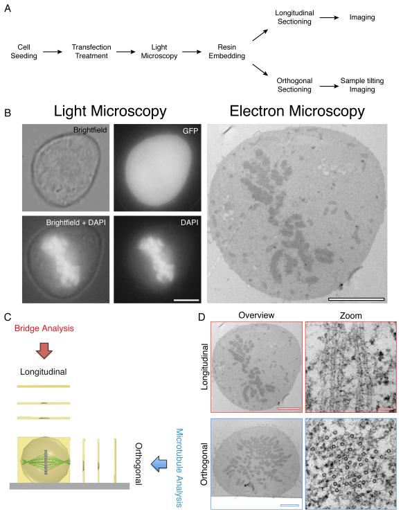
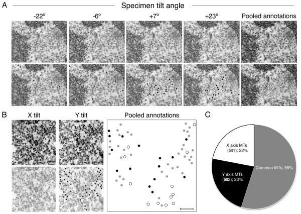
Electron microscopy (EM) has dominated high-resolution cellular imaging for over 50 years, thanks to its ability to resolve on nanometer-scale intracellular structures such as the microtubules of the mitotic spindle. It is advantageous to view the cell of interest prior to processing the sample for EM. Correlative light-electron microscopy (CLEM) is a technique that allows one to visualize cells of interest by light microscopy (LM) before being transferred to EM for ultrastructural examination. Here, we describe how CLEM can be applied as an effective tool to study the spindle apparatus of mitotic cells. This approach allows transfected cells of interest, in desirable stages of mitosis, to be followed from LM to EM. CLEM has often been considered as a technically challenging and laborious technique. In this chapter, we provide step-by-step pictorial guides that allow successful CLEM to be achieved. In addition, we explain how it is possible to vary the sectioning plane, allowing spindles and microtubules to be analyzed from different angles, and the outputs that can be obtained from these methods when applied to the study of kinetochore fiber ultrastructure.

Keywords: Correlative electron microscopy; Kinetochore-fiber; Microtubule; Mitosis; Mitotic spindle.


Figures

Figure 1
Figure 1. CLEM performed on mitotic cells.
(A) A workflow to achieve CLEM using longitudinal or orthogonal sectioning. (B) A transfected mitotic HeLa observed by LM (Brightfield, GFP and DAPI) and by electron microscopy. Scale bar 5 μm. (C) Schematic of Longitudinal and orthogonal EM sectioning, and examples of output analysis. (D) Representative electron micrographs of cells sectioned longitudinally (above) and orthogonally (below) with high magnification of microtubules (right). Scale bar 4 μm (overview) and 50 nm (zoom).
Figure 2
Figure 2. Optimization of mitotic spindle and cell structure preservation.
Orthogonal sections of cells fixed with 280, 440 or 1100 mOsm. Representative high magnification electron micrographs of the cytosol in each condition are shown below. Scale bars 5 μm (overview) and 100 nm (bottom).
Figure 3
Figure 3. Pictorial guide to CLEM processing for sectioning longitudinally to the spindle axis.
Following polymerization, resin was separated from the CLEM dish. Unwanted plastic was removed from the edges of the dish using pliers (A-C) allowing a razor to be inserted between the resin and the dish base (D). Following the separation of resin and dish (E) excess resin was removed using pliers (F) until just the capsule remained (G). The cell of interest was marked (H) with the aid of LM images (M) and resin coordinates (N). Unwanted resin was removed using a junior hacksaw (I) and a razor (J). Resin was trimmed using a microtome and a glass knife (K) until a neat block was generated at the top of a pyramid (L). Blocks were sectioned using a diamond knife (O) and ribbons collected using 100 mesh copper grids (P & Q), coated with formvar.
Figure 4
Figure 4. A pictorial guide to orthogonal CLEM processing.
Following polymerization, resin was separated from the CLEM dish (A-D). Unwanted plastic was removed from the edges of the CLEM dish using pliers (B) allowing a razor to be inserted between the resin and the dish base (C). Following separation (D) the position of the spindle was estimated using the reference LM images (E-G). These images allowed the re-orientation of the resin (H & I) so that an appropriate block could be marked (J) before excision using a junior hacksaw and a mitre block (K & L). The excised block (M) was inserted into a microtome chuck (N & O) and fine trimmed using a glass knife (P) before serial sections were taken of the cell, in the desired orientation (Q & R).
Figure 5
Figure 5. Sample tilting is necessary to achieve full coverage of microtubules in orthogonal sections.
(A) Example electron micrographs taken from a −45° to +45° tilt series of a K-fiber. Observable microtubules are marked with dots (bottom row). Black dots represent microtubules that are unique to that tilt frame. White dots represent the accumulating microtubules identified in previous tilt frames. The total number of microtubule annotations were pooled together onto one frame (far right) giving a fair overview of the whole K-fiber. Scale bar 100 nm. (B & C) A dual tilt series of one K-fiber was carried out. (B) Representative electron micrographs taken from the central region of both X and Y tilts (A-top). All microtubules observed in each tilt series were annotated (bottom; X axis in white, Y axis in black). The sum of microtubules from both tilts were pooled together on to a single blank image, any microtubules that were common to both tilts were marked grey. Scale bar, 100 nm. (C) A pie chart showing the percentage of total microtubules that were unique to each tilt and also the common ones.

References

    1. Booth DG, Hood FE, Prior IA, Royle SJ. A TACC3/ch-TOG/clathrin complex stabilises kinetochore fibres by inter-microtubule bridging. Embo Journal. 2011;30:906–919. DOI: 10.1038/emboj.2011.15. - PMC - PubMed
    1. Cheeseman LP, Booth DG, Hood FE, Prior IA, Royle SJ. Aurora A kinase activity is required for localization of TACC3/ch-TOG/clathrin inter-microtubule bridges. Communicative & integrative biology. 2011;4:409–12. - PMC - PubMed
    1. Ding R, McDonald KL, McIntosh JR. Three-dimensional reconstruction and analysis of mitotic spindles from the yeast, Schizosaccharomyces pombe. J Cell Biol. 1993;120:141–51. - PMC - PubMed
    1. Engelborghs Y, Heremans KAH, Demaeyer LCM, Hoebeke J. Effect of temperature and pressure on polymerization equilibrium of neuronal microtubules. Nature. 1976;259:686–689. DOI: 10.1038/259686a0. - PubMed
    1. Euteneuer U, McIntosh JR. Structural polarity of kinetochore microtubules in Ptk1-cells. Journal of Cell Biology. 1981;89:338–345. DOI: 10.1083/jcb.89.2.338. - PMC - PubMed

Publication types

LinkOut - more resources