. 2013 Oct 25:554:167-71.
doi: 10.1016/j.neulet.2013.08.025. Epub 2013 Aug 21.

Contribution of CA3 and CA1 pyramidal neurons to the tonic α7 nAChR-dependent glutamatergic input to CA1 pyramidal neurons

Affiliations

Contribution of CA3 and CA1 pyramidal neurons to the tonic α7 nAChR-dependent glutamatergic input to CA1 pyramidal neurons

Jyotirmoy Banerjee et al. Neurosci Lett. .

Abstract

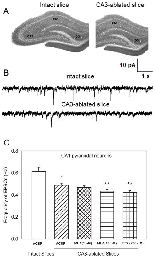
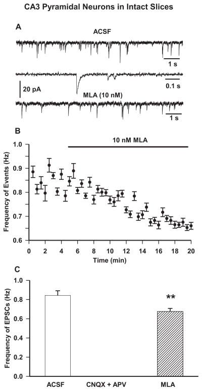
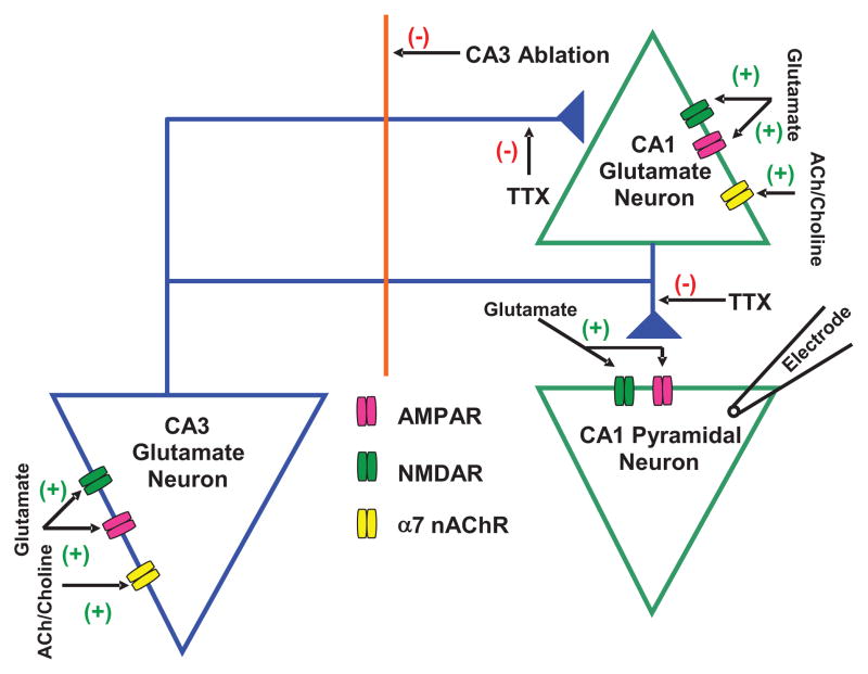
The Schaffer collaterals are among the major glutamatergic inputs to CA1 pyramidal neurons, the primary output of the hippocampus, which also receive sparse recurrent inputs from pyramidal neurons in the CA1 field. Although tonically active α7 nicotinic acetylcholine receptors (nAChRs) have been shown to sustain spontaneous glutamate transmission to CA1 pyramidal neurons in hippocampal slices under resting conditions, it remains to be determined whether these receptors are those expressed by CA3 or CA1 pyramidal neurons. This study was designed to test the hypothesis that the CA3 field of the hippocampus is a significant source of α7 nAChR-sustained glutamatergic transmission to CA1 pyramidal neurons. To this end, spontaneous excitatory postsynaptic currents (EPSCs) were recorded from CA1 and CA3 pyramidal neurons in intact rat hippocampal slices as well as from CA1 pyramidal neurons in CA3-ablated slices under various experimental conditions. Surgical removal of the CA3 region from the slices reduced by 20% the frequency of spontaneous EPSCs recorded from CA1 pyramidal neurons. This finding is in agreement with the concept that the CA3 field contributes significantly to the maintenance of spontaneous glutamatergic synaptic activity in CA1 pyramidal neurons. In addition, the α7 nAChR antagonist methyllycaconitine (MLA, 10nM) reduced the frequency of spontaneous EPSCs recorded from CA1 pyramidal neurons by 30% in intact slices and 12% in CA3-ablated slices. Taken together, these results demonstrate that tonically active α7 nAChRs in CA3 pyramidal neurons and/or in the Mossy fibers that innervate the CA3 pyramidal neurons do in fact contribute to the maintenance of glutamatergic synaptic activity in CA1 pyramidal neurons of hippocampal slices under resting conditions.

Keywords: 2-amino-5-phosphonovaleric acid; 6-cyano-7-nitroquinoxaline-2,3-dione; ACSF; APV; CNQX; EPSC; Hippocampus; IPSC; MLA; Methyllycaconitine; Pyramidal neuron; TTX; Tetrodotoxin; artificial cerebrospinal fluid; excitatory postsynaptic current; inhibitory postsynaptic current; methyllycaconitine; nAChR; nicotinic acetylcholine receptor; tetrodotoxin; time constant of decay; α7 nAChR; τ(d).


Conflict of interest statement

Conflict of Interest

The authors report no conflict of interest.

Figures

Figure 1
Figure 1. Effect of CA3 ablation on the effects of MLA and TTX on the frequency of spontaneous EPSCs in CA1 pyramidal neurons
A. Images of hippocampal slices representing the intact and CA3-ablated preparation. B. Sample traces of spontaneous EPSCs recorded from CA1 pyramidal neurons of 30-day-old rats in intact and CA3-ablated slices. C. Comparison of spontaneous EPSCs recorded from CA1 pyramidal neurons under various conditions. Graph shows mean ± S.E.M frequency of EPSCs from intact slices (ACSF) and CA3-ablated slices under control (ACSF), 15-min superfusion with MLA (1 nM), 15-min superfusion with MLA (10 nM), and 1-h incubation followed by superfusion with 200 nM TTX. Data were obtained from ten neurons from seven rats for intact slices, twelve neurons from seven rats in CA3-ablated slices (control and MLA), and eight neurons from five rats for TTX. # p < 0.001 compared to intact slices by one-way ANOVA followed by Dunnett post-hoc test; ** p < 0.01 compared to ACSF in CA3-ablated slices by one-way ANOVA followed by Dunnett post-hoc test.
Figure 2
Figure 2. Effect of pharmacological agents on spontaneous EPSCs in CA3 pyramidal neurons in intact hippocampal slices
A. Sample recordings of spontaneous EPSCs recorded from CA3 pyramidal neurons of 30-day-old rats at −70 mV under control (first trace) and in presence of α7 nAChR antagonist MLA (10 nM) for 15 min (third trace). The second trace shows EPSCs at an expanded time scale. B. Scatter plot showing effect of superfusion of MLA (10 nM) was derived from averaging the number of events in 30-s intervals. Results are presented as mean ± S.E.M.. Baseline rates were recorded for 5 min before bath application of MLA (thick black bar). Plot represents data from eight neurons from four rats. C. Mean frequency of EPSCs in control condition, 15 min superfusion with ACSF containing glutamate receptor antagonists APV (50 μM) and CNQX (10 μM) and 15 min superfusion with ACSF containing 10 nM MLA. Graph and error bars represent mean and S.E.M., respectively, of data obtained from fourteen neurons from nine rats for control, five neurons from three rats for APV+CNQX, and eight neurons from four rats for MLA. (** p < 0.01 by paired t-test).
Figure 3
Figure 3. Schematic representation of glutamatergic transmission to CA1 pyramidal neuron
Proposed model shows tonic regulation of glutamatergic input to CA1 pyramidal neurons by α7 nAChRs located on CA3 and CA1 pyramidal neurons. Spontaneous EPSCs were recorded from CA1 pyramidal neurons that received glutamate inputs from CA3 and CA1 pyramidal neurons. The α7 nAChR is reported to be present on CA3 and CA1 pyramidal neurons. The decrease in EPSC frequency by CA3 ablation suggests that CA3 pyramidal neurons regulate the excitability of CA1 pyramidal neurons. The magnitude of effect of MLA (10 nM) on EPSCs was larger in intact slices compared to that in CA3-ablated slices, which suggests that endogenous ACh or choline activates α7 nAChRs located on both CA3 and CA1 regions and controls glutamate activity in CA1 pyramidal neurons. (+) represents activation and (−) represents inhibition in the neurocircuitry.

References

    1. Albuquerque EX, Pereira EFR, Alkondon M, Rogers SW. Mammalian nicotinic acetylcholine receptors: from structure to function. Physiol Rev. 2009;89:73–120. - PMC - PubMed
    1. Alkondon M, Pereira EFR, Eisenberg HM, Kajii Y, Schwarcz R, Albuquerque EX. Age dependency of inhibition of α7 nicotinic receptors and tonically active N-methyl-D-aspartate receptors by endogenously produced kynurenic acid in the brain. J Pharmacol Exp Ther. 2011;337:572–582. - PMC - PubMed
    1. Bekkers JM, Stevens CF. Presynaptic mechanism for long-term potentiation in the hippocampus. Nature. 1990;1990(346):724–729. - PubMed
    1. Banerjee J, Alkondon M, Pereira EF, Albuquerque EX. Regulation of GABAergic inputs to CA1 pyramidal neurons by nicotinic receptors and kynurenic acid. J Pharmacol Exp Ther. 2012a;341:500–509. - PMC - PubMed
    1. Banerjee J, Alkondon M, Albuquerque EX. Kynurenic acid inhibits glutamatergic transmission to CA1 pyramidal neurons via α7 nAChR-dependent and -independent mechanisms. Biochem Pharmacol. 2012b;84:1078–1087. - PubMed

Publication types

MeSH terms

LinkOut - more resources