Review
. 2013 Nov;145(5):954-65.
doi: 10.1053/j.gastro.2013.08.038. Epub 2013 Aug 21.

The spectrum of achalasia: lessons from studies of pathophysiology and high-resolution manometry

Affiliations
Review

The spectrum of achalasia: lessons from studies of pathophysiology and high-resolution manometry

Peter J Kahrilas et al. Gastroenterology. 2013 Nov.

Abstract

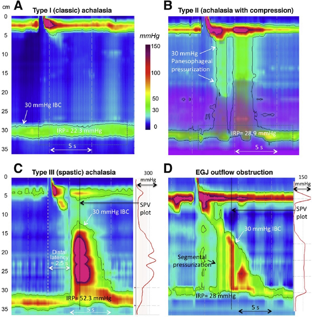
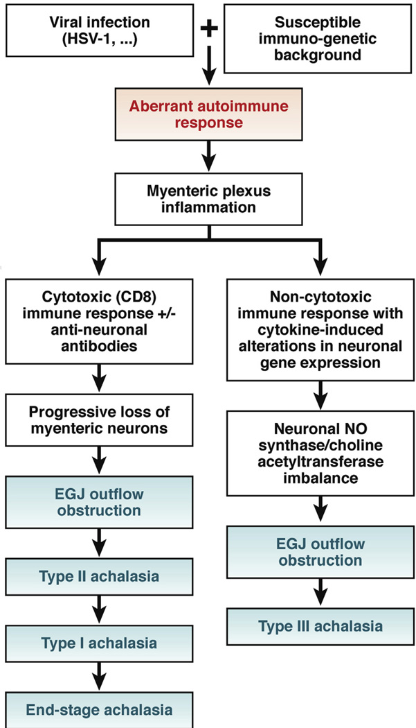
High-resolution manometry and recently described analysis algorithms, summarized in the Chicago Classification, have increased the recognition of achalasia. It has become apparent that the cardinal feature of achalasia, impaired lower esophageal sphincter relaxation, can occur in several disease phenotypes: without peristalsis, with premature (spastic) distal esophageal contractions, with panesophageal pressurization, or with peristalsis. Any of these phenotypes could indicate achalasia; however, without a disease-specific biomarker, no manometric pattern is absolutely specific. Laboratory studies indicate that achalasia is an autoimmune disease in which esophageal myenteric neurons are attacked in a cell-mediated and antibody-mediated immune response against an uncertain antigen. This autoimmune response could be related to infection of genetically predisposed subjects with herpes simplex virus 1, although there is substantial heterogeneity among patients. At one end of the spectrum is complete aganglionosis in patients with end-stage or fulminant disease. At the opposite extreme is type III (spastic) achalasia, which has no demonstrated neuronal loss but only impaired inhibitory postganglionic neuron function; it is often associated with accentuated contractility and could be mediated by cytokine-induced alterations in gene expression. Distinct from these extremes is progressive plexopathy, which likely arises from achalasia with preserved peristalsis and then develops into type II achalasia and then type I achalasia. Variations in its extent and rate of progression are likely related to the intensity of the cytotoxic T-cell assault on the myenteric plexus. Moving forward, we need to integrate the knowledge we have gained into treatment paradigms that are specific for individual phenotypes of achalasia and away from the one-size-fits-all approach.

Keywords: Achalasia; Dysphagia; EGJ; EPT; Esophageal Motility; HSV-1; IRP; LES; Pathogenesis; TLESR; esophageal pressure topography; esophagogastric junction; herpes simplex virus 1; integrated relaxation pressure; lower esophageal sphincter; transient lower esophageal sphincter relaxation.


Conflict of interest statement

Conflicts of interest

The authors disclose no conflicts.

Figures

Figure 1
Figure 1
Averaged pressure topography plot of peristalsis created by superimposing 10 successive swallows of a single healthy subject to illustrate the stereotypic features of peristalsis. The peristaltic contraction is composed of 3 contractile segments (S1, S2, and S3) and the LES contraction separated by 3 relative pressure troughs: proximal (P), middle (M), and distal (D). The latency of contraction at each esophageal locus is a function of centrally mediated deglutitive inhibition (1) persisting for the duration of the S1, centrally programmed contraction and (2) the balance of postganglionic excitation (depolarization) and inhibition (hyperpolarization) from cholinergic and nitrergic myenteric neurons, respectively. LES relaxation (3) is a function of the same myenteric plexus influences, albeit in this case superimposed on a resting myogenic contraction not present in the remainder of the esophagus.
Figure 2
Figure 2
With the adoption of HRM and EPT, 3 distinct subtypes of achalasia were defined using pressure topography metrics. All have impaired EGJ relaxation and absent peristalsis, but the differentiating features are in the patterns of esophageal pressurization; type I has 100% failed peristalsis (aperistalsis), type II exhibits panesophageal pressurization ≥30 mm Hg in ≥20% swallows, and type III exhibits 2 or more premature (spastic) contractions. Note that the impedance data in the type II patient (purple overlay) indicates fluid retention in the esophagus. D is from a patient who did not meet the criterion of absent peristalsis but clearly shows impaired EGJ relaxation with segmental pressurization between the peristaltic contraction and the EGJ; hence, EGJ outflow obstruction was diagnosed. The differentiation between a spastic contraction and segmental pressurization is evident from the spatial pressure variation (SPV) plots to the right of C and D, indicating the top-to-bottom intraluminal pressure profile at the time of the black vertical dotted line. With segmental pressurization, the bolus is trapped between high-pressure zones. The patient in D was treated with a Heller myotomy with relief of dysphagia. Her postmyotomy study showed weak peristalsis. Several recent reports have shown differences in the prognostic value of these achalasia subtypes, supporting the classification scheme
Figure 3
Figure 3
Proposed model of immunotoxicity patterns leading to distinct achalasia phenotypes. Although other possibilities have been proposed, the model assumes that spastic achalasia is an immune-mediated functional aberration that is not part of the progression to aganglionosis. It is also proposed that the specifics of the antibody response vary among patients and may participate in the cytotoxic attack and/or result in cytokine-mediated alterations in neuron function. The pathogenesis of muscle hypertrophy is unknown, but this is consistently observed to accompany hypercontractility.
Figure 4
Figure 4
Incomplete TLESR in a patient with type III achalasia. Similar to patients without achalasia, patients with achalasia frequently exhibit TLESR in the course of a manometry protocol after performing a series of swallows in the supine position and then sitting up. In the case of achalasia, the resultant motor response, shown in this figure, contains all elements of a TLESR (prolonged esophageal shortening with concomitant inhibition of the crural diaphragm and strong after-contraction) except LES relaxation. In fact, the LES paradoxically contracts, consistent with the hypothesis that type III achalasia is a consequence of immune-mediated dysregulation of postganglionic inhibitory nerve function, often with an accentuation of excitatory postganglionic nerve function.
Figure 5
Figure 5
A patient with type II achalasia before and after laparoscopic Heller myotomy. After myotomy, a remnant of peristalsis with normal latency and likely extending to the proximal margin of the myotomy is clearly evident. This is a weak contraction based on the large break in the isobaric contour (IBC) between the first and second segments.

References

    1. Lendrum FC. Anatomic features of the cardiac orifice of the stomach with special reference to cardiospasm. Arch Intern Med. 1937;59:474–451.
    1. Pandolfino JE, Kahrilas PJ. The second American Gastroenterological Association technical review on the clinical use of esophageal manometry. Gastroenterology. 2005;128:209–229. - PubMed
    1. Pandolfino JE, Kahrilas PJ. American Gastroenterological Association medical position statement: clinical use of esophageal manometry. Gastroenterology. 2005;128:207–208. - PubMed
    1. Bredenoord AJ, Fox M, Kahrilas PJ, et al. Chicago Classification criteria of esophageal motility disorders defined in high resolution esophageal pressure topography. Neurogastroenterol Motil. 2012;24(Suppl 1):57–65. - PMC - PubMed
    1. Kahrilas PJ, Kishk SM, Helm JF, et al. A comparison of pseudoachalasia and achalasia. Am J Med. 1987;82:439–446. - PubMed

Publication types