. 2013 Nov;195(21):4844-53.
doi: 10.1128/JB.00671-13. Epub 2013 Aug 23.

Novel feature of Mycobacterium avium subsp. paratuberculosis, highlighted by characterization of the heparin-binding hemagglutinin adhesin

Affiliations

Novel feature of Mycobacterium avium subsp. paratuberculosis, highlighted by characterization of the heparin-binding hemagglutinin adhesin

Louise H Lefrancois et al. J Bacteriol. 2013 Nov.

Abstract

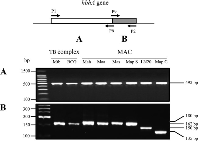
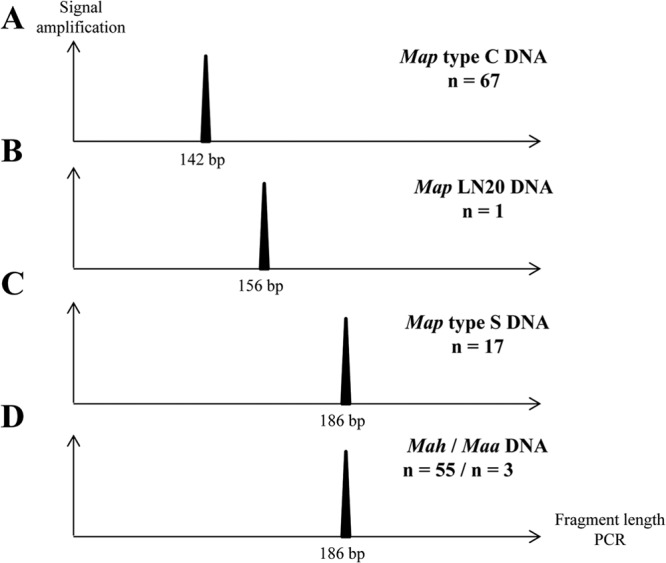
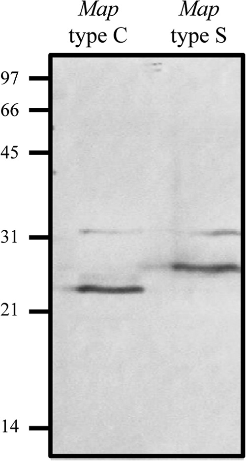
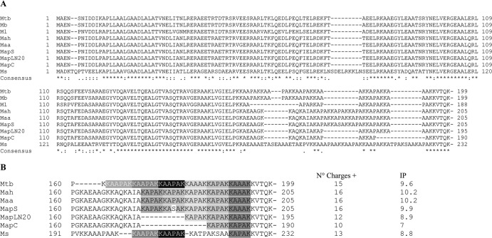
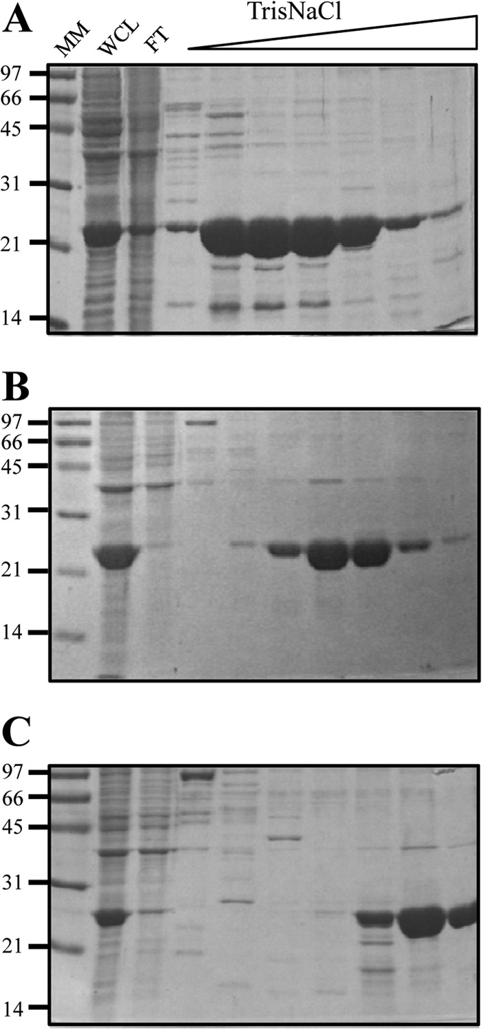
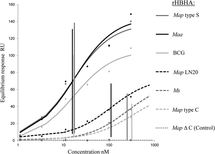
Mycobacterium avium subsp. paratuberculosis comprises two genotypically defined groups, known as the cattle (C) and sheep (S) groups. Recent studies have reported phenotypic differences between M. avium subsp. paratuberculosis groups C and S, including growth rates, infectivity for macrophages, and iron metabolism. In this study, we investigated the genotypes and biological properties of the virulence factor heparin-binding hemagglutinin adhesin (HBHA) for both groups. In Mycobacterium tuberculosis, HBHA is a major adhesin involved in mycobacterium-host interactions and extrapulmonary dissemination of infection. To investigate HBHA in M. avium subsp. paratuberculosis, we studied hbhA polymorphisms by fragment analysis using the GeneMapper technology across a large collection of isolates genotyped by mycobacterial interspersed repetitive-unit-variable-number tandem-repeat (MIRU-VNTR) and IS900 restriction fragment length polymorphism (RFLP-IS900) analyses. Furthermore, we analyzed the structure-function relationships of recombinant HBHA proteins of types C and S by heparin-Sepharose chromatography and surface plasmon resonance (SPR) analyses. In silico analysis revealed two forms of HBHA, corresponding to the prototype genomes for the C and S types of M. avium subsp. paratuberculosis. This observation was confirmed using GeneMapper on 85 M. avium subsp. paratuberculosis strains, including 67 strains of type C and 18 strains of type S. We found that HBHAs from all type C strains contain a short C-terminal domain, while those of type S present a long C-terminal domain, similar to that produced by Mycobacterium avium subsp. avium. The purification of recombinant HBHA from M. avium subsp. paratuberculosis of both types by heparin-Sepharose chromatography highlighted a correlation between their affinities for heparin and the lengths of their C-terminal domains, which was confirmed by SPR analysis. Thus, types C and S of M. avium subsp. paratuberculosis may be distinguished by the types of HBHA they produce, which differ in size and adherence properties, thereby providing new evidence that strengthens the genotypic differences between the C and S types of M. avium subsp. paratuberculosis.


Figures

Fig 1
Fig 1
Interspecies and intraspecies variability of the hbhA gene. Shown are PCR amplification products of the DNA coding for the N-terminal domain (A) and the C-terminal domain (B) of HBHA, obtained by using genomic DNA of M. tuberculosis (Mtb), BCG, M. avium subsp. hominissuis (Mah) (1 sample representative of 28 strains analyzed), M. avium subsp. avium (Maa), M. avium subsp. silvaticum (Mas), M. avium subsp. paratuberculosis type S (Map S) (1 sample representative of 17 strains analyzed), M. avium subsp. paratuberculosis strain LN20, and M. avium subsp. paratuberculosis type C (Map C) (1 sample representative of 34 strains analyzed). The arrows in the diagram at the top indicate the positions and directions of primers P1, P2, P6, and P9, the sequences of which were deduced from the HBHA-encoding gene of M. tuberculosis. Positions on a 100-bp ladder (Promega) (A) and a low-molecular-weight DNA ladder (New England BioLabs) (B), used as molecular weight markers, are indicated on the left. The sizes of the PCR products are indicated on the right.
Fig 2
Fig 2
Distinct hbhA genes within the subspecies M. avium subsp. paratuberculosis were examined with a large collection of isolates. The gene coding for the C-terminal domain of HBHA was amplified by PCR on genomic DNA using primers labeled at their 5′ ends with the fluorophore 6-carboxyfluorescein. Results were visualized using ABI GeneMapper software, version 4.0, with 600 LIZ size standards (Applied Biosystems, USA). The expected sizes of the PCR products are 142 bp with M. avium subsp. paratuberculosis type C genomic DNA (A), 156 bp with M. avium subsp. paratuberculosis LN20 genomic DNA (B), and 186 bp with M. avium subsp. paratuberculosis type S (C), M. avium subsp. avium, or M. avium subsp. hominissuis (D) genomic DNA. This analysis was performed on 67 strains of M. avium subsp. paratuberculosis type C, 18 strains of M. avium subsp. paratuberculosis type S, including strain LN20, 55 M. avium subsp. hominissuis strains, and 3 strains of M. avium subsp. avium. n, number of strains analyzed.
Fig 3
Fig 3
Immunoblot analysis of native HBHAs produced by M. avium subsp. paratuberculosis types C and S. Whole-cell lysates of M. avium subsp. paratuberculosis type C (left lane) and type S (right lane) strains were separated by 12% SDS-PAGE and then electroblotted onto nitrocellulose membranes. The blots were incubated with the anti-HBHA monoclonal antibody 3921E4. The positions of molecular mass markers (in kilodaltons) are indicated on the left.
Fig 4
Fig 4
Alignment of HBHA sequences from different mycobacterial species. (A) The amino acid sequences of M. tuberculosis HBHA were aligned with those of its homologues from other mycobacterial species by using the ClustalW program with the BLOSUM64 matrix allowing gaps. Asterisks represent identical residues; colons, conserved substitutions; periods, semiconserved substitutions. (B) Multiple alignment of the C-terminal heparin-binding domain of HBHA. The lysine-rich hexa-repeats (R1) (white letters on a gray ground) and penta-repeats (R2) (black letters on a gray ground) defined by Lebrun et al. (25) are indicated. The numbers of charges distributed throughout the C-terminal domains of different HBHA homologues (amino acids 158 to 232) and their isoelectric points (IP) are given to the right of the sequences. Mb, M. bovis; Ml, M. leprae; Ms, M. smegmatis; C, consensus.
Fig 5
Fig 5
Differences between rHBHA proteins from M. avium subsp. paratuberculosis type C, LN20, and type S in the strength of binding to heparin-Sepharose. Lysates of E. coli expressing recombinant HBHA of M. avium subsp. paratuberculosis type C (A), M. avium subsp. paratuberculosis LN20 (B), and M. avium subsp. paratuberculosis type S (C) were sonicated. The soluble material was subjected to heparin-Sepharose chromatography and was eluted using a 0 to 1 M NaCl gradient. SDS-PAGE analyses were performed on whole-cell lysates (WCL), flowthrough (FT), and elution fractions of rHBHA. The positions of molecular mass markers (MM), expressed in kilodaltons, are indicated on the left.
Fig 6
Fig 6
Quantification by SPR analysis of the heparin-binding activities of the rHBHA proteins of M. avium subsp. paratuberculosis type C, LN20, and type S. SPR analysis of the binding of recombinant HBHA to heparin was performed on a BIAcore T100 system (GE Healthcare). Biotinylated heparin was immobilized on a streptavidin-coated CM4 sensor chip. Recombinant HBHA proteins purified by heparin-Sepharose chromatography were injected over a range of concentrations at a flow rate of 30 μl min−1. Recombinant mapS-HBHAΔCter was used as a negative control. KD were determined by the equilibrium method. The KD results (nM) are indicated at the intersections of the vertical lines with the concentration axis.
Fig 7
Fig 7
Reactivity of a serum panel with the rHBHA proteins from M. avium subsp. paratuberculosis strains of types C and S. ELISA were performed with plates coated with either PPDj, recombinant BCG HBHA, mapS-HBHA, mapS-HBHAΔCter, or mapC-HBHA. The panel of sera tested included a M. avium subsp. paratuberculosis-negative commercial antiserum (lane 1), a M. avium subsp. paratuberculosis-positive commercial serum (lane 2), and sera obtained from ruminants infected by M. avium subsp. paratuberculosis type C (lane 3) or type S (lane 4). Each serum sample was diluted at 1:100 in 50 μl PBS/T/G and was incubated for 2.5 h at 37°C. The plates were then washed and were incubated for 90 min at 37°C with 50 μl peroxidase-conjugated anti-ruminant IgG diluted at 1:600 in PBS/T/G. After washing, the plates were developed with 50 μl of a peroxidase substrate for 30 min at 37°C. The reaction was stopped with 50 μl 1 N HCl, and the plates were read photometrically at 490 nm. The results shown are averages for triplicate samples from one experiment representative of three independent experiments. Statistical analysis was performed with Tukey's multiple comparison tests. **, P < 0.001; ns, not significant (P > 0.05).

References

    1. Alexander DC, Turenne CY, Behr MA. 2009. Insertion and deletion events that define the pathogen Mycobacterium avium subsp. paratuberculosis. J. Bacteriol. 191:1018–1025 - PMC - PubMed
    1. Turenne CY, Collins DM, Alexander DC, Behr MA. 2008. Mycobacterium avium subsp. paratuberculosis and M. avium subsp. avium are independently evolved pathogenic clones of a much broader group of M. avium organisms. J. Bacteriol. 190:2479–2487 - PMC - PubMed
    1. Biet F, Sevilla IA, Cochard T, Lefrancois LH, Garrido JM, Heron I, Juste RA, McLuckie J, Thibault VC, Supply P, Collins DM, Behr MA, Stevenson K. 2012. Inter- and intra-subtype genotypic differences that differentiate Mycobacterium avium subspecies paratuberculosis strains. BMC Microbiol. 12:264.10.1186/1471-2180-12-264 - DOI - PMC - PubMed
    1. Castellanos E, Romero B, Rodriguez S, de Juan L, Bezos J, Mateos A, Dominguez L, Aranaz A. 2010. Molecular characterization of Mycobacterium avium subspecies paratuberculosis types II and III isolates by a combination of MIRU-VNTR loci. Vet. Microbiol. 144:118–126 - PubMed
    1. Castellanos E, Aranaz A, Gould KA, Linedale R, Stevenson K, Alvarez J, Dominguez L, de Juan L, Hinds J, Bull TJ. 2009. Discovery of stable and variable differences in the Mycobacterium avium subsp. paratuberculosis type I, II, and III genomes by pan-genome microarray analysis. Appl. Environ. Microbiol. 75:676–686 - PMC - PubMed

Publication types

MeSH terms

LinkOut - more resources